Antiskin Cancer and Antioxidant Activities of Formulated Agar from Brown Seaweed Laminaria digitata (Hudson) in Dimethyl Benzanthracene-Induced ...
←
→
Page content transcription
If your browser does not render page correctly, please read the page content below

Hindawi
International Journal of Polymer Science
Volume 2021, Article ID 9930777, 12 pages
https://doi.org/10.1155/2021/9930777

Research Article
Antiskin Cancer and Antioxidant Activities of Formulated
Agar from Brown Seaweed Laminaria digitata (Hudson) in
Dimethyl Benzanthracene-Induced Swiss Albino Mice

 Jeneesha George,1 A. Thabitha,1 N. Vignesh,1 V. Manigandan,1 R. Saravanan ,2
 Ghaji Daradkeh,3 and M. Walid Qoronfleh 4
 1
 Department of Medical Biotechnology, Chettinad Academy of Research and Education (Deemed to be University), Kelambakkam,
 603 103 Tamil Nadu, India
 2
 Native Medicine and Marine Pharmacology Laboratory, Faculty of Allied Health Sciences, Chettinad Academy of Research and
 Education (Deemed to be University), Kelambakkam, 603 103 Tamil Nadu, India
 3
 Department Nutrition, Hamad Medical Corporation–Al-Khor Branch, Doha, Qatar
 4
 Research & Policy Department, World Innovation Summit for Health (WISH), Qatar Foundation, P.O. Box 5825, Doha, Qatar

 Correspondence should be addressed to R. Saravanan; saran_prp@yahoo.com and M. Walid Qoronfleh; walidq@yahoo.com

 Received 23 March 2021; Revised 29 April 2021; Accepted 3 June 2021; Published 16 June 2021

 Academic Editor: Antonio Caggiano

 Copyright © 2021 Jeneesha George et al. This is an open access article distributed under the Creative Commons Attribution
 License, which permits unrestricted use, distribution, and reproduction in any medium, provided the original work is
 properly cited.

 This study explores the antiskin cancer effect of formulated agar (FA) from Laminaria digitata on dimethyl benzanthracene-
 (DMBA-) induced skin cancer mice. The agar was extracted and formulated (emulgel), and FA was biochemically characterized.
 The in vitro cytotoxicity of FA was tested using NTT 3T3 mice fibroblast cells. The mice were divided into 5 groups: group 1
 served as control mice, group 2 mice were considered as DMBA-induced cancer control, group 3 mice were FA pretreated (low
 dose) + DMBA-induced mice, group 4 mice were FA pretreated (high dose) + DMBA-induced mice, and group 5 were positive
 control + DMBA-induced mice. The behaviour and biochemical markers of cancer were significantly decreased in group 2
 (DMBA-induced) mice, which were brought to near normalcy by FA pretreated mice (groups 3 and 4). The levels of p53 and
 keratin were significantly elevated in group 2 mice and these levels were decreased in 3 and 4 mice as well. The histopathological
 examination of DMBA-induced mice was shown degenerated cervical patches in the skin, cirrhosis in liver, oedema in the renal
 tissue, and swollen and damage in cardiac tissue, which were reduced for the mice applied with FA. This confirms that FA
 pretreatment offered potential antiskin cancer property.

1. Introduction cutaneous melanomas off the last few years. Despite lack of
 complete prevalence data, different cancer athenaeum in
Worldwide, 18.1 million new cases and 9.6 million deaths are India announced an increasing incidence of skin cancer
due to skin cancer [1]. In India, 63% of death is due to non- changing from 0.5 to 2 per 100 000 communities [3, 4].
communicable diseases, and cancer is one of the dominating Surgery and radiation therapy are the standard available
causes, accounting for 9%. These figures will nearly double by treatments for cancer. However, the main drawback of these
2040; skin cancer is almost egregious malignancies among therapies includes damage to normal cells with a high
other common cancers though not ranked among the top proliferate index. Most chemotherapeutics are dispensed
ten common cancers [2]. There has been a continuous methodically and are cytotoxic to normal cells; hence, cancer
increase in the prevalence of skin cancers, especially that of case must undergo considerable morbidity. The traditional-
2 International Journal of Polymer Science

based topical administration of anticancer natural drug is a triethanolamine (5.6 ml) with stirring. This was followed by
fascinating flipside for increasing drug directing and reme- the addition of methylparaben (0.6 g) and propylparaben
dial benefits [5]. (0.07 g) with continuous exhaustive stirring until medium
 The dimethyl Benz (a) anthracene (DMBA) is a polycy- components were homogeneous, then mannitol (17.5 g),
clic aromatic hydrocarbon (PAHs), branded as an effective lysine (0.88 g), and glycerol (8.75 ml) were added; finally,
carcinogen and/or endocrine disruptors. PAHs are widely the gelated agar (100 g) core component of the formulation
distributed in the environment as a result of incomplete com- was added. The mixture was mixed well, and it was stirred
bustion of fossil fuels and other organic molecules and are for 30 min (Magnetic stirrer, PCE Pvt. Ltd. India) until a clear
common contaminants of terrestrial and aquatic ecosystems consistent gel base was obtained. The formulated agar was
[6]. Seaweeds are attractive sources of biomolecules such as stored in an air-tight container under freeze conditions [11].
proteins, polysaccharides, minerals, vitamins, steroids, and
dietary fibers which are profitable resources for nutraceutical 2.4. Characterization of FA. Organoleptic characteristics of
and pharmaceutical applications [7]. The brown seaweeds the FA, such as color, consistency texture, sterility, and homo-
are the largest marine sources for polysaccharide extraction geneity, were assessed by visual examination. Besides, several
(44-53%) globally. Some of the valuable bioactivities revealed physicochemical characteristics were also analyzed [12–16].
by the polysaccharides and their derivatives, both in vivo and The data represented here was for triplicate determinations.
in vitro, include immunomodulatory ability, anticancer, anti-
viral, anticoagulant, and/or antithrombotic properties. They 2.5. FT-IR Spectroscopy of FA. The FT-IR spectroscopic anal-
are also proved to have potent antidislipidaemic, hypoglycae- ysis of FA from brown seaweed was performed using a Bru-
mic antioxidants, antibiotics, and anti-inflammatory effects. ker Alpha instrument (USA) measured in the range of 4000
Previously, we have reported the antioxidant, anticoagulant, to 400 cm-1 at 30 scans at RT. The samples were thoroughly
and mosquitocidal properties of phloroglucinol and some mixed up with KBr, and the spectra were taken [8].
water-soluble polysaccharides from Indian brown seaweeds
[8, 9]. But till now, there is no report on the anticancer
activity of FA from seaweed (IP No. E-101/17997/dated 2.6. Microstructure Studies of FA by Scanning Electronic
01.07.2019). Hence, the present study is aimed at finding out Microscope (SEM). The morphological features of the FA
the antiskin cancer property of formulated agar from brown were studied with a JSM-5600 LV scanning electron micro-
seaweed (L. digitata) against DMBA-induced mice model. scope (JEOL, Tokyo, Japan). The dried sample was mounted
 on a metal stub, and the images were taken at an accelerating
 temperature (200°C, 400°C, 600°C, and 1200°C) with magni-
2. Materials and Methods
 fications 5.00 KX, 2.50X, 2.52 KX, and 1.00 KX [17].
The study was conducted in accordance with the Basic &
Clinical Pharmacology & Toxicology policy for experimental 2.7. MTT Assay of FA. The cell viability of FA was deter-
and clinical studies [9]. mined by MTT assay. Freshly cultured NIT 3T3 mouse fibro-
 blast cell line cells were trypsinized, collected, counted, and
2.1. Seaweed Collection. Brown seaweed (L. digitata) was col- seeded in a 96-well plate. After 24 h incubation, cells were
lected from Kanyakumari, (Lat. 8.0866 N and Long. 77.5544 treated with various concentrations of FA. Then, the cells
E), Tamil Nadu, India, during December 2018–January 2019. were incubated in 5% CO2 at 37°C for 24 h. Later, 100 μl of
The collected brown seaweed species were identified using MTT was added, and incubation was extended for more 4
the Central Marine Fisheries Research Institute (CMFRI) h. Finally, the medium was removed and 100 μl of DMSO
taxonomic manuals and bulletin. The collected samples were were used to solubilize the formazan crystals. The samples
cleaned assiduously with seawater and distilled water and were read at 570 nm (Tecan Multimode Multiwall Plate
finally washed with distilled water. The processed seaweeds Reader, Austria). Percentage cytotoxicity was calculated as
were shadow dried, powdered, and stored at -20°C for described earlier [10].
further study.
2.2. Extraction of Agar. A 10 g of powdered seaweed sample 2.8. In Vivo Anticancer Assay. The skin cancer was experimen-
was transferred to the beaker and mix up with 100 ml of tally induced in female Swiss albino mice following the ethical
high-pure Milli-Q water then autoclaved at 121°C for 1 h clearance obtained (IAEC4/Proposal: 31/A.Lr:13/Dated:
for extraction of water-soluble polysaccharides. Further, the 20.12.18). The animals were divided into 5 groups as men-
water-soluble polysaccharides were filtered through What- tioned in Table 1. Control mice received normal food and
mann No. 1 filter paper to eliminate seaweed debris present water ad libitum. The 5-FU (100 mg/100 ml acetone)-treated
in the extract, and then, it was centrifuged at 5000 rpm for mice served as a positive control. The FA was applied topically
15 min at RT. The filtered extract was kept at RT, and it gets weekly twice 30 min before exposure for 63 days. The physical
gelated. The gelated samples were dried out at 60°C overnight parameters like measurement of tumor size, body weight, and
and stored in -20°C for further experiments [10]. food and water intake of the experimental mice groups were
 measured during the entire experimental period. At the end
2.3. Formulation of Agar (FA). The carbopol 934 was lique- of the experimental period, the mice were sacrificed and the
fied slowly while stirring in 60 ml of Milli-Q water for 1 h to skin, liver, kidney, and heart were dissected, then the organs
evade agglomeration. Carbopol was then neutralized with were stored under -20°C until further use.

International Journal of Polymer Science 3

 Table 1: Experimental design.

Group 1 Normal control mice (normal diet and water)
Group 2 Cancer-induced mice (100 nM DMBA dissolved in 200 μl acetone was applied topically weekly twice for four weeks)
 FA (low dose)—60 mg/ml in 200 μl acetone was applied topically weekly twice for four weeks, 30 min before DMBA
Group 3
 exposure (100 nM DMBA dissolved in 200 μl acetone was applied topically weekly twice)
 FA (high dose)—120 mg/ml in 200 μl acetone was applied topically weekly twice for four weeks, 30 min before DMBA
Group 4
 exposure (100 nM DMBA dissolved in 200 μl acetone was applied topically weekly twice)
 Positive control: 5-FU-100 mg/ml in 100 ml acetone was applied topically weekly twice for four weeks, 30 min before
Group 5
 DMBA exposure (100 nM DMBA dissolved in 200 μl acetone was applied topically weekly twice)

 Table 2: Stability of FA.

 Stability of FA storage conditions
S. no. Parameters/tests 25° C ± 2° C 32° C ± 2° C 40° C ± 2° C
 Months (0–3) Months (0–3) Months (0–3)
1 Color No change in color No change in color No change in color
2 Odor No change No change No change
3 Homogeneity Good Good Good
4 Consistency Smooth Smooth Smooth
5 pH 6 6 6
6 Viscosity (m2/s) 0.385 0.382 0.381
 No microbial growth is observed No microbial growth is observed No microbial growth is observed
7 Microbial load (bacteria)
 at 24, 48, and 72 h at 24, 48, and 72 h at 24, 48, and 72 h
8 Sterility No microbial contamination No microbial contamination No microbial contamination
9 Vibrational test No phase of separation No phase of separation No phase of separation
10 Centrifugation test No phase of separation No phase of separation No phase of separation
Time period (three months).

2.9. Measurement of Tumor in Experimental Mice. Measure- Table 3: Texture analysis of FA.
ment of tumor development and the effect of FA on tumor
development was evaluated by using Vernier caliper. The Moisture (%) 48:28 + 0:21
length, width, and height of the tumor were measured for Hardness (g) 243:43 ± 0:60
during the experimental days. Adhesiveness (N/mm) 61:50 ± 0:40
2.10. p53 Protein Level Measurement. Determination of p53 Cohesiveness 0:952 ± 0:0:12
protein level in mice skin tissues and isolated cells were esti- Springiness (mm) 3:520 ± 0:012
mated by using mouse p53 ELISA kit. Gumminess (N) 3:699 ± 0:10
2.11. Isolation and Determination of Primary Keratinocytes.
The epidermis of experimental mice were isolated by incuba- Both soluble and covalently crosslinked insoluble collagen
tion in dispase for up to 72 h that was cut subsequently into fractions were obtained by successive treatment of the skin
small pieces with sterilized scissors then incubated for 30 with 10 volumes of 5 types of a buffer. The collagen extracted
minutes in 0.025% DNase in DMEM at RT with gentle agita- with buffers A and B was categorized as soluble collagen and
tion. Afterwards, the mixture was filtered through a 100 mM that extracted with buffers C, D, and E were categorized as
cell strainer, and the cells were pelleted by centrifugation. insoluble collagen [19, 20].
Cells were resuspended in 3 : 1 defined keratinocyte serum-
free medium containing appropriate growth supplements, 2.13. Enzymatic and Nonenzymatic Assessments. The levels of
seeded on plates previously coated with collagen, then char- reduced glutathione (GSH), superoxide dismutase (SOD),
acterized by spectroscopy [18]. catalase (CAT), and glutathione peroxidase (GPx) were mea-
 sured in mice skin tissues determined by using commercial
2.12. Collagen and Elastin Contents Determination. The kits from Sigma, USA.
amount of collagen and elastin in the dermis was measured
using a Sircol assay kit based on the fact that the sirius red 2.14. Histopathology. The skin, liver, kidney, and heart of
dye binds to the side chains of the amino acids in collagen. each group of mice were fixed in 10% formalin for
4 International Journal of Polymer Science

 100 m 20 m

 10 m 2 m

Figure 1: SEM of FA from brown seaweed (The FA (2-10 μm) exhibits regular, layered, patched and smooth, and flat continuous sheet
appearance).

haematoxylin and eosin staining (H&E). Serial sections of 20 yield difference is largely due to the species, season period,
μm thickness were cut and mounted on slides and stained. and geographical location. A cost-effective hot water
The fixed samples were trimmed and dissected in the extraction method with comparable yield was successfully
crosssections and were processed. The samples were fixed developed for agar extraction and its formulation to
in stretches of formalin and alcoholic formalin, 95% develop antiskin cancer property of FA from brown sea-
alcohol, absolute alcohol, xylene, and liquid paraffin wax. weed (L. digitata) [23]. Screening for new drugs from natu-
Crosssections were selected from the three plates per ral sources mainly from marine environment to discover
sample. The morphological changes were detected under anticancer activities is valuable as they are likely to be more
the microscope [21]. safe, robust, and effective. Since agents of synthetic origin
 may have various adverse effects. Natural products have been
2.15. Statistical Analysis. All the statistical analyses were pre- used as remedies for skin cancer prevention and other tradi-
sented by using software using the SPSS software (SPSS, IBM, tional treatments as well. Sulfated polysaccharides (SP) from
version 20.0). Each test is performed from all animals, and all marine sources were investigated to have good antioxidant,
the results were expressed as the means plus-minus their cor- antitumor, and immunomodulating activities. It was sug-
responding standard deviation (SD). A value of p < 0:05 was gested that, next to the pharmacological activities of SP, their
measured to specify a significant difference between groups. physicochemical properties, like good water solubility,
 thickening, and solidity over a wide pH range, offer extra
3. Results and Discussion benediction for their use as a preservative in topical applica-
 tion products [10].
3.1. Extraction Yield and FA. The agar yield from L. digitata
was found to be 40%. This is considered the maximum yield 3.2. Characterization of FA. Stability studies of FA were
when compared to a previous report [22], which indicated essential to ensure product quality, efficacy, and safety. Thus,
that the agar yield from brown seaweeds could vary. The these studies tend to give the development and improvement
International Journal of Polymer Science 5

 1.0

 0.5

 Transmittance (%)
 0.0 Standard agar

 1.0

 0.5 Formulated agar

 1852
 2362

 908
 2927

 1075
 1408
 0.0

 1639
 –0.5
 4000 3500 3000 2500 2000 1500 1000 500
 Wavelength (cm–1)

 Figure 2: FT-IR spectrum of formulated agar.

 Cell viability
 0.8
 0.7
 0.6
 OD at 570 nm

 0.5
 0.4
 0.3
 0.2
 0.1
 0
 Control 50 100 200 250 300
 Concentration ( g/ml)
 (a)

 Control 50 g/ml 100 g/ml 200 g/ml 300 g/ml
 (b)

Figure 3: MTT assay of FA (Cells treated above 300 μg/ml concentration showed a significant (p < 0:05) death of cell; compared with control
(a, b)).

of formulations, creating the product validity and observing eters such as no color and odor change, good consistency,
its physical, chemical, and microbiological characteristics. and homogeneity were smooth, and there was no microbial
The FA extracted from the L. digitata is slightly soluble in contamination. All these features indicate the sterility of FA
distilled water and its dispersal acquiesce a light brown color, in Table 2. The pH value of FA remains substantial during
slick solution. The FA was virtually insoluble in polar and storage which was considered adequate to avoid the risk of
nonpolar solvents like acetone, chloroform, and ethanol. Sta- irritation upon application to the skin. Hence, there was no
bility may be affected by environmental factors such as pH, change in the pH; the FA gel could be suitable for topical
light, temperature, air, and transport which can induce harsh application. Centrifugation tests and vibrational tests are
damage on the product constituents. The FA stability param- the main tools for estimating and predicting the shelf life of

6 International Journal of Polymer Science

 25.00 25.00

 20.00 20.00

 Mean water consumed (g)
 Mean food consumed (g)

 15.00 15.00

 10.00 19.44
 10.00 19.00
 19.89
 18.89 18.22 18.67 18.33
 17.78 17.33 16.89

 5.00 5.00

 .00 .00
 Group I Group II Group III Group IV Group V Group I Group II Group III Group IV Group V
 Group Group
 Error bars: +/– 2 SE Error bars: +/– 2 SE
 Experimental groups Experimental groups
 (a) (b)

Figure 4: (a, b) Food and water intake of experimental mice (DMBA-induced mice significantly (p < 0:05) reduced the food and water intake;
FA-treated mice significantly (p < 0:05) increased the food and water intake).

the formulated product. From our data, we found that the FA 40.00
does not show any indication of phase separation during the
study period, which are consistent with the findings of
Andrade et al. [24]. 30.00
 Mean body weight (g)

3.3. Texture Analysis of FA. The texture analyses (moisture,
hardness, adhesiveness, cohesiveness, and springiness) of 20.00
FA were described in Table 3. The texture analysis results
 30.94
of FA are considered high when compared to the results of 29.21 27.76 29.57
 26.96
Reshma [23], who had extracted the agar by using hot water 10.00
from brown seaweed. The difference in the texture numbers is
likely due to the method of extraction and formulated nature
of the sample. Texture analysis is one of the most essential
 .00
unique inspection criteria for pharmaceuticals since it con- Group I Group II Group III Group IV Group V
tributes important identification and exclamation knowledge.
 Experimental groups
Characteristics with small intraclass variations and large
interclass variations of differing textures are an essential prod- Figure 5: Body weight of experimental mice (DMBA-induced mice
uct requirement. Here, we explored characteristics focused on significantly (p < 0:05) reduced the body weight; FA-treated mice
those techniques that have been a standard in texture analyses significantly (p < 0:05) increased the body weight).
or applied for repeated material examination.

3.4. Phytochemical Composition of FA. The phytochemical
composition of FA includes crude protein amount (3.79%), physiological conditions, various processing techniques,
total carbohydrates (12.33%), and element composition com- and methods of mineralization. The macrominerals for
prising of Ca (1.5%), P (0.18%), N (2.74%), K (2.99%), Mn example K, Ca, Na, and Mg are present in marine algae at
(16.50 ppm), Zn (50.40 ppm), Fe (30.0 ppm), Cu (75.41 very high levels than in land sources. Further, the level of
ppm), and Co (3.00 ppm), respectively. The element contents Fe and Cu were reported to be higher in seaweeds than spin-
of FA were somewhat high when compared to the previous ach and meat. The polysaccharides are major components of
report of Fleurence et al. [25]. The observed difference in brown seaweed, accounting for 40% of the dry weight. The
the mineral contents is owing to species difference and hot water extract of agar biomass is loaded in low molecular
method selection for quantification. The mineral composi- weight, water-soluble polysaccharides, (WSC) and insoluble
tion of FA grants antioxidant properties which could be in aqueous medium [10].
partially responsible for its anticancer effect. Mineral compo-
sition typically is alerted by unusual environmental and 3.5. Structural Characterization

International Journal of Polymer Science 7

 10.00

 12.50

 8.00

 10.00
 Mean tumor length

 Mean tumor width
 6.00
 7.50

 4.00
 5.00

 2.00
 2.50

 .00 .00

 1 2 3 4 5 6 7 8 9 1 2 3 4 5 6 7 8 9
 Week Week
 Group Group
 Group I Group IV Group I Group IV
 Group II Group V Group II Group V
 (a) (b)

 4.00

 3.00
 Mean tumor height

 2.00

 1.00

 .00

 1 2 3 4 5 6 7 8 9
 Week
 Group
 Group I Group IV
 Group II Group V
 (c)

Figure 6: (a–c) Tumor measurement of experimental mice (DMBA-induced mice significantly (p < 0:05) increased the tumor size;
FA-treated mice significantly (p < 0:05) decreased tumor size).

3.5.1. Analytical Method. The FA was dissolved in MilliQ bance of the present study was similar to the result of Azizi
water to bring about a homogeneous solution for scanning et al. [26], who have reported the aqueous extract polysaccha-
in a range of wavelengths between 200 and 700 nm, and it ride from S. muticum recorded the same UV absorbance at
showed maximum absorbance at 280 nm. The UV absor- 280 nm. UV-visible spectroscopy is widely used for the

8 International Journal of Polymer Science

investigation of chromophore associations of atoms identi- 1.7000
fied by energetically absorbing electronic transitions. The
UV-visible absorption spectrum of the FA calculated in the
 1.6000
present study is displayed in Figure 1. Our data indicate that

 p53 level (ng/ml)
there was a significant amount of absorption in the UV
region, particularly, the maximum absorption ranged from 1.5000
280 to 350 nm. Further, there was a shoulder detected around
280 nm. 1.4000

3.5.2. FT-IR Spectroscopy of FA. FT-IR spectroscopy is a 1.3000
promising method for the biochemical analysis of FA and
other cellular materials. It affords objective assessment on
 1.2000
the holistic biochemistry of a cell or formulated sample and
has been practiced in many areas of biomedical research.

 Gr.I - control

 Gr.II - DMBA
 induced

 Gr.III - FA

 Gr.IV - FA

 Gr.V - PC
 60 mg/nl

 120 mg/nl

 120 mg/nl
The FT-IR spectrum of FA in the range 4000-500 cm−1 is
depicted in Figure 2. Analysis of the given spectra, the peaks
obtained were identified as C-H stretch (alkanes) that
appeared in the range of 3000-2850 cm−1, -C=C- stretch
 Group
(alkenes) in the range of 1680-1640 cm−1, C-N stretch in
the range of 1250-1020 cm−1 that contributes aliphatic Figure 7: p53 level in skin of experimental mice (DMBA
amines, and C=H bend (alkenes) in the range of 1000-650 significantly (p < 0:05) increased the p53 level; FA-treated mice
cm−1, when compared with standard agar. Sankar and Rhim significantly (p < 0:001) reduced the p53 level).
et al. [27] reported similar types of FT-IR analyses conducted
on both brown algae S. wightii and red algae G. corticata in
between the ranges 4000 and 500 cm−1. 800

3.5.3. SEM Analysis. SEM offers surface morphology evolu-
 Skin keratinocyte content ( g/ml)

tion information due to 2D island nucleation and amalgam 600
in FA and adsorbents. SEM images affirm various mount of
layer-by-layer arrangement of FA. The step proliferation
acceleration can be examined from these images. The vertical
verdict was highly adequate to distinguish an atomic layer, 400
 750.43
while the lateral resolution was 2–100 nm, depending on
the sheet appearance and dispatch intensity. The biological 456.23 645.98
 545.78 590.45
and natural source of a formulated material serves as a major
 200
determining factor on the basis of granule shape, morphol-
ogy size, and morphology. The SEM of FA is shown in
Figure 3; it exhibits regular, layered, patched, and smooth,
flat continuous sheet appearance. It has an irregular geomet- 0
rical shape with certain gaps on the surface and loosely Group I Group II Group III Group IV Group V
superimposed. These properties could give significance when
 Experimental groups
considering applications based on surface characteristics.
These SEM pictures revealed the structure of the FA network. Figure 8: Keratinocyte contents in skin of experimental mice
This intimate connection could also assist the physical stabil- (DMBA-induced mice significantly (p < 0:05) lowers the
ity of the formulated product. On comparison with a recent keratinocytes, but it was increased in FA-treated groups).
study by Kim et al. [7], fucoidan present in brown algae
induces apoptosis of human colon cancer. In this study, the
FA from brown seaweed was fractioned and evaluated for the skin. The result indicated that above 500 μg/ml concen-
surface features and structural forms compound with tration, significant cell death was observed. UVA and UVB
smooth, flat continuous sheet appearance. influence the skin surface and are moderately rejected by
 the stratum corneum of the epidermis and engrossed by epi-
3.6. In Vitro Cell Viability Study (MTT Assay). The in vitro dermal melanin. Conversely, significant volumes run off the
cell viability effects of FA on NIT 3T3 mouse fibroblast cells cell walls can activate DNA and cause damage to skin cells,
were assessed. The set of cells that were administrated with inducing skin cancer.
concentrations above 300 μg/ml exhibited significant death
when compared to control group Figures 3(a) and 3(b). From 3.7. Skin Irritation Test. The FA was evaluated for its skin
this result, the LD50 of FA was calculated as 300 μg/ml. Fon- irritant effect. No skin irritation was observed even after 10
seca et al. [28], reported the cytotoxicity effect of Calendula days of study, demonstrating that the FA could be a safe
officinalis extract against UV-B-induced oxidative stress in material. Daniel et al. [29] reported that the UV-A sunscreen

International Journal of Polymer Science 9

 12.0000 50.0000

 10.0000

 Catalase (IU/mg protein)
 40.0000
 SOD (IU/mg protein)

 8.0000
 30.0000
 6.0000
 20.0000 36.9996
 36.7614 35.5693
 4.0000 7.5776
 28.1393
 5.7253 5.5069
 2.0000
 5.1288 10.0000 18.9550
 3.9407

 .0000 .0000
 Gr.I - control

 Gr.II - DMBA
 induced

 Gr.III - FA

 Gr.IV - FA

 Gr.V - PC
 60 mg/nl

 120 mg/nl

 120 mg/nl

 Gr.I - control

 Gr.II - DMBA
 induced

 Gr.III - FA

 Gr.IV - FA

 Gr.V - PC
 60 mg/nl

 120 mg/nl

 120 mg/nl
 Group Group
 (a) (b)

 50.0000 40.0000

 40.0000
 GPx (IU/mg protein)

 30.0000
 GSH (mg/dl)

 30.0000
 20.0000
 34.6620 33.9431
 20.0000
 36.3881 27.7604
 34.1325 32.9348
 24.1136
 27.5480
 22.9439
 10.0000 20.7506
 10.0000

 .0000 .0000
 Gr.I - control

 Gr.II - DMBA
 induced

 Gr.III - FA

 Gr.IV - FA
 60 mg/nl

 120 mg/nl

 Gr.V - PC
 120 mg/nl

 Gr.I - control

 Gr.II - DMBA
 induced

 Gr.III - FA

 Gr.IV - FA
 60 mg/nl

 120 mg/nl

 Gr.V - PC
 120 mg/nl
 Group Group
 (c) (d)

Figure 9: (a–d) In vivo antioxidant enzymes in skin tissues of experimental mice (The DMBA significantly (p < 0:05) decreased levels of SOD,
CAT, GPx, and GSH, whereas, the FA enhanced the antioxidant enzymes activity of SOD, GSH, GPx, and CAT).

gel from red algae-formulated product did not show any skin ment, which continued up to nine weeks. The body weights
irritation which corroborates our current data. of DMBA-induced mice were significantly decreased
 (p < 0:05) in group II when compared with control group
3.8. Physiological Observation Tumour Measurement. Exper- (group I) and FA-treated groups (group III (60 mg/ml))
imental mice exhibited significant variations (p < 0:05) in and group IV (120 mg/ml) mice Figure 5. Additionally, body
food and water intake Figures 4(a) and 4(b). The water intake weights of group II mice are significantly decreased
was significantly increased (p < 0:05) after DMBA adminis- (p < 0:001) when compared to other groups.
trated mice (group II, (from weeks 3 to 9)) on association Measurements of tumor length, width, and height in
with group I, II, and III mice. Later, the water intake becomes group II mice were significantly maximum (p < 0:001) on
near normal after treatment with FA to mice groups III and comparison with groups III and IV (Figure 6). The present
IV Figure 4(b). Group II mice food intake is significantly results confirm Swallow et al. [30] study, which stated that
reduced (p < 0:05) after induction of DMBA (weeks 3 to 9), food intake, water intake, body weight measurement, lack
and the food intake becomes near normal after topical treat- of physical activities, physiological changes, and tumor mea-
ment of FA to mice groups III and IV Figure 4(a). surement potentially play a major role in body composition.
 Before animals were sacrificed, no mice died during the This study results suggested that all of the above-mentioned
full experimental course. From the third week onwards, mice behaviors and detected changes were due to DMBA-
induced with DMBA (group II) presented evident cancer induced mice skin cancer, whereas this was normalized in
effects such as listlessness, lag in response, and slow move- the FA topically applied mice.

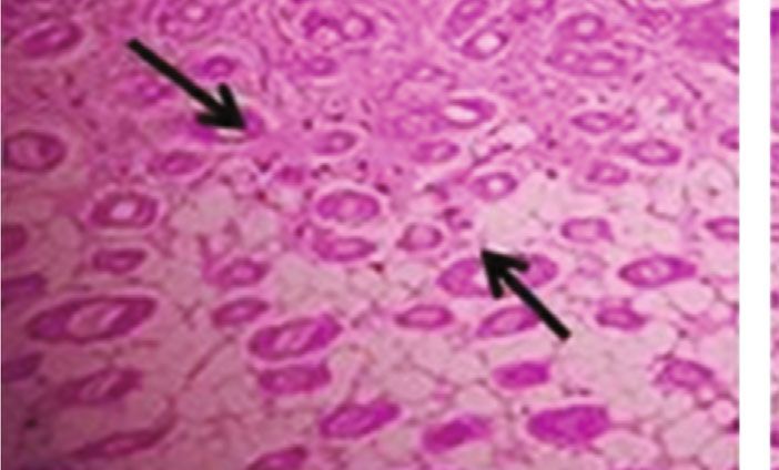
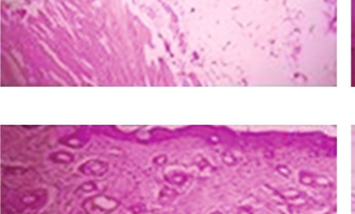
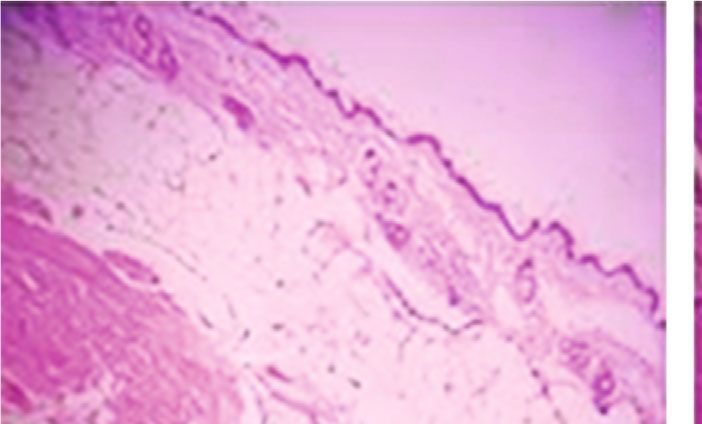
10 International Journal of Polymer Science

 (a) (b)

 (c) (d)

 (e)

 Indicates the cervical patches

Figure 10: (a–e) Histological evaluation of skin tissue corresponding untreated, DMBA-induced group, FA-treated group, and positive
control (The DMBA induced the degenerated cervical patches of the skin of mice; it was brought to normal by FA-treated mice).

3.9. p53 Level Analysis. The p53 protein level in group II mice 3.10. Keratinocytes Contents of the Skin. In the present inves-
showed significant elevation (p < 0:001) when compared with tigation, significant loss of keratinocytes was found in the
control and which was significantly reduced (p < 0:001) in DMBA-induced mice (p < 0:05) than control mice. The FA
other treated groups Figure 7. High expression of p53 protein administrated groups III and IV displayed significant
and p53 alterations have been noticed in a great proportion of improvement (p < 0:05) in keratinocyte content when com-
SCC and premalignant abrasions in renal allograft recipient pared to induced group mice Figure 8. Epidermis said to
cases. The p53 tumor suppressor gene was reported to have abide of multistate constantly renovating keratinocytes most
association with the cell cycle capture and inauguration of of the time, and this structure forms the epidermal barrier
programmed cell death [31]. which need to donate the self-protective responses against
International Journal of Polymer Science 11

various environmental factors (for example, cold, heat, Data Availability
radiation, trauma, and microbial infection to name a few).
Increased level of keratinocytes indicates keratinocyte- The data used to support the findings of this study are avail-
connected cutaneous disorders, covering CSC, zoster herpes, able from the authors upon request.
and psoriasis [32].
 Additional Points
3.11. In Vivo Antioxidant Enzymes Levels. The in vivo antiox- Animal Welfare. Animal use complied with institutional,
idant defence system like SOD, CAT, GPx, and GSH in national, and international guidelines including the Commit-
DMBA-induced mice exhibited a significant reduction tee for the Purpose of Control and Supervision of Experi-
(p < 0:05) in the skin tissues when compared to treated ments on Animals (CPCSEA), Govt. of India guidelines.
groups Figures 9(a) and 9(d). Cell life in an oxygenated back- Guidelines of “Guide for the Care and Use of Laboratory Ani-
ground has necessitated the progression of effective cellular mals” (Institute of Laboratory Animal Resources, National
tactics to identify and detoxify metabolites of free radicals. Academic Press 1996; NIH publication number #85-23,
These effects reported to disturb significantly the host of revised 1996) were strictly followed throughout the study.
physiological practices and metabolic pathway locally Institutional Animal Ethical Committee (IAEC) approved
bounded up with the skin cancer of the animal and skin or this study. Research ethical clearance was obtained (IAEC4/-
the change of its related disorders [33]. The results revealed Proposal: 31/A.Lr:13/Dated: 20.12.18).
significant rise (p < 0:05) in the activities of the primary anti-
oxidant defence enzymes SOD, CAT, GPx, and GSH in the Ethical Approval
samples of topically applied mice group (groups III and IV)
suggesting that the FA could enhance the antioxidant system This research does not involve human participants, human
in the skin of experimental mice. In particular, GSH estima- material, or human data. This research involves experimental
tion is an important indicator for quantifying the level of animals approved by the Institutional Animal Ethics Com-
antioxidant activity. The GSH enzyme can be as curtained mittee, Chettinad Academy of Research and Education
in almost every cell of the body and have tremendous antiox- (Deemed to be University), Kelambakkam-603 103, Tamil
idant and detoxifying activity. GSH not only eradicates free Nadu, India, and adhered to the Committee for the Purpose
radicals in vivo but also boosts the immunity level [34]. of Control And Supervision of Experiments on Animals
 (CPCSEA), Govt. of India guidelines.
3.12. Histopathological Analysis. The paraffin sections of the
skin, the liver, the heart, and the kidney of experimental mice Conflicts of Interest
are represented in Figure 10. From this result, the degener-
 All authors declare that there are no conflicts of/or compet-
ated cervical patches of the skin tissue of the (DMBA) group
 ing interests.
II was more than that of groups I, III, IV, and V. The cervical
patches of the skin were degenerated with skin cancer forma-
tion, whereas, these changes were regenerated in the skin Acknowledgments
cervical patches of formulated agar (group III and group The authors want to thank their respective institutions for
IV)-treated mice. The DMBA-induced mice kidney showed their continued support. The technical and language editing
oedema formation and treated mice seen as decrease of done by The Editing Refinery, MD, USA, is highly appreci-
oedema. In the heart, the myofibrils were collapsed in the ated. The Indian authors gratefully acknowledge the Depart-
induced group, and in the treated group, showed the normal- ment of Biotechnology, Ministry of Science and Technology,
ized myofibrils. The DMBA-induced mice showed significant Govt. of India, New Delhi (BT/PR15676/AAQ/03/794/2016),
liver cirrhosis, and these changes were nearly normalized in for their financial support.
FA-treated mice.
 References
4. Conclusion [1] S. Labani, S. Asthana, K. Rathore, and K. Sardana, “Incidence
Both the in vitro and in vivo findings of our present study of melanoma and nonmelanoma skin cancers in Indian and
 the global regions,” Journal of Cancer Research and Therapeu-
revealed that FA from brown seaweed could act as a potent
 tics, 2020.
antiskin cancer agent and antioxidant. All these outcomes
 [2] P. Mathur, K. Sathishkumar, M. Chaturvedi et al., “Cancer
seem to be proposing that pretreatment of FA from L. digitata statistics, 2020: report from National Cancer Registry
has a great development perspective for mitigation of the skin Programme, India,” American Society of Clinical Oncology,
cancer induced by DMBA. However, there is a need to find the vol. 16, pp. 1063–1075, 2020.
exact mechanism of action of FA, which warrants the need for [3] G. Singh, P. W. Wong, J. Pecoriello et al., “Characterizing
extensive biochemical and molecular studies. The microbial index keratinocytic carcinomas in commercially insured adults
load and UV-induced skin cancer activities of FA from brown younger than age 50 years in the United States,” Journal of the
seaweed are not analysed in this study, which could be tested American Academy of Dermatology, vol. 83, no. 5, pp. 1458–
in the future. 1460, 2020.
12 International Journal of Polymer Science

 [4] S. V. Deo, S. Hazarika, N. K. Shukla, S. Kumar, M. Kar, and tumors in mice,” Journal of Investigative Dermatology,
 A. Samaiya, “Surgical management of skin cancers: experience vol. 137, no. 4, pp. 921–930, 2017.
 from a regional cancer centre in North India,” Indian Journal [19] S. Möllers, I. Heschel, L. H. Damink et al., “Cytocompatibility
 of Cancer, vol. 42, no. 3, p. 145, 2005. of a novel, longitudinally microstructured collagen scaffold
 [5] G. Bertino, G. Sersa, F. De Terlizzi et al., “European Research intended for nerve tissue repair,” Tissue Engineering Part A,
 on Electrochemotherapy in Head and Neck Cancer vol. 15, no. 3, pp. 461–472, 2009.
 (EURECA) project: results of the treatment of skin cancer,” [20] I. Stoilov, B. C. Starcher, R. P. Mecham, and T. J. Broekelmann,
 European Journal of Cancer, vol. 63, pp. 41–52, 2016. “Measurement of elastin, collagen, and total protein levels in
 [6] K. Bernard, C. Forest, and X. Coumoul, “Dimethyl-Benz (a) tissues,” Methods in Cell Biology, vol. 143, pp. 133–146, 2018.
 anthracene: a mammary carcinogen and a neuroendocrine [21] J. Sullivan-Brown, M. E. Bisher, and R. D. Burdine, “Embed-
 disruptor,” Biochimie Open, vol. 3, pp. 49–55, 2016. ding, serial sectioning and staining of zebrafish embryos using
 [7] S. K. Kim and I. Wijesekara, “Development and biological JB-4 resin,” Nature Protocols, vol. 6, no. 1, pp. 46–55, 2011.
 activities of marine-derived bioactive peptides: a review,” [22] R. Ahmad, M. Surif, N. Ramli, N. Yahya, A. R. Nor, and
 Journal of Functional Foods, vol. 2, no. 1, pp. 1–9, 2010. L. Bekbayeva, “A preliminary study on the agar content and
 [8] K. Ramachandran, V. Manigandan, R. Sheeba, R. Saravanan, agar gel strength of Gracilaria manilaensis using different agar
 and P. R. Rajaian Rajesh, “Structural characterization and extraction processes,” World Applied Sciences Journal, vol. 15,
 comparative biomedical properties of phloroglucinol from no. 2, pp. 184–188, 2011.
 Indian brown seaweeds,” Journal of Applied Phycology, [23] B. S. Reshma, Antioxidant and Antiaging Properties of Aque-
 vol. 28, no. 6, pp. 3561–3573, 2016. ous Extract from Laminaria digitata (Hudson) in D-Galactose
 [9] P. Tveden-Nyborg, T. K. Bergmann, and J. Lykkesfeldt, “Basic Induced Aging Mice, M.Sc., Dissertation, Chettinad University,
 & clinical pharmacology & toxicology policy for experimental 2017.
 and clinical studies,” Basic & Clinical Pharmacology & Toxicol- [24] K. A. Martínez Andrade, C. Lauritano, G. Romano, and
 ogy, vol. 123, no. 3, pp. 233–235, 2018. A. Ianora, “Marine microalgae with anti-cancer properties,”
[10] M. Venkatesan, V. Arumugam, R. Pugalendi et al., “Antioxi- Marine Drugs, vol. 16, no. 5, p. 165, 2018.
 dant, anticoagulant and mosquitocidal properties of water [25] J. Fleurence, “Seaweed proteins,” Trends in Food Science &
 soluble polysaccharides (WSPs) from Indian seaweeds,” Technology, vol. 10, no. 1, pp. 25–28, 1999.
 Process Biochemistry, vol. 84, pp. 196–204, 2019. [26] S. Azizi, M. B. Ahmad, F. Namvar, and R. Mohamad, “Green
[11] E. A. Yapar, O. İnal, and M. S. Erdal, “Design and in vivo eval- biosynthesis and characterization of zinc oxide nanoparticles
 uation of emulgel formulations including green tea extract and using brown marine macroalga Sargassum muticum aqueous
 rose oil,” Acta Pharmaceutica, vol. 63, no. 4, pp. 531–544, extract,” Materials Letters, vol. 116, pp. 275–277, 2014.
 2013. [27] S. Shankar and J. W. Rhim, “Preparation of nanocellulose from
[12] J. P. Jadhav, S. S. Phugare, R. S. Dhanve, and S. B. Jadhav, micro-crystalline cellulose: the effect on the performance and
 “Rapid biodegradation and decolorization of Direct Orange properties of agar-based composite films,” Carbohydrate Poly-
 39 (Orange TGLL) by an isolated bacterium Pseudomonas mers, vol. 135, pp. 18–26, 2016.
 aeruginosa strain BCH,” Biodegradation, vol. 21, no. 3, [28] Y. M. Fonseca, C. D. Catini, F. T. Vicentini, A. Nomizo, R. F.
 pp. 453–463, 2010. Gerlach, and M. J. Fonseca, “Protective effect of Calendula offi-
[13] A. N. Syad, K. P. Shunmugiah, and P. D. Kasi, “Seaweeds as cinalis extract against UVB-induced oxidative stress in skin:
 nutritional supplements: analysis of nutritional profile, physi- evaluation of reduced glutathione levels and matrix metallo-
 cochemical properties and proximate composition of G. acer- proteinase secretion,” Journal of Ethnopharmacology,
 osa and S. wightii,” Biomedicine & Preventive Nutrition, vol. 127, no. 3, pp. 596–601, 2010.
 vol. 3, no. 2, pp. 139–144, 2013. [29] S. Daniel, S. Cornelia, and Z. Fred, “UV-A sunscreen from red
[14] V. C. Deuschle, R. A. Deuschle, M. R. Bortoluzzi, and M. L. algae for protection against premature skin aging,” Cosmet
 Athayde, “Physical chemistry evaluation of stability, spread- Toiletries Manufacture Worldwide, vol. 2004, pp. 139–143, 2004.
 ability, in vitro antioxidant, and photo-protective capacities [30] J. Swallow, P. Koteja, P. Carter, and T. Garland, “Food con-
 of topical formulations containing Calendula officinalis L. leaf sumption and body composition in mice selected for high
 extract,” Brazilian Journal of Pharmaceutical Sciences, vol. 51, wheel-running activity,” Journal of Comparative Physiology
 no. 1, pp. 63–75, 2015. B, vol. 171, no. 8, pp. 651–659, 2001.
[15] M. Muehlbach, R. Brummer, and R. Eggers, “Study on the [31] C. L. Benjamin and H. N. Ananthaswamy, “p53 and the path-
 transferability of the time temperature superposition principle ogenesis of skin cancer,” Toxicology and Applied Pharmacol-
 to emulsions,” International Journal of Cosmetic Science, ogy, vol. 224, no. 3, pp. 241–248, 2007.
 vol. 28, no. 2, pp. 109–116, 2006. [32] K. Douroudis, K. Kingo, T. Traks et al., “Polymorphisms in the
[16] M. Tomczykowa, M. Wróblewska, K. Winnicka et al., “Novel ATG16L1 gene are associated with psoriasis vulgaris,” Acta
 gel formulations as topical carriers for the essential oil of Dermato-Venereologica, vol. 92, no. 1, pp. 85–87, 2012.
 bidens tripartita for the treatment of candidiasis,” Molecules, [33] H. Ye, K. Wang, C. Zhou, J. Liu, and X. Zeng, “Purification,
 vol. 23, no. 10, p. 2517, 2018. antitumor and antioxidant activities in vitro of polysaccha-
[17] S. Tajane, P. Dadhe, S. Mandavgane, and S. Mehetre, “Kinetic rides from the brown seaweed Sargassum pallidum,” Food
 and thermodynamic azadirachtin extraction from whole neem Chemistry, vol. 111, no. 2, pp. 428–432, 2008.
 fine powder formulation,” Indian Journal of Chemical Tech- [34] G. Lavanya, S. P. Voravuthikunchai, and N. H. Towatana,
 nology, vol. 24, pp. 218–222, 2017. “Acetone extract from Rhodomyrtus tomentosa: a potent nat-
[18] M. Dahlhoff, S. Muzumdar, M. Schäfer, and M. R. Schneider, ural antioxidant,” Evidence-based Complementary and Alter-
 “ERBB2 is essential for the growth of chemically induced skin native Medicine, vol. 2012, 8 pages, 2012.
You can also read