Association Pyoderma Gangrenosum and Systemic Lupus Erythematosus
←
→
Page content transcription
If your browser does not render page correctly, please read the page content below
ISSN: 2474-3682
Rkiouak et al. Clin Med Img Lib 2021, 7:177
DOI: 10.23937/2474-3682/1510177
Volume 7 | Issue 4
Clinical Medical Image Library
Open Access
IMAGE ARTICLE
Association Pyoderma Gangrenosum and Systemic Lupus
Erythematosus
A Rkiouak, PhD*, I El Kassimi, MD, N Sahel, MD, M Zaizae, MD and Y Sekkach, PhD
Check for
updates
Department of Internal Medicine A, Mohammed V Military Hospital, Medical School of Rabat, Morocco
*Corresponding author: Adil Rkiouak, PH.D, Department of Internal Medicine A, Mohammed V Military Hospital, Medical
School of Rabat, Morocco, Tel: +212-66-179-44-04, Fax: +212-66-179-44-04
Introduction any recurrence of PG to date, with a 1-year follow-up.
19-year-old patient, follow-up for 8 years for SLE Pyoderma Gangrenosum (PG) is part of the spec-
treated with hydroxychloroquine 400 mg/day and who trum of the neutrophilic dermatoses, which is usually
had been in remission for a year. SLE diagnosis was idiopathic. PG typically presents with single or multiple
based on the association of a malar rash, photosensi- skin ulcers with undetermined erythematous-viola-
tivity, lymphopenia, autoimmune hemolytic anemia, ceous borders.
antinuclear antibodies positivity, anti-DNA antibodies Although PG has been associated with several sys-
identified and lupus nephritis. temic diseases, it has rarely been reported in associa-
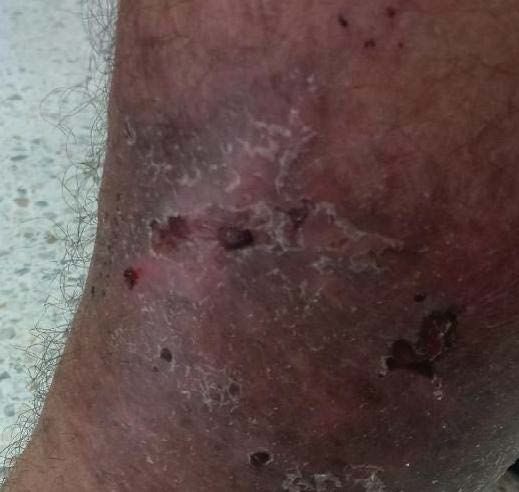
On December 2019, patient noticed a well-defined tion with Systemic Lupus Erythematosus (SLE).
irregular popular lesion on left leg (Figure 1) associat- No absolute criteria for the diagnosis of PG are avail-
ed with ulcerations appeared 1 month later. Treatment able, but diagnostic criteria, to which our patient re-
with local antibiotics was without improvement. plied, have been proposed [1].
Both lesions had well-defined purplish undermined The diagnosis of PG was made based on features of
borders and a necrotic base with surrounding erethey- rapid onset and progression, resistance to treatment
ma, and there was marked tenderness on palpation. with antibiotics, characteristic of lesion (ulcerating,
Skin examination found a deep ulceration making 3 well-defined purple undermined borders) and histolog-
cm of diameter of the anterior face left leg with inflam- ical study.
matory border. More than 50% of patients with PG have an asso-
A Doppler ultrasound of right leg showed no evi- ciated systemic disease, most commonly inflammatory
dence of venous obstruction. An arterial sonography bowel disease (14-34%), arthropathies (11-25%) and
scan showed no evidence of arterial insufficiency. hematologic disease or hematologic malignancy (20%)
[2].
Histologic examination of the lesion showed a dense
infiltration of dermis by leukocytoclastic or intact neu- Systemic Lupus Erythematosus (SLE) is not a com-
trophils, without granuloma or vascular damage. The monly recognized associate of PG.
diagnosis of ulcerative variant of PG was retained con- This association may be supported by common in-
cluding in pyoderma gangrenosum. No pathogens in- nate immunity dysregulation and abnormal neutrophil
cluding atypical mycobacteria or yeast were evidenced. activation.
A treatment with prednisone 1 mg/kg/day orally was Among the reported cases of patients with PG in a
started up, and complete resolution of PG was obtained context of SLE, a large majority were women [3,4].
within 6 months (Figure 2). The patient did not present
Citation: Rkiouak A, El Kassimi I, Sahel N, Zaizae M, Sekkach Y (2021) Association Pyoderma Gangreno-
sum and Systemic Lupus Erythematosus. Clin Med Img Lib 7:177. doi.org/10.23937/2474-3682/1510177
Accepted: April 24, 2021; Published: April 26, 2021
Copyright: © 2021 Rkiouak A, et al. This is an open-access content distributed under the terms of the
Creative Commons Attribution License, which permits unrestricted use, distribution, and reproduction
in any medium, provided the original author and source are credited.
Rkiouak et al. Clin Med Img Lib 2021, 7:177 • Page 1 of 3 •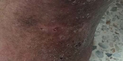
DOI: 10.23937/2474-3682/1510177                                                                         ISSN: 2474-3682

                                 Figure 1: Deep ulcerations with inflammatory border of the left leg.

                                       Figure 2: Healing ulcerations after corticosteroid therapy.

Rkiouak et al. Clin Med Img Lib 2021, 7:177                                                               • Page 2 of 3 •
DOI: 10.23937/2474-3682/1510177                                                                            ISSN: 2474-3682

   The diagnosis of lupus usually precedes the onset of     References
PG, as in our patient.                                      1. Maverakis E, Ma C, Shinkai K, Fiorentino D, Callen JP, et
    Classically, the treatment of PG is based on (colchi-      al. (2018) Diagnostic Criteria of Ulcerative Pyoderma Gan-
                                                               grenosum. JAMA Dermatology 154: 461-466.
cine, dapsone, corticosteroids, minocycline). However,
SLE treatments (hydroxychloroquine, methotrexate,           2. Binus AM, Qureshi AA, Li VW, Winterfield LS (2011) Pyo-
                                                               derma gangrenosum: a retrospective review of patient
mycophenolate mofetil, and cyclophosphamide) ap-
                                                               characteristics, comorbidities and therapy in 103 patients.
pear to be effective in PG. Our patient was treated with       Br J Dermatol 165: 1244-1250.
general corticosteroid therapy with regression of lupus
                                                            3. Gonzalez-Moreno J, Ruiz-Ruigomez M, Callejas Rubio
lesions and PG lesions.                                        JL,Ríos Fernández R, Ortego Centeno N, et al. (2015) Pyo-
   The association between LES and PG is rare. The             derma gangrenosum and systemic lupus erythematosus: a
                                                               report of five cases and review of the literature. Lupus 24:
prognosis of the disease does not appear to be wors-           130-137.
en by PG. corticosteroid therapy is usually sufficient to
                                                            4. Hamzi AM, Bahadi A, Alayoud A, El Kabbaj D, Benyahia M
treat a possible association.
                                                               (2013) Skin ulcerations in a lupus hemodialysis patient with
                                                               hepatitis C infection: what is your diagnosis? Iran J Kidney
                                                               Dis 7: 191.

Rkiouak et al. Clin Med Img Lib 2021, 7:177                                                                   • Page 3 of 3 •
You can also read