Atlas of Dermatoses in Pigmented Skin - Ranthilaka R. Ranawaka Ajith P. Kannangara Ajith Karawita Editors
←
→
Page content transcription
If your browser does not render page correctly, please read the page content below
Atlas of Dermatoses
in Pigmented Skin
      Ranthilaka R. Ranawaka
      Ajith P. Kannangara
      Ajith Karawita
      Editors

      123
Atlas of Dermatoses in Pigmented Skin
Ranthilaka R. Ranawaka
Ajith P. Kannangara • Ajith Karawita
Editors

Atlas of Dermatoses
in Pigmented Skin
Editors
Ranthilaka R. Ranawaka                             Ajith P. Kannangara
Consultant Dermatologist                           Consultant Dermatologist
General Hospital Kalutara                          Base Hospital Balapitiya
Kalutara                                           Balapitiya
Sri Lanka                                          Sri Lanka

Ajith Karawita
Consultant Venereologist
Teaching Hospital Anuradhapura
Anuradhapura
Sri Lanka

ISBN 978-981-15-5482-7    ISBN 978-981-15-5483-4                           (eBook)
https://doi.org/10.1007/978-981-15-5483-4

© Springer Nature Singapore Pte Ltd. 2021
This work is subject to copyright. All rights are reserved by the Publisher, whether the whole or
part of the material is concerned, specifically the rights of translation, reprinting, reuse of
illustrations, recitation, broadcasting, reproduction on microfilms or in any other physical way,
and transmission or information storage and retrieval, electronic adaptation, computer software,
or by similar or dissimilar methodology now known or hereafter developed.
The use of general descriptive names, registered names, trademarks, service marks, etc. in this
publication does not imply, even in the absence of a specific statement, that such names are
exempt from the relevant protective laws and regulations and therefore free for general use.
The publisher, the authors, and the editors are safe to assume that the advice and information in
this book are believed to be true and accurate at the date of publication. Neither the publisher nor
the authors or the editors give a warranty, expressed or implied, with respect to the material
contained herein or for any errors or omissions that may have been made. The publisher remains
neutral with regard to jurisdictional claims in published maps and institutional affiliations.

This Springer imprint is published by the registered company Springer Nature Singapore Pte Ltd.
The registered company address is: 152 Beach Road, #21-01/04 Gateway East, Singapore
189721, Singapore

Preface

This book has been written by clinicians for clinicians to provide a clear and
easy guide to identify skin diseases in pigmented skin. This book discusses
Over 500 Skin Problems with Over 2700 Colour Illustrations, and more than
300 Picture-Based Questions with Answers.
    Common skin diseases such as psoriasis, lichen planus, eczema and ery-
thrasma are darker in pigmented skin. For example in PASI (psoriasis area
and severity index), erythema is always 0 or 1 in pigmented skin; lichen pla-
nus is blackish rather than purplish; erythrasma is never erythematous; and
lichen amyloidosis is blackish in darker skin. Therefore, it is important that
dermatology trainees and general practitioners treating patients of pigmented
skin (Fitzpatrick type V) use an atlas with their patients to get familiar with
these diverse clinical presentations.
    Tropical diseases such as cutaneous tuberculosis, leishmaniasis, fungal
infections, oral submucous fibrosis and leprosy are not uncommon in our
routine skin clinics. Nevertheless, some of the diseases which were frequent
in our clinical practice twenty years ago are rarely encountered today such as
sporotrichosis, mycetoma, rhinosporidiosis, lobomycosis and subcutaneous
zygomycosis. Urbanization, change of farming practices and using pesticides
may play a role here. Therefore, it is imperative to document these disappear-
ing diseases for the future.
    Hypopigmented disorders such as pityriasis alba, vitiligo, guttate hypomel-
anosis, progressive macular hypomelanosis and chemical or physical induced
depigmentation are more marked and a cosmetic concern in pigmented skin
and are visible without any instrumental assistance, such as Wood’s light.
Interestingly, hyperpigmentary changes are more a cosmetic issue in aging
pigmented skin than wrinkles. Wrinkles appear ten years later in pigmented
skin compared to the white skin.
    This atlas is meant to stimulate the interest in tropical dermatology and
skin problems in pigmented skin, among medical students, postgraduate
trainees, general practitioners and dermatologists from non-tropical
countries.

Kalutara, Sri Lanka                                 Ranthilaka R. Ranawaka
Balapitiya, Sri Lanka                                   Ajith P. Kannangara
Anuradhapura, Sri Lanka                                      Ajith Karawita

                                                                             v
Acknowledgements

We wish to pay tribute to all the contributors for spending their invaluable
time and knowledge to the success of this atlas. We take this opportunity to
show our heartiest acknowledgements to all the patients, majority Sri
Lankans, who generously gave clinical photographs to this book. Finally, our
thanks go to Springer for accepting this book for publication.
   Dr Ranthilaka R. Ranawaka
   Dr Ajith P. Kannangara
   Dr Ajith Karawita

                                                                         vii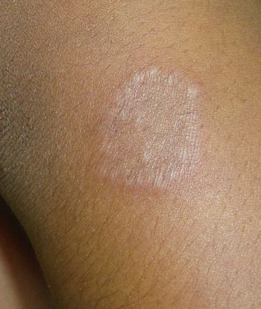
Contents

Part I Neonatal and Paediatric Dermatoses

 1	Dermatoses of the Neonate and Infancy��������������������������������������    3
     Ranthilaka R. Ranawaka
 2	Congenital Naevi and Melanocytic Naevi������������������������������������   45
     Ranthilaka R. Ranawaka
 3	Vascular Tumours and Malformations����������������������������������������   65
     Ranthilaka R. Ranawaka
 4	Neurocutaneous Syndromes����������������������������������������������������������   81
     Anuruddha Padeniya and Fous Lebbe

Part II Inflammatory Dermatoses

 5	Psoriasis������������������������������������������������������������������������������������������   91
     Kanchana Mallawaarachchi
 6	Eczematous Disorders ������������������������������������������������������������������ 107
     Ranthilaka R. Ranawaka
 7	Inherited Disorders of Keratinization������������������������������������������ 123
     Priyanka Karagaiah and Varsha M. Gowda
 8	Allergic and Irritant Contact Dermatitis������������������������������������ 151
     Ranthilaka R. Ranawaka
 9	Occupational Dermatoses�������������������������������������������������������������� 169
     Ajith P. Kannangara
10	Photodermatosis���������������������������������������������������������������������������� 183
     Hiromel de Silva
11	Cutaneous Reactions���������������������������������������������������������������������� 201
     Ajith P. Kannangara
12	Neutrophilic Dermatoses �������������������������������������������������������������� 213
     Binari K. S. Wijenayake

                                                                                                           ix
x                                                                                                                Contents

Part III Infections and Infestations

13	Cutaneous Tuberculosis���������������������������������������������������������������� 227
     Sujay Khandpur and Neha Taneja
14	Mycobacterial Infections in Sri Lanka���������������������������������������� 237
     Ranthilaka R. Ranawaka
15	Leprosy�������������������������������������������������������������������������������������������� 257
     Ranthilaka R. Ranawaka
16	Viral Infections������������������������������������������������������������������������������ 297
     Premini Rajendiran
17	Superficial Fungal Infections�������������������������������������������������������� 319
     Ranthilaka R. Ranawaka
18	Subcutaneous and Systemic Mycoses������������������������������������������ 359
    S. N. Arseculeratne, Archana Singal,
    and Ranthilaka R. Ranawaka
19	Onychomycosis������������������������������������������������������������������������������ 381
     Ranthilaka R. Ranawaka
20	Diseases Caused by Arthropods and Parasites���������������������������� 397
     Hiran Gunasekera
21	Leishmaniasis in Sri Lanka���������������������������������������������������������� 417
     Ranthilaka R. Ranawaka, Yamuna Siriwardana,
     and Shalindra Ranasinghe
22	Sexually Transmitted Infections �������������������������������������������������� 445
     Ajith Karawita

Part IV Systemic Dermatoses

23	Sarcoidosis�������������������������������������������������������������������������������������� 469
     Katerina Damevska, Snejina Vassileva, Kossara Drenovska,
     Slavica Kostadinova-Kunovska, and Valeria Mateeva
24	Autoimmune Blistering Diseases�������������������������������������������������� 481
     Binari K. S. Wijenayake
25	Lichen Planus and Lichenoid Dermatoses���������������������������������� 497
     Ajith P. Kannangara
26	Acne������������������������������������������������������������������������������������������������ 503
     Sanjeewani Fonseka
27	Rosacea�������������������������������������������������������������������������������������������� 511
     Sanjeewani Fonseka
28	Disorders of Connective Tissue���������������������������������������������������� 515
     Chathurarya Siriwardena
Contents                                                                                                               xi

           29	Leg Ulcers �������������������������������������������������������������������������������������� 529
                Ranthilaka R. Ranawaka
           30	Cutaneous Vasculitis���������������������������������������������������������������������� 547
                Binari K. S. Wijenayake
           31	Autoimmune Connective Tissue Diseases������������������������������������ 563
                Kanchana Mallawaarachchi

           Part V The Skin and the Organs

           32	Benign Lesions in the Oral and Maxillofacial Region���������������� 587
                K. M. S. Kosgoda
           33	Skin and the Ear���������������������������������������������������������������������������� 613
                Ranthilaka R. Ranawaka
           34	Psychocutaneous Disorders���������������������������������������������������������� 627
                Ranthilaka R. Ranawaka
           35	Disorders of Hair���������������������������������������������������������������������������� 637
                Lidia Rudnicka and Anna Waśkiel-Burnat
           36	Trichoscopy: Dermoscopy Hair���������������������������������������������������� 663
                Deepani Rathnayake
           37	Disorders of Nails�������������������������������������������������������������������������� 723
                Eckart Haneke and Ranthilaka R. Ranawaka
           38	Genital Dermatoses������������������������������������������������������������������������ 765
                Ajith Karawita and Ranthilaka R. Ranawaka

           Part VI Pigmentary and Cosmetic Dermatoses

           39	Cultural Dermatoses���������������������������������������������������������������������� 797
                Felicia Srisaravanapavananthan
           40	Facial Melanosis ���������������������������������������������������������������������������� 803
                Ranthilaka R. Ranawaka
           41	Vitiligo �������������������������������������������������������������������������������������������� 823
                Ranthilaka R. Ranawaka
           42	Hypopigmentary Disorders���������������������������������������������������������� 837
                Ranthilaka R. Ranawaka
           43	Hyperpigmentary Disorders �������������������������������������������������������� 849
                Premini Rajendiran

           Part VII Benign and Malignant Skin Tumours

           44	Oral Potentially Malignant Disorders (OPMDs)������������������������ 879
               W. M. Tilakaratne and Ruwan D. Jayasinghe
           45	Oral Cancer������������������������������������������������������������������������������������ 903
               Ruwan D. Jayasinghe and W. M. Tilakaratne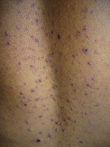
xii                                                                                                     Contents

46	Hypopigmented Mycosis Fungoides�������������������������������������������� 921
     Ranthilaka R. Ranawaka
47	Tumours of the Skin Appendages ������������������������������������������������ 941
     V. G. Abeywickrama
48	Benign Skin Proliferations������������������������������������������������������������ 955
     Ranthilaka R. Ranawaka
49	Precursors of Skin Carcinoma������������������������������������������������������ 971
     Ranthilaka R. Ranawaka
50	Skin Carcinomas���������������������������������������������������������������������������� 989
     Ranthilaka R. Ranawaka, Kanishka de Silva,
     and Priyanka H. Abeygunasekara

Part VIII Therapy and Complications

51	Cutaneous Adverse Reactions to Drugs �������������������������������������� 1017
     Binari K. S. Wijenayake

Part IX Invited Chapter

52	Dermatoses from Brazil���������������������������������������������������������������� 1049
    Sinésio Talhari and Carolina Chrusciak Talhari Cortez
List of Contributors
Priyanka H. Abeygunasekara, D Path, MD Path (Histopath) National
Cancer Institute, Maharagama, Sri Lanka
V. G. Abeywickrama, MBBS, MD District General Hospital, Matale, Sri
Lanka
S. N. Arsecularatne, MBBS, Dip. Bact. D. Phil. Faculty of Medicine,
University of Peradeniya, Peradeniya, Sri Lanka
Katerina Damevska, MD, MSc, PhD Clinic of Dermatology, Medical
Faculty, Ss. Cyril and Methodius University, Skopje, Macedonia
Kossara Drenovska, MD, PhD Department of Dermatology and
Venereology, Medical Faculty, University of Medicine, Sofia, Sofia, Bulgaria
Varsha M. Gowda, MD Bangalore Medical College and Research Centre,
Bangalore, Karnataka, India
Hiran Gunasekara, MBBS, MD Teaching Hospital, Kuliyapitiya,
Kuliyapitiya, Sri Lanka
Eckhart Haneke, MD, PhD Clinical professor of dermatology, consultant
dermatologist (Germany), Visiting professor, Nail Diseases, Nail Surgery,
Nail Pathology, Department of Dermatology, Inselspital, Universitätsspital
Bern, Bern, Switzerland
R. D. Jayasinghe, BDS, MS Department of Oral Medicine and
Periodontology, Faculty of Dental Sciences, University of Peradeniya,
Peradeniya, Sri Lanka
Ajith P. Kannangara, MBBS, MD Consultant Dermatologist, Base
Hospital Balapitiya, Balapitiya, Sri Lanka
Priyanka Karagaiah, MD Bangalore Medical College and Research
Centre, Bangalore, Karnataka, India
Ajith Karawita, MBBS, Pg Dip Ven, MD, FSLCoSHH Sexual Health
Centre, Teaching Hospital Anuradhapura, Anuradhapura, Sri Lanka
Sujay Khandpur, MD, DNB, MNAMS Department of Dermatology and
Venereology, All India Institute of Medical Sciences, New Delhi, India
xiiixiv List of Contributors K. M. S. Kosgoda, BDS, MS Consultant Oral and Maxillofacial Surgeon, Teaching Hospital Anuradhapura, Anuradhapura, Sri Lanka Slavica Kostadinova-Kunovska, MD, PhD Institute of Pathology, Medical Faculty, Ss. Cyril and Methodius University, Skopje, Macedonia Fous Lebbe, MBBS, MD Lady Ridgeway Hospital for Children, Colombo, Sri Lanka Kanchana Mallawarachchi, MBBS, MD Base Hospital Balangoda, Balangoda, Sri Lanka Valeria Mateeva, MD, PhD Department of Dermatology and Venereology, Medical Faculty, University of Medicine, Sofia, Sofia, Bulgaria Anuruddha Padeniya, MBBS, MD Lady Ridgeway Hospital for Children, Colombo, Sri Lanka Teaching Hospital Kandy, Kandy, Sri Lanka Department of Paediatrics, University of Rajarata, Mihintale, Sri Lanka Premini Rajendrian, MBBS, MD District General Hospital, Chilaw, Chilaw, Sri Lanka Shalindra Ranasinghe, MBBS, M.Phil (Keele), PhD Department of Parasitology, Faculty of Medical Sciences, University of Sri Jayewardenepura, Nugegoda, Sri Lanka Ranthilaka R. Ranawaka, MBBS, MD Consultant Dermatologist, General Hospital Kalutara, Kalutara, Sri Lanka Deepani Ratnayake, MBBS, MD, FACD Sinclair Dermatology, East Melbourne, VIC, Australia Lidia Rudnicka, MD, PhD Department of Dermatology, Medical University of Warsaw, Warsaw, Poland Hiromel de Silva, MBBS, MD Consultant Dermatologist, Base Hospital Tangalle, Tangalle, Sri Lanka Kanishka de Silva, MS, FRCS National Cancer Institute, Maharagama, Sri Lanka Archana Singal, MD, FAMS University College of Medical Sciences & GTB Hospital, New Delhi, India Faculty of Medical Sciences, University of Delhi, New Delhi, India Chaturyaya Siriwardena, MBBS, MD (Derm), MRCP (UK) District General Hospital Nuwara Eliya, Nuwara Eliya, Sri Lanka Yamuna Siriwardena, MBBS, PhD Parasitic Disease Research Unit, Department of Parasitology, Faculty of Medicine, University of Colombo, Colombo, Sri Lanka Felicia Srisaravanapavananthan, MBBS, MD Teaching Hospital Jaffna, Jaffna, Sri Lanka
List of Contributors                                                                             xv

                       Sinesio Talhari, PhD Heitor Vieira Dourado Foundation of Tropical
                       Medicine, Manaus, Amazonas, Brazil
                       Carolina Chrusciak Talhari Cortez, PhD Department of Dermatology,
                       Heitor Vieira Dourado Foundation of Tropical Medicine, Manaus, Amazonas,
                       Brazil
                       Neha Taneja, MD, DNB Department of Dermatology and Venereology, All
                       India Institute of Medical Sciences, New Delhi, India
                       W. M. Tilakaratne, BDS, MS, FDSRCS, FRCPath, PhD Department of
                       Oral Pathology, Faculty of Dental Sciences, University of Peradeniya,
                       Peradeniya, Sri Lanka
                       Department of Oral and Maxillofacial Clinical Sciences, Faculty of Dentistry,
                       University of Malaya, Kuala Lumpur, Malaysia
                       Snejina Vassileva, MD, PhD Department of Dermatology and Venereology,
                       Medical Faculty, University of Medicine, Sofia, Sofia, Bulgaria
                       Anna Waskiel-Burnat, MD, PhD Department of Dermatology, Medical
                       University of Warsaw, Warsaw, Poland
                       Sanjeewani Fonseka, MBBS, MD (Dermatology), PhD Department of
                       Pharmacology, Faculty of Medicine, University of Peradeniya, Peradeniya,
                       Sri Lanka
                       Binari K. S. Wijenayake, MBBS, MD Teaching Hospital Karapitiya, Galle,
                       Sri Lanka
About the Editors
Ranthilaka R. Ranawaka, MBBS, MD, from
the University of Colombo, Sri Lanka. She has had
extensive training towards conducting clinical tri-
als in psoriasis and acne at St John’s Institute of
Dermatology, London, UK, in 2006. She is cur-
rently working as a consultant dermatologist at
General Hospital Kalutara, Sri Lanka. Dr
Ranawaka has published more than 26 scientific
papers in national and international journals
including six clinical trials. She is an author of six
dermatology books and three chapters, academic
editor in two journals and reviewer in a number of
indexed journals. She was awarded a number of
travel awards and scholarships (AAD 2006, EADV
2006, WCD 2007, ICD 2009, EADV 2020) and
research awards including European Nail Research
Grant in 2008 and SLMA-FairMed research grant
in 2012 and 2014. She was awarded President’s
Award for Scientific Publications in 2009, 2010,
2011, 2015, 2016 and 2017. She was the Co-Chair
and Guest Speaker at the “Tropical dermatoses”
symposium at the World Congress of Dermatology
2019, in Milan, Italy.
Ajith P. Kannangara, MBBS, MD, Consultant
Dermatologist, specialist in anti-aging medicine
and cosmetic dermatology, is currently attached
to the Ministry of Health, Sri Lanka. He obtained
his MBBS from the University of Peradeniya and
MD from the University of Colombo, Sri Lanka.
Dr Kannangara gained Fellowship from National
Skin Centre, Singapore and postdoctoral
International Dermatology Fellowship from
Wake Forest University, Baptist Medical Centre,
North Carolina, USA. He also earned Diploma
from the American Academy of Anti-Aging and
xviixviii About the Editors
Regenerative Medicine and American Academy
of Aesthetic Medicine.
Dr Kannangara also serves as editor and
reviewer for various national and international
journals. His research papers are published in
most renowned national and international journals
of dermatology. He is one of the pioneers of the
proposed classification of cutaneous reactions:
Koebner, Wolf isotopic, Renbok, Koebner nonre-
action, isotopic nonreaction, immunocompro-
mised district and other related phenomena, and
introducing the concept of Sparing phenomena.
Ajith Karawita, MBBS, PgDip and MD
(Venereology), is currently serving as a consultant
venereologist in a teaching hospital at the Ministry
of Health, Sri Lanka. He qualified in MBBS at the
University of Peradeniya in 1997 and subsequently
obtained his postgraduate diploma and MD in
Venereology from the Postgraduate Institute of
Medicine (PGIM), University of Colombo, Sri
Lanka. He is a fellow and past president (2015–
2016) of the Sri Lanka College of Sexual Health
and HIV Medicine. Further, he is a member of the
board of study, board of examinations and research
proposal review committee on venereology at the
PGIM. He is a Visiting Lecturer at Rajarata
University of Sri Lanka and the PGIM.
He is a member of the Australasian Society of
HIV Medicine (ASHM); honorary member of the
Research Institute of Tuberculosis and Anti-
tuberculosis Association, Japan; life member of
the Sri Lanka Medical Association; and a member
of the Asia Pacific Association of Medical Journal
Editors (APAME) and the editor-in-chief of the
Sri Lanka Journal of Sexual Health and HIV
Medicine (Sri Lanka JoSHH). He has authored
numerous research papers, invited articles, book
chapters, reports and guidelines. He has been a
guest speaker for multiple forums and presented
many research papers at local and international
conferences.Part I Neonatal and Paediatric Dermatoses
Dermatoses of the Neonate
                            and Infancy                                                                            1
                            Ranthilaka R. Ranawaka

                                                            1.
Introduction

More than 95% of newborns have cutaneous find-
ings, which often are distressing to parents but
frequently are benign and self-limited. Among
them are milia, cutis marmorata, congenital der-
mal melanocytosis and the benign neonatal pus-
tular eruptions (Rayala and Morrell 2017; Zuniga
and Nguyen 2013). The majority of the dermato-
logic alterations in neonates are physiological
and transient and do not require any treatment;
thus the parents can be reassured about the good
prognosis (Patrizi et al., 2017). An overview of
the most common or important dermatoses pre-
senting in neonatal and infant age group is illus-
trated in this chapter.

                                                                     A 6 months old baby was brought with this
                                                                 skin lesion which has been there since birth,
                                                                 the mother is worried that it is being progres-
                                                                 sively enlarging.
                                                                   (a) What is the diagnosis?
                                                                   (b) What is the sequelae of the disease?

The clinical photographs in this chapter are photographed
by Dr. Ranthilaka R. Ranawaka, consultant dermatologist,
General Hospital Kalutara, Sri Lanka

R. R. Ranawaka (*)
General Hospital Kalutara, Kalutara, Sri Lanka

© Springer Nature Singapore Pte Ltd. 2021                                                                     3
R. R. Ranawaka et al. (eds.), Atlas of Dermatoses in Pigmented Skin,
https://doi.org/10.1007/978-981-15-5483-4_1
4 R. R. Ranawaka
2. 3.
A 1-year-old child came with the above
lesions which were asymptomatic. He was
getting these lesions frequently. His 3-year-
old sister also was getting few lesions on the
face. What is the diagnosis?
4.
A 3 months old baby was brought with
mildly erythematous hypopigmented
patches on the groin, back of the trunk, neck
and upper chest. They were mostly asymp-
tomatic. What is the diagnosis?
A 4-month-old baby was brought with itchy
skin lesions on the trunk, buttocks and geni-
talia for 3 weeks.
(a) What is the diagnosis?
(b) Who else do you want to examine?1 Dermatoses of the Neonate and Infancy 5
5. 7.
The mother of this 6 months old baby is wor-
ried about these scaly lesions on the scalp
which are mildly itchy. A 4 days old neonate was brought with this
(a) What is the diagnosis? skin eruption. What is the diagnosis?
(b) What advice would you give the mother?
8.
6.
A 5-year-old boy has these asymptomatic
hypopigmented patches on both cheeks for 2
months which are progressively enlarging.
What is the diagnosis?
A 2-year-old child has a distractible itching 9.
more pronounced during the night. On direct
inquiry revealed that his whole family has
generalized itching without any visible skin
rash.
(a) What is the diagnosis?
(b) What is the condition shown in this
picture?
This painless boggy mass has appeared and
enlarged within 1 week despite antibiotics
prescribed by the family physician. What is
the diagnosis?6 R. R. Ranawaka
10. 11.
A 6-year-old child developed these patchy
hair loss over 3 weeks which were treated
with native remedies. What is the diagnosis?
12.
These asymptomatic lesions had appeared
over 3 weeks. What is the diagnosis?
13.
This 4-year-old child’s mother is worried
about these asymptomatic lesions which
appeared 4 weeks ago.
(a) What is the diagnosis?
(b) What advice would you give the mother?
A 4-year-old child came with these painful
pustules localized to shown area.1 Dermatoses of the Neonate and Infancy 7
(a) What are the differential diagnoses? (a) What are the possible causes?
(b) What was the diagnosis in this child?
17. T h e
14.
A 7-year-old boy had developed these lesions
on the face over 3 months.
(a) What are the possible diagnoses?
(b) What are the other features you want to
look for?
(c) How do you treat this skin problem?
15.
mother of this 7-year-old child complains
The mother of this 6 months old baby is wor- foul smell from his feet when he removes
ried about these linear hypopigmented socks after school.
patches on the trunk. (a) What is the diagnosis?
(a) What is the diagnosis? (b) What are the predisposing factors?
(b) What advice would you give the mother?
18.
16.
A newborn had this patch of alopecia.8 R. R. Ranawaka
A 4-year-old child was brought with this (a) What is the diagnosis?
nail abnormality. After few weeks these nails
fall spontaneously.
(a) What is the diagnosis?
(b) What are the predisposing factors?
(c) How do you manage this?
19.
21.
A 4-month-old baby was brought with this
inguinal rash for 1 month.
(a) What are the differential diagnoses?
22.
A 20 days old baby girl who was normal at
birth had developed these blisters and ero-
sions for the last 3 days.
(a) What are the differential diagnoses?
20.
A 12-year-old child came with this linear
itchy lesion on the dorsum of the hand. They
noticed that the lesion is extending forward.
(a) What is the diagnosis?
Answers
1. Infantile haemangioma (Chap. 3).
It will slowly grow over the first 12 months
and then regress gradually over the next
2–3 years which almost disappears leaving
some slack skin.
No treatment is given unless it obstructs
the function of vital organs. But there is a
A 5-year-old child developed this skin erup- tendency to easy bleeding and secondary
tion with fever, malaise and body aches. infection.1 Dermatoses of the Neonate and Infancy 9
2. Infantile seborrhoeic dermatitis. 17. Pitted keratolysis.
3. Staph impetigo. Hyperhidrosis and frequent emersion of
4. Tinea infection (Chap. 17). feet in water are the predisposing factors.
Close relatives who look after the child, 18. Onychomadesis (Chap. 37).
e.g. parents, grandparents, nanny, etc. This not uncommon in children after viral
Usually infants acquire this infection from infection, most commonly hand, foot and
adults. You should treat the adult who has mouth disease.
tinea infection too, to avoid reinfection. 19. Bullous impetigo and dystrophic epidermol-
5. Cradle cap. ysis bullosa (DEB).
This will spontaneously clear over In DEB the child is clinically well in spite
months. Treat symptomatically. of large erosions, and lesions are confined to
6. Scabies (Chap. 20). areas of friction.
Scabietic nodules which have predilec- 20. Chickenpox (Chap. 16).
tion to genitalia, which are very itchy and 21. Infantile psoriasis, napkin dermatitis and
can persist for weeks to months after suc- infantile seborrhoeic dermatitis.
cessful treatment of scabies. Involvement of the napkin area (psoriatic
7. Folliculitis. napkin eruption) may be the first presenta-
8. Polymorphic light eruption (Chap. 10). tion of psoriasis in infancy (Burden and
9. Kerion (Chap. 17). Kirby 2016). Edges of the lesions are well
10. Granuloma annulare. defined unless altered by medicaments.
These are benign lesions which resolve 22. Cutaneous larva migrans.
spontaneously with time.
11. Alopecia areata (Chap. 35).
12. Molluscum contagiosum (Chap. 16). 1.1 Inflammatory Conditions
13. Staph impetigo and herpes infection.
Herpes infection (HSV I). 1.1.1 Cradle Cap
14. Acneiform eruption due to topical or sys-
temic steroids, drug-induced acne and ade- Cradle cap may be seen in the neonate; the condi-
noma sebaceum (angiofibromas) in tuberous tion is most common between the ages of 4 and
sclerosis complex (TCS). 16 weeks. It can occur in isolation or in associa-
Hypomelanotic macules (ash-leaf mac- tion with seborrhoeic dermatitis. Most cases of
ules) and angiofibromas are pathognomonic cradle cap resolve spontaneously after a few
in TCS, also look for periungual fibromas weeks (Das and Das 2014).
(Koenen tumours) and shagreen patch Clinical presentation: Thick scales adherent
(Chap. 4). to scalp since birth. Mild inflammation with itch-
This is adenoma sebaceum (angiofibromas) ing in some, but most are asymptomatic (Fig. 1.1).
in tuberous sclerosis complex (TCS) which can Management: An emollient to lift the scale,
be treated with electrocautery or laser. in combination with a shampoo.
15. Pigmentary mosaicism, streaks and whorls
of hypo- or hyperpigmentation follow-
ing Blaschko’s lines. Infants with pig- 1.1.2 Infantile Seborrhoeic
mentary mosaicism should be thoroughly Dermatitis
assessed for the development, the internal
organs and skeletal and ophthalmological Infantile seborrhoeic dermatitis (ISD) occurs
abnormalities. between the ages of 4 and 12 weeks but most
16. This lesion is circumscribed alopecia of con- commonly before the age of 2 months.
genital origin. This can be due to several Clinical presentation: macerated erythema
causes, sebaceous naevus, aplasia cutis or on the skin folds, neck, axillae, inguinal region,
sutural alopecia which will be obvious later. elbows and skin creases. ISD may associate with10 R. R. Ranawaka
cradle cap, rarely symptomatic (Sarkar and Garg rhoeic dermatitis or infantile psoriasis where skin
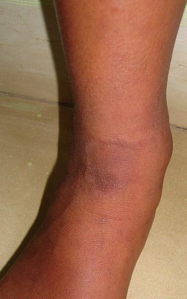
2010). Typically inflammation resolves with folds are affected) (Figs. 1.8, 1.9 and 1.10).
postinflammatory hypopigmentation which is Differential diagnosis: allergic contact der-
marked in pigmented skin and is a great concern matitis, intertrigo, psoriasis and atopic and sebor-
to parents (Figs. 1.1, 1.2, 1.3, 1.4 and 1.5). rhoeic dermatitis (Tüzün et al. 2015).
Differential diagnosis: napkin dermatitis, Management: the mainstay of management is
atopic eczema, Langerhans cell histiocytosis to keep the skin dry and use barrier creams or
(Leung et al. 2019). emollients to restore normal epidermis. Mild topi-
Management: a combination of steroid–anti- cal steroids are used if inflamed or itchy. Treat sec-
fungal creams in cases where inflammation is ondary bacterial or candida infection appropriately.
marked, intermittently for short periods. Advise to wear disposable diapers which are much
Hypopigmentation resolves spontaneously more absorbent, especially during the night.
within weeks to months (Victoire et al. 2019).
1.1.5 Infantile Psoriasis
1.1.3 Atopic Eczema
Napkin psoriasis with dissemination is the most
In infants, AE characteristically begins on the face common pattern in infants (question 21). All pat-
with subsequent spread to involve the torso and terns of psoriasis have been described in children:
limbs. A more nummular (discoid) pattern occurs, guttate, chronic plaque, pustular and erythroder-
particularly on the back and legs, especially in mic, but severe disease and joint involvement are
toddlers. AE in the majority of infants clears over relatively rare (Burden and Kirby 2016).
time. Nearly half of children with early AE are in Differential diagnosis: napkin dermatitis,
complete remission by age 3 years. A significant infantile seborrhoeic dermatitis and irritant con-
number of children were found to develop AD tact dermatitis.
shortly after their ISD diagnosis. This finding Treatments: mild topical steroids, often in
demonstrates a strong association in the clinical combination with an anticandidal agent, and
course between the two diseases or indicates that emollients usually suffice in most children.
the two diseases may be in the same clinical spec-
trum (Alexopoulos et al. 2014).
Clinical presentation: Mildly itchy scales 1.1.6 Miliaria
mainly on the cheeks and on extensors, which
change to flexural involvement when the child Miliaria is due to blockage of eccrine sweat
grows (Figs. 1.6 and 1.7). ducts. Immature sweat ducts are an important
Management: Exclusive breastfeeding for factor in neonates although high levels of heat
the first 6 months is advisable. This is a chronic and humidity are important at any age. It is sub-
recurrent problem in many. Topical steroid should divided into three subtypes dependent on the
be used sparsely with frequent emollients. level of blockage: miliaria crystallina (stratum
corneum), miliaria rubra (mid-epidermal) and
miliaria profunda (dermal–epidermal junction)
1.1.4 Napkin Dermatitis (Paige 2016; Dixit et al. 2012).
Prolonged contact with urine induces an irritant Clinical Presentation
erythema, which may break down to form ero-
sions if untreated. Miliaria crystallina presents as crops of clear,
Clinical presentation: dermatitis is limited to thin-walled, superficial vesicles 1–2 mm in diame-
the areas in contact with the irritant, while the ter, without associated erythema, resembling drops
skin folds may be spared (in contrast to sebor- of water. These are exceedingly delicate and gener-1 Dermatoses of the Neonate and Infancy 11
a b
Fig. 1.1 (a, b) Cradle cap. Large flakes of brownish scale crust. There is usually minimal inflammation, but the eye-
are seen on the scalp, especially over the vertex and fron- brows may be involved
tal regions, and may become matted into large plaques of
Fig. 1.2 Infantile seborrhoeic dermatitis. More macer- Fig. 1.3 Infantile seborrhoeic dermatitis. Typically the
ated erythema occurs in the skin folds, especially the neck inflammation resolves with transient hypopigmentation,
and the inguinal regions which is pronounced in children with pigmented skin12 R. R. Ranawaka
Fig. 1.5 Infantile seborrhoeic dermatitis. Postin
flammatory depigmentation is marked since this
Fig. 1.4 Infantile seborrhoeic dermatitis. Note skin folds child’s skin colour is very dark
are affected (in contrast to napkin dermatitis where skin
folds are spared)
a b
Fig. 1.6 (a, b) Atopic eczema. In infants, atopic eczema characteristically begins on the face1 Dermatoses of the Neonate and Infancy 13
a b
Fig. 1.7 (a, b) Atopic eczema on flexures in a 9-month-old baby girl
Fig. 1.9 Napkin dermatitis in an infant. This dermatitis is
usually confined to nappy areas
Fig. 1.8 Napkin dermatitis in a 3-month-old infant.
Note skin folds are spared in contrast to seborrhoeic der-
matitis or infantile psoriasis. Dermatitis can spread up to
lower back or abdomen when infants sleep on a rubber
mattress14 R. R. Ranawaka
ally rupture within 24 h and are followed by bran-
like desquamation. Lesions are asymptomatic.
Miliaria rubra (prickly heat, sweat rash) com-
prises erythematous papules and papulovesicles
about 1–4 mm in diameter, on a background of
macular erythema. Lesions can be itchy or sore.
Miliaria pustulosa: Some of the miliaria
rubra lesions are pustular, but not infected.
Miliaria profunda (granulomatous giant cen-
trifugal variant) is very uncommon in neonates as
it usually occurs in adults where there have been
repeated episodes of miliaria rubra.
Management: Miliaria crystalline improves
spontaneously when sweat ducts are mature.
Remove from conditions of high heat/humidity
Fig. 1.10 Napkin dermatitis with candida superinfection
in a preterm neonate (photographed by Dr. Maduranga and any occlusive clothing or bedding (Figs. 1.11,
Mendis, medical officer, neonatology unit, General 1.12 and 1.13).
Hospital Kalutara, Sri Lanka)
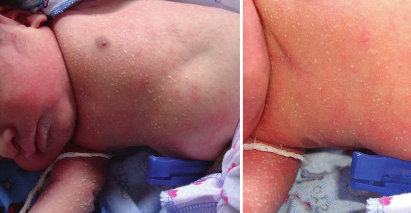
Fig. 1.11 Miliaria crystallina resembling drops of water in a 10-day-old baby on the face and the trunk1 Dermatoses of the Neonate and Infancy 15
Fig. 1.12 Miliaria crystallina in a new born baby
1.2 Infections and Infestations
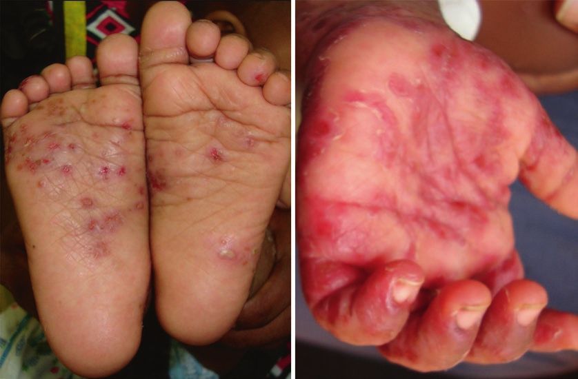
1.2.1 and, Foot and Mouth
H
Disease
Coxsackie A viral infection, most usually A16,
and infection with A6, Coxsackie B and enterovi-
rus 71 have also been described. Highly conta-
gious and widespread outbreaks occur every year.
Clinical presentation: Mild malaise, oral
ulcers and hand and foot vesicular eruption.
Crops of vesicles are grouped in elbows and
knees. Very rarely, a widespread vesicular erup-
tion occurs over the buttocks, trunk and perioral
area. In some, the eruption may be papular or
maculopapular without vesicles. When it resolves
it may form marked scaling or postinflammatory
hyperpigmentation which settles spontaneously
(Sterling 2016; Higgins and Glover 2016)
(Figs. 1.14, 1.15, 1.16 and 1.17).
Fig. 1.13 Miliaria pustulosa in an older child16 R. R. Ranawaka
Fig. 1.14 Hand, foot and mouth disease showing small vesicles with surrounding erythema on the sole and palms
a b
Fig. 1.15 (a, b) Hand, foot and mouth disease, vesicles are commonly crops around knees and elbows, but it can spread
to all over the bodyYou can also read