Complications in Lasers, Lights, and Radiofrequency Devices
←
→
Page content transcription
If your browser does not render page correctly, please read the page content below
340

      Complications in Lasers, Lights, and
      Radiofrequency Devices
      Naief AlNomair, M.D. 1         Rachel Nazarian, M.D. 1           Ellen Marmur, M.D. 1

      1 Mount Sinai–Dermatology, New York, New York                        Address for correspondence and reprint requests Naief AlNomair,
                                                                           M.D., Mount Sinai, School of Medicine, Dermatology Department,
       Facial Plast Surg 2012;28:340–346                                   5 East 98th St., 5th Floor, New York, NY 10029
                                                                           (e-mail: naif35@yahoo.com).

        Abstract                           Lights, lasers, and radiofrequency are unique sources of energy that are increasingly
                                           utilized for therapeutic and cosmetic purposes. As the indications for these tools
                                           continue to increase and their use expands beyond physicians to aestheticians,
        Keywords                           physician-extenders, and technicians, the incidence of complications has also risen. It
        ►   lasers                         is imperative that operators of these tools be as familiar with the management of
        ►   complications                  potential complications as they are with their usage and indications. This article serves
        ►   radiofrequency                 as a review of potential complications encountered with usage of lasers, lights, and
        ►   side effects                   radiofrequency devices in dermatology.

                                                                                                                                               Downloaded by: NYU. Copyrighted material.
      The use of lasers, lights, and radiofrequency in dermatologic        pose a higher risk of nonspecific tissue damage and thermal
      surgery has rapidly advanced since the device was first               injury. The pulsed and Q-switched (QS) systems adhere most
      introduced more than four decades ago.1 Continued innova-            closely to the principles of selective photothermolysis and
      tions in optical technology and refinement of existing devices        result in the highest degree of selective destruction with the
      have allowed for new developments to meet growing con-               lowest risk of scarring from excessive thermal diffusion.4
      sumer demands for effective and safe laser therapies.2 Such             One of the greatest advances in laser surgery in recent years
      innovations include the expanding use of specific wave-               has been the development of skin-cooling technologies, which
      lengths, pulse durations, and cooling strategies; the introduc-      protect the epidermis from heat injury and allow for the use of
      tion of nonablative rejuvenation techniques, including               higher fluences.5,6 Failure of skin cooling, whether due to device
      radiofrequency, intense pulsed light, and other light sources;       or operator error, poses risks of thermal injury: misalignment of
      the use of fractional resurfacing; and the combinations of           the cryogen spray tip, angling of the hand piece, and inadequate
      laser, light, and radiofrequency technologies.                       duration of cryogen spray have been described.7,8
          Continued refinement in laser, lights, and radiofrequency            Specific eye protection is essential for all treatments. The
      technology and technique has made safe treatment of photo-           retina and cornea are targets for laser damage. Also, corneal
      induced facial rhytides, dyschromias, lentigines, and atrophic       abrasions may occur due to contact with topical numbing
      scars possible, with low incidences of adverse sequelae.             preparations, or inadvertent rubbing of the eye by the patient
      Derived from the principle of selective photothermolysis             when the eyelid is numb. Management of all eye complica-
      elucidated by Anderson and Parrish in 1983, an entire gener-         tions should involve a timely consultation with an
      ation of lasers has been developed to improve tissue specific-        ophthalmologist.
      ity.1 Selective photothermolysis is accomplished through the            Although complication rates reported in association with
      use of appropriate wavelength and pulse duration best ab-            cutaneous lasers, light, and radiofrequency are consistently
      sorbed by specific chromophores such as melanin or hemo-              low, many potential adverse reactions may occur; even in the
      globin. However, not all lasers, light, and radiofrequency           hands of an experienced laser surgeon, unexpected side
      sources adhere to this principle. Continuous wave lasers are         effects may result. Most complications are due to problems
      least selective and may produce unwanted tissue damage and           arising from these three categories: technique, mechanical
      scarring through heat conduction to neighboring normal skin.         defect, or patient compliance. Complicating factors include
      Quasi-continuous wave lasers limit excessive thermal de-             poor intraoperative technique, failure to adhere to a strict
      struction by delivery of a series of brief laser pulses but still    postoperative recovery regimen, the internal operations and

                                Issue Theme Avoiding and Managing          Copyright © 2012 by Thieme Medical      DOI http://dx.doi.org/
                                Complications in Facial Plastic Surgery;   Publishers, Inc., 333 Seventh Avenue,   10.1055/s-0032-1312701.
                                Guest Editor, Steven J. Pearlman, M.D.,    New York, NY 10001, USA.                ISSN 0736-6825.
                                F.A.C.S.                                   Tel: +1(212) 584-4662.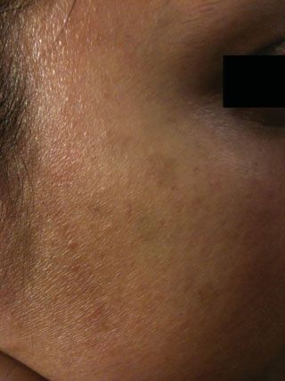
Complications in Lasers, Lights, and Radiofrequency Devices AlNomair et al. 341
tanning; this technique has recently fallen out of favor in
many demographics.
Transient acneiform eruptions and milia are relatively
common for nonfractionated laser resurfacing, with up to
80% of cases developing with the former and more than 14%
developing with the latter.9
Rate of reactivation of HSV infection with traditional
resurfacing laser treatment range from 2 to 7%, and requires
a prophylactic antiviral treatment for patients with positive
history of HSV. The rate of bacterial infection tends to be low
(0.5 to 4.5% of cases) and includes the likelihood of pathogen
overgrowth, primarily Staphylococcus aureus, Pseudomonas
aeruginosa, and Enterobacter cloacae.8,9
The risk of these untoward side effects are significantly
reduced when appropriate pretreatment patient selection is
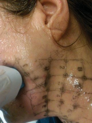
Figure 1 Thermage operational defect (missing a metal contact
inside clip on right side). May have led to eyelid burn. Other major made, proper surgical technique is used, and the post-treat-
errors include failure to use coupling fluid, failure to ensure four ment recovery period occurs under optimal healing
corners of hand piece are in full contact with skin, treating areas of conditions.4
broken skin.
Fractional Photothermolysis
mechanical status of the equipment used (►Fig. 1), and the The concept of fractional photothermolysis, coined in 2004 by
Downloaded by: NYU. Copyrighted material.
individual characteristics of each patient undergoing treat- Manstein and colleagues, has revolutionized the field of laser
ment (e.g., Fitzpatrick skin phototype, degree of ultraviolet skin resurfacing by providing the ability to obtain significant
light exposure, pretreatment, and medical condition).3 clinical results with minimal posttreatment recovery.9,11 This
technique generates microthermal treatment zones in the
dermis, which are columns of thermally denatured skin of
Resurfacing Lasers
controlled width and depth.9
The introduction of ablative laser skin resurfacing techniques Side effects and complications of fractional laser resurfac-
with high-energy, pulsed carbon dioxide (CO2) and erbium- ing have been shown to be distributed among different age
doped yttrium aluminum garnet (Er:YAG) devices in the mid- groups, body locations, cutaneous conditions, and skin
1990s was met with great enthusiasm. They displayed excel- phototypes.
lent clinical outcomes in the treatment of atrophic scars and Immediate posttreatment erythema or urticaria is an
photodamaged facial skin, including rhytides, lentigines, and expected consequence of fractionated laser skin resurfacing
dermal elastosis, but the prolonged recovery and risk of that usually resolves within 3 to 4 days (►Figs. 2, 3). Pro-
potential side effects ultimately made them less attractive longed erythema is defined as posttreatment erythema that
treatment alternatives.9 persists longer than 4 days with nonablative resurfacing and
Resurfacing with either the high-energy, pulsed, or beyond 1 month with ablative treatment. It has been re-
scanned CO2 or Er:YAG laser results in complete epidermal ported in fewer than 1% of nonablative patients and more
ablation and upper papillary dermal destruction with colla- than 12.5% of ablative laser-treated patients, although ery-
gen remodeling. As a result, newly resurfaced skin lacks an thema typically resolves in these latter cases within
intact epithelium, producing an exposed weeping wound 3 months.9
with copious serous discharge. Complete reepithelialization Viral, bacterial, and fungal infections are rare, but usually
occurs within an average of 8.5 days for CO2 laser-resurfaced occur during the first postoperative week and require proper
skin, compared with 5.5 days for Er:YAG laser-treated skin. identification and treatment to avoid further complications.9
Complications of cutaneous laser resurfacing range from The rate of HSV infection, the most common type of infection
transient side effects to permanent disfigurement.3 after fractional laser skin resurfacing, has been reported in 0.3
Mild complications include prolonged erythema, acne, to 2% of cases.9,12,13 Patients may not present with classic
milia formation, contact dermatitis, and pruritus. Complica- herpetiform vesicopustules but instead may demonstrate
tions of moderate severity include reactivation of herpes only superficial erosions that develop during the first week
simplex virus (HSV), superficial bacterial and fungal infec- after treatment.9,13 To minimize the risk of HSV reactivation
tions, postinflammatory hyperpigmentation, and delayed- with fractional resurfacing, antiviral prophylaxis should be
onset hypopigmentation. The most severe complications administered when a prior history of facial HSV is docu-
associated with resurfacing include hypertrophic scar forma- mented or if full-face ablative laser procedures are
tion, ectropion, and disseminated infection.3,4,8,10 Resurfac- performed.9
ing treatment limited to the face, and excluding the neck and Bacterial infection is rarely observed after fractionated
chest, was noted to leave a visible hypopigmented line of skin resurfacing, with only 0.1% of all treated cases docu-
demarcation above the angle of the mandible unresponsive to mented to develop impetigo.9,12,13 Excessive wound
Facial Plastic Surgery Vol. 28 No. 3/2012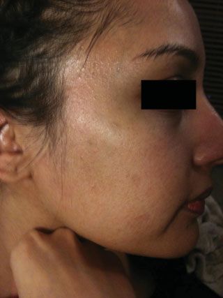
342   Complications in Lasers, Lights, and Radiofrequency Devices          AlNomair et al.

                                                                        remains primarily gentle acne medications or a medrol dose
                                                                        pack in more severe cases.
                                                                           Postinflammatory hyperpigmentation is much less fre-
                                                                        quent with fractional laser skin resurfacing than with other
                                                                        ablative procedures, but is observed in 1 to 32% of patients,
                                                                        depending on the system used, parameters applied, and skin
                                                                        phototypes treated.9,12 Patients with darker skin phototypes
                                                                        (Fitzpatrick III to VI) have a higher likelihood of developing
                                                                        postinflammatory hyperpigmentation. In general, fractional
                                                                        resurfacing of darker skin should use higher fluencies, lower-
                                                                        density settings, and longer treatment intervals. Hypopig-
                                                                        mentation is an extremely uncommon complication of frac-
                                                                        tional laser skin resurfacing.4,9
                                                                           Hypertrophic scarring is another known and rare compli-
                                                                        cation of fractional ablative resurfacing. There are several
      Figure 2 Normal post-Fraxel erythema.
                                                                        potential explanations for hypertrophic scarring, including
                                                                        the use of excessively high-energy densities, postoperative
                                                                        infection of the skin, and lack of technical finesse. The neck is a
      occlusion during the early postoperative period can enhance       well-recognized site that is especially susceptible to the
      the likelihood of pathogen overgrowth, primarily S. aureus        development of scarring because of the small number of
      and P. aeruginosa.9 Increased pain, focal intense erythema,       pilosebaceous units and poor vasculature in this region,
      increased exudate, and erosions with crusting should alert        which are essential for wound healing. In addition, the thin

                                                                                                                                             Downloaded by: NYU. Copyrighted material.
      the physician to the possibility of bacterial superinfection      skin of the neck renders it more susceptible to thermal injury.
      that usually presents 1 to 3 days after treatment.9,13 Treat-     Other scar-prone anatomic locations that require more con-
      ment of bacterial infection includes topical antibiotics, such    servative treatment protocols include the periorbital and
      as mupirocin, or oral antibiotics, including antistaphylococcal   mandibular regions and other areas over bony
      penicillins, amoxicillin/clavulanate, cephalosporins, and         prominences.9,14
      macrolides.                                                          Other rare complications of fractional ablative resurfacing
          Although rarely seen, cutaneous candidiasis induced by        include cicatricial ectropion, multiple eruptive keratoacan-
      Candida albicans is the most common fungal infection re-          thomas, and delayed purpura.15,16
      ported after fractional laser skin resurfacing (usually 7 to
      14 days after treatment) and should be treated with antifun-
                                                                        Vascular-Specific Lasers
      gal medications, such as topical ketoconazole, to prevent
      scarring.9                                                        Vascular-specific laser systems target intravascular oxyhe-
          Acneiform eruptions have been documented (2 to 10%)           moglobin to effect destruction of various congenital and
      with fractional skin resurfacing, and the incidence of milia      acquired vascular lesions. Lasers and light sources that have
      development has been reported in as many as 19% of treated        been used to treat vascular lesions include the argon (488/
      patients.4,9,12 In some cases, this may represent a simple        514 nm), argon-pumped tunable dye (577/585 nm), potassi-
      folliculitis due to the wound-healing response. Treatment         um-titanyl-phosphate (KTP; 532 nm), krypton (568 nm),
                                                                        copper vapor/bromide (578 nm), pulsed dye laser (PDL;
                                                                        585 to 595 nm), and neodymium:yttrium-aluminum-garnet
                                                                        (Nd:YAG; 532/1064 nm) lasers and the intense pulsed light
                                                                        (IPL) source (515 to 1200 nm).4
                                                                           The most common complications of KTP are linear crusting
                                                                        of skin overlying treated blood vessels, transient pigmentary
                                                                        changes, mild fibrosis, cutaneous depressions, and
                                                                        hypopigmentation.4,8
                                                                           The most common complications of PDL include transient
                                                                        purpura, erythema, edema, and dyspigmentation.4 These
                                                                        complications are typically self-resolving and last days to
                                                                        months.
                                                                           Because absorption of both oxyhemoglobin and melanin is
                                                                        reduced in the near-infrared region of the electromagnetic
                                                                        spectrum, significantly higher fluences are required for
                                                                        photocoagulation of deeper vessels. The use of proper cooling
                                                                        methods and avoidance of overlapping pulses is essential
                                                                        with long-pulsed 1064-nm lasers to prevent injury associated
      Figure 3 Normal post-Fraxel urticaria.                            with inadequate dissipation of heat.2 The most common

      Facial Plastic Surgery   Vol. 28   No. 3/2012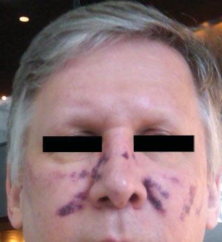
Complications in Lasers, Lights, and Radiofrequency Devices                      AlNomair et al.         343

complications of Nd:YAG are purpura, vesiculation, superfi-                Patients should be informed of all potential and oft-seen
cial thrombosis, transient hyperpigmentation, and atrophic             complications before they are anaesthetized. It is also recom-
scarring, most notably over the perinasal area.4                       mended to discuss postprocedural care instructions before
   To minimize the risk while using different kinds of vascular        any numbing or procedures are performed to ensure under-
lasers, the lasers should be used with extra caution on                standing by the patient and to address questions they may
patients with recent sun exposure or in those with intrinsi-           have regarding postprocedural practice.
cally dark skin tones, particularly with KTP and IPL. Avoid
applications of excessive energy densities and/or pulse stack-
                                                                       Pigment-Specific Lasers
ing except when indicated (e.g., PDL stacking while treating
warts).                                                                High-energy, ultrashort pulsed laser systems can successfully
                                                                       lighten or eradicate a wide variety of benign epidermal
                                                                       and dermal pigmented lesions and tattoos with minimal
Intense Pulsed Light
                                                                       risk of untoward effects. Epidermal lesions (solar lentigines,
The IPL system emits noncoherent broadband light in the                ephelides, café au lait macules, and seborrheic keratoses),
range of 515 to 1200 nm. Indications for IPL treatment are             dermal and mixed epidermal/dermal lesions (melanocytic
numerous and include pigmented and vascular lesions, pho-              nevi, blue nevi, nevi of Ota/Ito, infraorbital hyperpigmenta-
toaging, and photoepilation.17                                         tion, drug-induced hyperpigmentation, Becker's nevi, and
   Common side effects after treatment with IPL include mild           nevus spilus), and tattoos (amateur, professional, and trau-
postoperative erythema, edema, and ecchymosis (►Fig. 4).               matic) have all been shown to be amenable to laser
More serious superficial burns and scarring can occur with              treatment.4
aggressive treatment parameters.4,17 Because of the potential              The three most common types of QS lasers used in practice
for competition between different chromophores with IPL                today are the 694-nm ruby, 755-nm alexandrite, and 532/

                                                                                                                                                             Downloaded by: NYU. Copyrighted material.
systems, this device should be used cautiously in type III to VI       1064-nm Nd:YAG. Each produces an immediate ash-white
skin and suntanned skin.8,17                                           epidermal tissue response when targeting cutaneous pig-
   Care should be taken to maintain slight overlap between             ment or tattoo ink. Other possible treatment side effects
pulses to ensure confluent treatment of the target area and             include punctuate bleeding, tissue splatter, edema, pruritus,
avoid “skip” (untreated) areas. These “skip” areas may leave a         vesiculation, textural changes, skin crusting, and transient
footprint of dyspigmentation in contrast to neighboring                hypopigmentation.8
treated areas.17                                                           Potential complications reported from QS laser irradiation
   Most complications can be minimized by use of appropri-             of tattoos include generalized cutaneous allergic reactions (to
ate treatment settings, cooling methods, and proper patient            cadmium- or mercury-containing inks), hemorrhagic bullae
selection. Patients should also be advised regarding photo-            formation (particularly in the distal extremities), irreversible
protection with sun avoidance and sunscreen to minimize                darkening of cosmetic tattoos (particularly white, flesh-tone,
postinflammatory hyperpigmentation after an erythematous                pink/red inks due to presence of iron oxide or titanium
treatment response.8,17                                                dioxide pigments that undergo a laser-induced oxidation
                                                                       reduction reaction), and rarely scarring and tissue fibrosis
                                                                       (particularly with inadequate wound management).8 Laser
                                                                       conversion of ferric oxide to ferrous oxide in tattoo ink leads
                                                                       to formation of the insoluble black pigment. Patients who
                                                                       have received gold therapy in the past and undergo QS laser
                                                                       treatment may also experience hyperpigmentation due to
                                                                       alteration of gold particles still present in the skin.
                                                                           Treatment of allergic reactions may be managed with oral
                                                                       or midpotency topical corticosteroids and oral antihist-
                                                                       amines. Rarely, a granulomatous allergic reaction can occur
                                                                       with subsequent hypertrophic scar formation. Intralesional
                                                                       steroid injections or occlusion and pressure therapy can be
                                                                       used to reduce the bulky nature of such lesions without
                                                                       further worsening of the inciting allergic reaction. Skin
                                                                       dyspigmentation that results from QS laser irradiation is
                                                                       managed in much the same way as dyspigmentation caused
                                                                       by other forms of laser treatment.4

                                                                       Laser Hair Removal
                                                                       The most common lasers used for the reduction of hair
Figure 4 Ecchymosis after a recently calibrated intense pulsed light   include the long-pulsed ruby (694 nm), alexandrite
laser. Resolved in 10 days.                                            (755 nm), diode (800 nm), and Nd:YAG (1064 nm) lasers,

                                                                                                      Facial Plastic Surgery    Vol. 28   No. 3/2012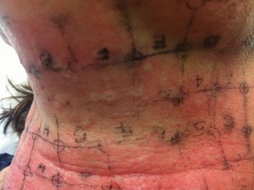
344   Complications in Lasers, Lights, and Radiofrequency Devices            AlNomair et al.

                                                                                                                                              Downloaded by: NYU. Copyrighted material.
      Figure 5 Postinflammatory hyperpigmentation 3 months after ruby      Figure 6 Close-up of postinflammatory hyperpigmentation after ruby
      laser treatment due to failure to wear sunscreen.                   laser treatment.

      as well as IPL (515 to 1200 nm) sources. These devices are          phenomenon has been reported when low (subthreshold)
      most effective for hair removal because of their ability to         fluences are delivered to a susceptible patient, triggering hair
      target melanin in the hair shaft and follicle and to penetrate to   induction rather than follicular destruction. Although laser-
      the appropriate dermal depth to effect selective follicular         induced paradoxical hair growth responds well to subsequent
      destruction. Complications after photoepilation are influ-           laser treatments at moderate to high fluences, all women
      enced by skin type, body location, seasonal variations, and         undergoing laser-assisted hair removal on the face or neck
      patient history of recent sun exposure. The extremities tend        should be informed of the possibility of this unwanted
      to suffer the most side effects, where as sun-protected areas,      reaction.4
      such as the axillary and inguinal regions, suffer the least. Side
      effects of laser-assisted hair removal are usually minor and
                                                                          Monopolar Radiofrequency
      transient (►Figs. 5 and 6).
         The most common reactions include pain during treat-             Radiofrequency tissue tightening is a unique nonsurgical
      ment, transient erythema, and perifollicular edema; vesicle         treatment of skin laxity and tissue contour. Controlled heat
      formation, pigmentary alteration, burning, and scarring have        modification of collagen stimulates a wound-healing
      also been documented (►Fig. 7). Less obvious reactions
      include reticulate erythema and ocular complications. Most
      of the latter complications have occurred in individuals who
      are either tanned or have darker skin phototypes (IV to
      VI).7,8,18 Because the 1064-nm wavelength is less efficiently
      absorbed by endogenous melanin, significantly fewer inci-
      dences of blistering, crusting, and dyspigmentation occur
      after Nd:YAG laser treatment of patients with darker or
      tanned skin. Complication rates will increase as skin pigment
      increases and as the power used increases. However, even in
      light-skinned individuals, with proper treatment parameters,
      complications and side effects can arise.19
         Paradoxical hair growth is a possible side effect of photo-
      epilation occurring in selected patient populations and body
      areas.15 It occurs predominately on the face and neck of
      women of Mediterranean ancestry with darker skin photo-
      types. The border of the treated area and the immediately           Figure 7 Postinflammatory hypopigmentation 1 week after diode
      adjacent untreated skin are most commonly affected. The             treatment.

      Facial Plastic Surgery   Vol. 28   No. 3/2012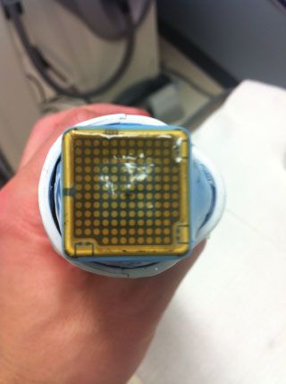
Complications in Lasers, Lights, and Radiofrequency Devices AlNomair et al. 345
response with immediate contraction of collagen fibrils and
delayed formation of new collagen.17 The complications
range from immediate transient erythema and swelling to
textural changes and cutaneous depressions.8 Late side ef-
fects such as cutaneous depressions can occur weeks or
months postoperatively and result from overheating of adi-
pose tissue and fibrous septae with monopolar radiofre-
quency devices (►Fig. 8). Caution must be used when
treating over bony prominences or thin skin such as the
forehead and temples; drawing skin away from bony prom-
inences during treatment is recommended to minimize
contour irregularities. Treating with lower fluences and
multiple passes is advised to decrease the risk of adverse Figure 9 Atrophic scar on left cheek due to Thermage over a cyst
1 year prior.
events. Mild dermal depressions often resolve spontaneously
over time (►Fig. 9). More severe contour irregularities and
scarring may improve with surgical subscision, autologous fat
transfer, or cosmetic fillers (►Fig. 10).17
As with other laser and light devices, malfunctioning of the
equipment may on occasion occur. The tools, including hand
pieces, should be routinely evaluated and inspected for areas
of damage or parts needing repair (►Figs. 11 and 12).
Downloaded by: NYU. Copyrighted material.
Conclusion
Although undesired, complications are a reality with clinical
application of lasers, lights, and radiofrequency. Understand-
ing the principles and mechanics of these tools can help
minimize unwanted side effects, and it is imperative that all
operators are aware of the spectrum of adverse effects and
complication profile specific to each laser and energy source
before use. Furthermore, physician and physician-extenders
should only utilize these tools after they have a full under-
standing of possible complications and are comfortable with
appropriate methods of treatment. These complications
should be discussed in detail with patients, before the influ-
ences of anesthesia or numbing are introduced, and a full
informed consent should be taken before the procedure.
Thorough documentation of all discussion, preoperative pho-
tos, and procedure risks and benefits is essential. It is recom- Figure 10 Thermage. Do not treat areas of acne excoriee.
mended to establish templates of these discussions for
completion and efficiency.
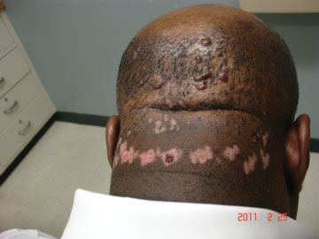
Figure 8 Thermage scar due to treatment by physician-extender over Figure 11 This patient developed spot blanching during treatment
an inflamed cyst. with a monopolar radiofrequency device.
Facial Plastic Surgery Vol. 28 No. 3/2012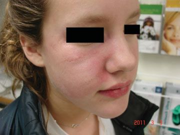
346 Complications in Lasers, Lights, and Radiofrequency Devices AlNomair et al.
References
1 Anderson RR, Parrish JA. Selective photothermolysis: precise
microsurgery by selective absorption of pulsed radiation. Science
1983;220:524–527
2 Alam M, Dover JS, Arndt KA. Energy delivery devices for cutaneous
remodeling lasers, sights, and radio waves. Arch Dermatol 2003;
139:1351–1360
3 Alster TS, Lupton JR. Prevention and treatment of side effects and
complications of cutaneous laser resurfacing. Plast Reconstr Surg
2002;109:308–316; discussion 317–318
4 Alster TS, Khoury RR. Treatment of laser complications. Facial Plast
Surg 2009;25:316–323
5 Nelson JS, Milner TE, Anvari B, et al. Dynamic epidermal cooling
during pulsed laser treatment of port-wine stain. A new method-
ology with preliminary clinical evaluation. Arch Dermatol
1995;131:695–700
6 Kelly KM, Nanda VS, Nelson JS. Treatment of port-wine stain
birthmarks using the 1.5-msec pulsed dye laser at high fluences
in conjunction with cryogen spray cooling. Dermatol Surg 2002;
28:309–313
7 Kelly KM, Svaasand LO, Nelson JS. Optimization of laser treatment
safety in conjunction with cryogen spray cooling. Arch Dermatol
2003;139:1372–1373
8 Willey A, Anderson RR, Azpiazu JL, et al. Complications of laser
dermatologic surgery. Lasers Surg Med 2006;38:1–15
9 Metelitsa AI, Alster TS. Fractionated laser skin resurfacing treat-
Downloaded by: NYU. Copyrighted material.
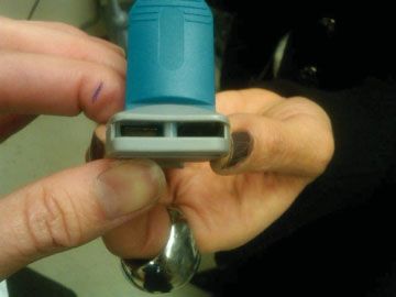
Figure 12 The tip was noted to have a black spot responsible for the ment complications: a review. Dermatol Surg 2010;36:299–306
dysfunction that developed midway through the treatment. The 10 Alster TS. Cutaneous resurfacing with CO2 and erbium: YAG lasers:
patient healed well within 2 weeks. preoperative, intraoperative, and postoperative considerations.
Plast Reconstr Surg 1999;103:619–632; discussion 633–634
11 Manstein D, Herron GS, Sink RK, Tanner H, Anderson RR. Fractional
If the operator is unfamiliar with maintenance or specific photothermolysis: a new concept for cutaneous remodeling using
upkeep required for these machines, individual companies microscopic patterns of thermal injury. Lasers Surg Med 2004;34:
426–438
should be contacted to determine a schedule of calibration
12 Graber EM, Tanzi EL, Alster TS. Side effects and complications of
and the recommended maintenance. Often the operator is fractional laser photothermolysis: experience with 961 treat-
familiar with settings, but not with the engineering of the ments. Dermatol Surg 2008;34:301–305; discussion 305–307
laser or device. Therefore, if an error occurs, the operator may 13 Setyadi HG, Jacobs AA, Markus RF. Infectious complications after
not recognize the cause such as water levels, ventilation, nonablative fractional resurfacing treatment. Dermatol Surg
2008;34:1595–1598
electrical contact, or cooling. Understanding the specific
14 Fife DJ, Fitzpatrick RE, Zachary CB. Complications of fractional
mechanics of the device—wavelength, energy source, power,
CO2 laser resurfacing: four cases. Lasers Surg Med 2009;41:179–
common mechanical errors—greatly reduces clinical side 184
effects. 15 Metelitsa AI, Alster TS. Fractionated laser skin resurfacing treat-
Awareness of the varying responses, and varying risks, ment complications: a review. Dermatol Surg 2010;36:299–306
between skin types may spare the physician and patient 16 Mamelak AJ, Goldberg LH, Marquez D, Hosler GA, Hinckley MR,
Friedman PM. Eruptive keratoacanthomas on the legs after frac-
unwanted side effects from these treatments, most especially
tional photothermolysis: report of two cases. Dermatol Surg
patients with darker skin types. Knowing which patients are 2009;35:513–518
ideal for each procedure and appropriately selecting from this 17 Dawson E, Willey A, Lee K. Adverse events associated with non-
patient population will also significantly decrease chances of ablative cutaneous laser, radiofrequency, and light-based devices.
undesired side effects. In addition, consistent use of proper Semin Cutan Med Surg 2007;26:15–21
surgical technique with every procedure and investing the 18 Vano-Galvan S, Jaen P. Complications of nonphysician-supervised
laser hair removal: case report and literature review. Can Fam
time to educate all patients of the posttreatment recovery
Physician 2009;55:50–52
period will ensure optimal healing conditions, and ultimately, 19 Weisberg NK, Greenbaum SS. Pigmentary changes after Alexan-
minimize risk of complications. drite laser hair removal. Dermatol Surg 2003;29:415–419
Facial Plastic Surgery Vol. 28 No. 3/2012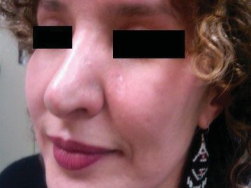
You can also read