Automated sequential chromogenic IHC double staining with two HRP substrates - bioRxiv
←
→
Page content transcription
If your browser does not render page correctly, please read the page content below
bioRxiv preprint first posted online Mar. 14, 2018; doi: http://dx.doi.org/10.1101/276501. The copyright holder for this preprint
       (which was not peer-reviewed) is the author/funder, who has granted bioRxiv a license to display the preprint in perpetuity.
                                        It is made available under a CC-BY 4.0 International license.

Automated sequential chromogenic IHC double
staining with two HRP substrates.
Kenneth H Petersen*, Jesper Lohse, Lasse Ramsgaard.
Agilent Technologies, Produktionsvej 42, Glostrup, Denmark
* Corresponding author

Abstract
Automated IHC double staining using DAB and HRP                                  However, there are drawbacks to the AP substrate system.
Magenta is illustrated utilizing a new acid block with                           First, Fast Red stains rapidly dissolve in ethanol and most
sulfuric acid to prevent cross-reactivity. Residual cross-                       organic mounting media. Second, the Fast Red diazonium
reactivity in double staining is determined to arise from                        salt can react with a range of other substances present
chromogenic-bound antibodies and amplification system                            in the tissue section. This includes the DAB stain which
during the first part of the double staining.                                    becomes darker after incubation with Fast Red. Third, Fast
                                                                                 Red chromogen must be used within 30 minutes of mixing.
Introduction                                                                     For automated staining systems, this requires onboard
                                                                                 mixing of the Fast Red reagents or alternatively the staining
A concern with many diagnostic procedures today is to                            is paused until the user can supply a freshly mixed Fast
get enough information from limited sample sizes to                              Red solution to the system.
reach an informed diagnostic decision. In pathology, using
immunohistochemistry (IHC), this is particularly important                       An alternative red chromogen, 3-amino-9-ethylcarbazole
when the biopsy is small and only a few different tests can                      (AEC) exists for the HRP enzyme system, but this is not
be run on the available tissue. The localization of different                    compatible with other peroxidase substrates due to poor
antigens in relation to each other can in some cases also be                     color contrast between reddish-brown AEC and brown DAB
important for a diagnosis. In these cases, staining the same                     (Nemes 1987).
tissue section for two antigens is a useful diagnostic tool.
                                                                                 Blue HRP substrates have previously been described.
To reduce the possibility of cross-staining between the two                      While they contrast well to DAB they are not optimal when
targets, antibodies derived from different species, usually                      hematoxylin is used as a counterstain (Petersen 2009).
rabbit and mouse, are commonly used (van der Loos 1993).
                                                                                 Recently a new magenta-colored HRP chromogen with
Additionally, the visualization of the antigens is performed
                                                                                 a sensitivity comparable to DAB has been described
using different enzymes in the visualization steps. Typically,
                                                                                 (Lohse 2016). This paper reports our findings with fully
brown and red visualization is performed using the enzymes
                                                                                 automated DAB/Magenta chromogen IHC double stains in
horseradish peroxidase (HRP) and alkaline phosphatase (AP)
                                                                                 combination with hematoxylin nuclear counterstain.
and the substrates diaminobenzidine (DAB) and Fast Red,
respectively (Malik 1982). No quenching of enzymatic activity
is required between the two stains as the two enzymes do not
function with each other’s substrate. Both colors contrast well
to the commonly used blue hematoxylin nuclear stain.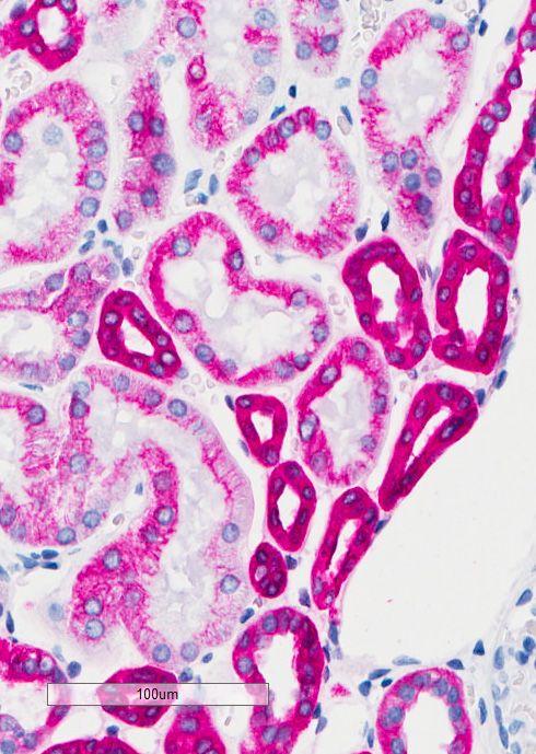
bioRxiv preprint first posted online Mar. 14, 2018; doi: http://dx.doi.org/10.1101/276501. The copyright holder for this preprint
(which was not peer-reviewed) is the author/funder, who has granted bioRxiv a license to display the preprint in perpetuity.
It is made available under a CC-BY 4.0 International license.
Results and discussion Next, we investigated if either of the two chromogens from
a completed staining was affected by a sulfuric acid block. A
When performing double staining, it is important that any Ki-67 stain of nuclei in tonsil and colon tissue was used for
cross-reactivity between reagents is eliminated. In this this experiment. Ki-67-stained nuclei can be counted using
study, we used two chromogens that are both substrates an image analysis algorithm. Slides stained with either DAB
for the HRP enzyme. This makes it important to efficiently or HRP Magenta were subjected to a subsequent block with
remove all enzyme activity from the first stain before the sulfuric acid, in concentrations ranging from 0 to 400 mM.
second stain. Peroxidase blocking is normally used to The percentage of Ki-67 positive nuclei in tonsil and colon
quench any endogenous peroxidase activity in the tissue was counted for each concentration of sulfuric acid. Figure 2
(Li 1987). However, this does not remove all HRP activity shows that the tested concentrations have no effect on the
when the tissue contains additional peroxidase activity staining of the slides, the chromogen already present on the
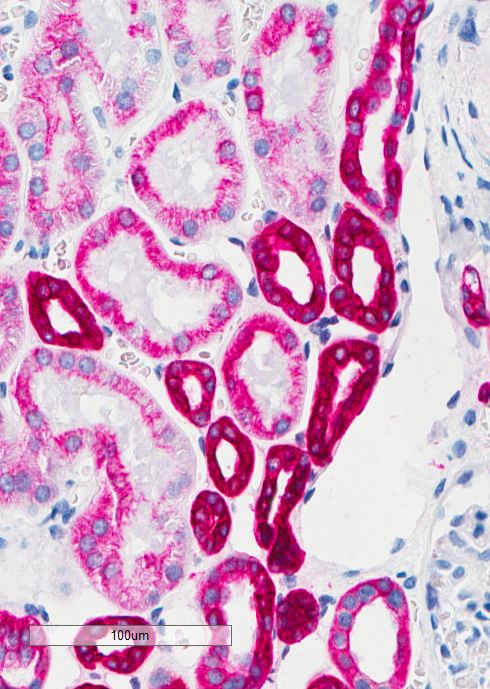
from the visualization conjugates as seen in Figure 1B, slides is not removed or faded by the acid block. We conclude
thus additional removal of peroxidase activity is needed. that sulfuric acid in the range 50-400 mM effectively removes
Sequential double staining using two HRP enzymes has residual HRP activity, without negatively impacting the
previously been described using an acidic block between intensity of a first DAB or HRP Magenta stain.
A B C D
100um 100um 100um 100um
Figure 1. Effect of different blocking washes on cytokeratin Pan staining in kidney tissue. A No wash, No DAB. B Peroxidase block then DAB. C 50 mM
H 2SO 4 then DAB. D 300 mM H 2SO 4 then DAB.
the two stains (Nakane 1968). An acid block works by Some epitopes may be sensitive to the sulfuric acid block
dissociating the antibodies from their targets so they can which could affect the intensity of the second staining. To
be washed away. investigate this, we applied an acid step prior to application
of the primary antibody and completed the staining as a
single stain with either DAB or HRP Magenta chromogen.
Sulfuric acid treatment Three different treatments before primary antibody
The efficiency of using an acidic block was tested using were compared: Block with deionized water, 50 mM or
sulfuric acid. Starting from 300 mM sulfuric acid a two-fold 300 mM sulfuric acid. A range of different antibodies
dilution series down to 20 mM sulfuric acid was made and were tested and the stains were evaluated using digital
tested with four different antibodies. The acid block step was image analysis. As seen in Figure 2B, we observed minor
set to 3 minutes, which is the minimum time on the automated changes in staining intensity which we consider to be
Dako Omnis staining system for a block or incubation step. within the uncertainty of the experiment. Here we use
The test was set up as a single chromogenic stain with HRP the stains qualitatively and the effect (if any) is therefore
Magenta chromogen followed by a 3-minute acid block, wash negligible. It is, however, important to be aware of any
buffer and then a DAB chromogen incubation. No DAB staining impact on staining intensity from either the first staining
was visible on any of the slides, the acid block removed all or the acid treatment if a double staining is to be used for
detectable enzyme activity and the morphology of the tissue quantification.
was not visibly impacted (Figure 1C+D).
2
bioRxiv preprint first posted online Mar. 14, 2018; doi: http://dx.doi.org/10.1101/276501. The copyright holder for this preprint
      (which was not peer-reviewed) is the author/funder, who has granted bioRxiv a license to display the preprint in perpetuity.
                                       It is made available under a CC-BY 4.0 International license.

                                                                                                                      Figure 2.A Percent Ki-67
 A                                                                                                                    positive nuclei in tonsil and
                                                                                                                      colon using either DAB or
                                                                                                                      Magenta as chromogen.
                                                                                                                      B Effect of sulfuric acid
                                                                                                                      block before IHC staining
                                                                                                                      and Magenta as chromogen
                                                                                                                      reported as either percent
                                                                                                                      stained nuclei or percent
                                                                                                                      stained complete membranes
                                                                                                                      (DI=deionized water).

 B

Moving forward with the double staining, we decided to                          Non co-localized targets
use a belt-and-braces approach with sulfuric acid first,                        We performed a double staining with monoclonal mouse
followed by a peroxidase block to ensure that all cross-                        antibodies against a nuclear (p63) and a membrane target
reactivity was eliminated. The two quenching reagents                           (carcinoembryonic antigen (CEA)), respectively. The guiding
are orthogonal; acid block dissociates the antibodies                           principle being that a first and a second IHC stain including the
from their targets while peroxidase block works by                              subsequent hematoxylin counterstain should ideally stand out
“overloading” the enzymes with hydrogen peroxide                                unaffected by each other since they are not co-localized. Staining
they cannot get rid of as no substrate is present. This                         using the same combination of antibodies were run both with
eventually quenches the enzyme activity. Sulfuric acid is                       HRP Magenta stain first then DAB and vice versa. Figure 3A-D
placed first in the second part of the staining protocol to                     shows these double stains.
remove as much bound antibody as possible before any
                                                                                Cross-reactivity can be seen when DAB is used as the first
remaining antibody-enzyme conjugates still in the tissue
                                                                                chromogen, either as a reddish ring around the nuclei (Figure
are quenched by treatment with the peroxidase block.
                                                                                3A) or as a change in the brown color (Figure 3B). Likewise,

                                                                                                                                                      3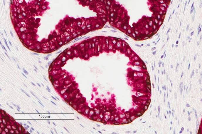
bioRxiv preprint first posted online Mar. 14, 2018; doi: http://dx.doi.org/10.1101/276501. The copyright holder for this preprint
        (which was not peer-reviewed) is the author/funder, who has granted bioRxiv a license to display the preprint in perpetuity.
                                         It is made available under a CC-BY 4.0 International license.

 A                                        B                                        C                                        D

        100um                                    100um                                    100um                                    100um

Figure 3. Carcinoembryonic antigen (CEA) and p63 double staining of tonsil tissue using monoclonal mouse antibodies. A 1st. p63/DAB 2nd. CEA/
Magenta. B 1st. CEA/DAB 2nd. p63/Magenta. C 1st. p63/Magenta 2nd. CEA/DAB. D 1st. CEA/Magenta 2nd. p63/DAB.

when the magenta color is used first, the magenta color                           peroxidase activity in the tissue. The reason for the cross-
becomes a darker red (Figure 3C+D). The purple color of the                       reactivity must be found by looking beyond the enzyme. We
nuclei in Figure 3B is expected since it is a combination of the                  believe an explanation for this unexpected color spillover is that
magenta and the blue hematoxylin colors.                                          reagents from the first stain remain in the tissue because the
                                                                                  chromogen has covalently bonded them to the tissue (Figure
The observed color spillover was not expected because our                         4A-E). When the second stain is performed the reagents are
first experiments demonstrated that we had removed all                            recognized by remaining amplification system (Figure 4C) and

                                                                                                                        Figure 4. Cross-reactivity through
 C                                                           D                                                          irreversible binding of antibodies.
                    D                                                                                                   A The first primary antibody
                                                                                                                        recognizes the first target and is
                                                                                                                        recognized by the amplification
                                                                                                                        system. B Chromogen is precipitated
                                                                                                                        and covalently binds antibodies
                                                                                                                        and amplification system to the
                                                                                                                        tissue, green circles. C The second
                                                                                                                        primary antibody is captured by
                                                                                                                        free antibodies of the amplification
                                                                                                                        system. D As the second layer of
                                                                                                                        amplification is applied, antibodies
 B                                                                                                                      captured by the first amplification
                                                                                                                        layer along with those covalently
                                                                                                                        bound to the tissue are captured.
                                                                                                                        E And color spillover results when
                                                                                                                        the next chromogen is applied.
                                                              E

 A

   4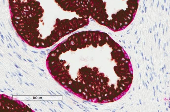
bioRxiv preprint first posted online Mar. 14, 2018; doi: http://dx.doi.org/10.1101/276501. The copyright holder for this preprint
(which was not peer-reviewed) is the author/funder, who has granted bioRxiv a license to display the preprint in perpetuity.
It is made available under a CC-BY 4.0 International license.
remaining primary antibodies are recognized by the amplification
system along with the primary antibodies captured by the
A B
remaining amplification system (Figure 4D). During the second
chromogen incubation the chromogen is then precipitated
(Figure 4E) and this gives rise to the color spillover.
To test our theory, we expanded on our initial sulfuric acid
and peroxidase block experiments. First a complete stain for
cytokeratin (CK) Pan with HRP Magenta was done and then
the slide was treated with 300 mM sulfuric acid and peroxidase
block to remove all remaining peroxidase activity. Next, we
incubated with the amplification system once more before a
final DAB incubation. As seen in Figure 5A-B, the magenta color
100um 100um
becomes visibly tainted by DAB (it is darker). We believe this
supports the theory.
Figure 5. Demonstration of residual covalently bound antibodies in kidney
The double staining of p63 and CEA was repeated; this time tissue after sulfuric acid and peroxidase block treatment by incubation
we used a rabbit antibody against CEA while the p63 antibody with DAB after block. A 300 mM sulfuric acid block. B 300 mM sulfuric
acid, peroxidase block then amplification.
was the same mouse antibody as in the initial experiment. As
seen in Figure 6A-D the colors of the double stainings became
used as the first antibody in the double staining. If the larger
untainted, regardless of which chromogen was used as the first
CK Pan area is stained first, a shielding of the co-localized
chromogen (compare Figure 3B with 6B and Figure 3D with 6D).
CK 18 target then comes into effect. This markedly lowers
the staining intensity of CK 18. In the case where DAB is
used together with CK Pan, the co-localization of the second
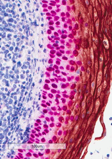
Co-localized targets
target is not easily seen (Figure 7B). The transparency of the
Our next step was to investigate how a double stain would
magenta color makes it comparably easier to see that the
look if the targets were co-localized. For this experiment, we
targets are co-localized. The HRP Magenta/DAB combination
used cytokeratin (CK) Pan and cytokeratin 18 antibodies.
is not ideal for co-localized targets, but using the more
CK 18 is not recognized by the CK Pan antibody, but is co-
transparent HRP Magenta as the first chromogen increases
localized with CK Pan in prostate glands. Figure 7A-D shows
the likelihood that co-localized targets are detected due to
the result from the cytokeratin double stainings. The CK Pan
tainting of the magenta color. When DAB was used as the
antibody stains a slightly larger area of the prostate epithelium
first chromogen only the expected areas for each antibody
than CK 18. This gives a rim of either HRP Magenta stained
were stained and the colors are not visibly tainted. We ascribe
CK Pan (Figure 7A) or DAB-stained CK Pan around the CK
the observed difference to the shielding effect that DAB
18 stain (Figure 7C). The rim is only visible when CK 18 is
A B C D
100um 100um 100um 100um
Figure 6. Rabbit polyclonal CEA and mouse monoclonal p63 antibody double staining of tonsil tissue. A 1st. p63/DAB 2nd. CEA/Magenta. B 1st. CEA/
DAB 2nd. p63/Magenta. C 1st. p63/Magenta 2nd. CEA/DAB. D 1st. CEA/Magenta 2nd. p63 DAB.
5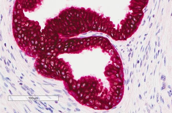
bioRxiv preprint first posted online Mar. 14, 2018; doi: http://dx.doi.org/10.1101/276501. The copyright holder for this preprint
          (which was not peer-reviewed) is the author/funder, who has granted bioRxiv a license to display the preprint in perpetuity.
                                           It is made available under a CC-BY 4.0 International license.

 A                                                                                    B

               100um                                                                            100um

 C                                                                                    D

               100um                                                                            100um

Figure 7. CK Pan/ CK 18 double staining of prostate tissue on Dako Omnis. A 1st. CK 18/DAB 2nd. CK Pan/Magenta. B 1st. CK Pan/DAB 2nd. CK 18/
Magenta. C 1st. CK 18/Magenta 2nd. CK Pan/DAB. D 1st. CK Pan/Magenta 2nd. CK 18/DAB.

has on epitopes that are stained with the DAB chromogen.                            Conclusion
The DAB precipitate can be so dense that access to
epitopes in the tissue by subsequent antibodies is almost                           We have demonstrated double staining using two HRP
impossible. This combined with the ability of the brown                             substrates as chromogens. The HRP activity from the
color to dominate other colors keeps the DAB stain brown                            first staining can be completely removed by a sulfuric
(Figure 7A) even after the second staining is complete. The                         acid block step. Using the HRP Magenta chromogen in
same is true in Figure 7B where the DAB stain keeps its                             combination with DAB makes fully automated protocols for
brown appearance but is lighter due to some HRP Magenta                             double staining possible.
precipitation. We ascribe this to different expression levels                       The best double staining in terms of no cross-reactivity
of cytokeratin, which affects how dense the DAB precipitate                         is still made with antibodies from two different species.
becomes.                                                                            However, in cases of densely packed epitopes using DAB
For the HRP Magenta chromogen the situation is the same,                            as the first chromogen, the spillover is practically hidden
a high shielding effect is observed when CK 18 is stained                           by the intense DAB stain. This makes it possible to do
first and less shielding when CK Pan is stained first.                              a double staining with two antibodies from the same

     6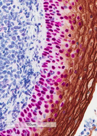
bioRxiv preprint first posted online Mar. 14, 2018; doi: http://dx.doi.org/10.1101/276501. The copyright holder for this preprint
       (which was not peer-reviewed) is the author/funder, who has granted bioRxiv a license to display the preprint in perpetuity.
                                        It is made available under a CC-BY 4.0 International license.

species. Using antibodies from different species the                             References
sequence of the chromogens is not important. Both DAB
and the HRP Magenta chromogen can be used as a first                             1. Li, c.-Y., Ziesmer, S. C. & Lazcano, Villareal, O. (1987).
stain. We have explained why cross-reactivity remains an                            Use of azide hydrogen peroxide as an inhibitor for
issue when using antibodies from the same species.                                  endogenous peroxidase in the immunoperoxidase
In our experience the use of DAB as the first chromogen                             method. J Histochem Cytochem; 35:1457-60. https://doi.
and HRP Magenta as the second chromogen have given                                  org/10.1177/35.12.2824601
consistently good results with minimal detectable cross-                         2. Lohse, J. Hansen, M. P. M. (2016). Chromogenic peroxidase
reactivity and this is now our standard approach when                               substrates. US20170175178A1
setting up double staining protocols.
                                                                                 3. Malik, N. J. and Daymon, M. E. (1982). Improved double
                                                                                    immunoenzyme labelling using alkaline phosphatase and
                                                                                    horseradish peroxidase. J Clin Pathol; 35:1092-94. http://
Materials and Methods                                                               dx.doi.org/10.1136/jcp.35.10.1092
                                                                                 4. Nakane, K. (1968). Simultaneous localization of multiple
All reagents were purchased from Sigma Aldrich and Agilent                          tissue antigens using the peroxidase-labeled antibody
Technologies. Antibodies were purchased from Agilent                                method: a study on pituitary glands of the rat. J Histochem
Technologies and used according to the manufacturer’s                               Cytochem; 16:557-60. https://doi.org/10.1177/16.9.557
specifications. Desmoglein-3, p40 and p16 were from
Abcam, Biocare and Santa Cruz Biotechnology. Automated                           5. Nemes, Z. (1987). Intensification of 3,3′-diaminobenzidine
staining was performed on either a Dako Omnis system or an                          precipitation using the ferric ferricyanide reaction, and its
Autostainer Link 48 system. On the Dako Omnis system the                            application in the double-immunoperoxidase technique
“IHC Double Stain Template” was used. For the Autostainer                        6. Histochem; 86:415-19. https://doi.org/10.1007/
Link 48, a protocol was defined and it can be found in the                          BF00495003
supplementary material.                                                          7. Petersen, K. H. (2009). Novel horseradish peroxidase
Images were captured on an Aperio Scanscope. Areas                                  substrates for use in immunohistochemistry. J
of interest were loosely circled and analyzed using the                             Immun Methods; 340:86-89. https://doi.org/10.1016/j.
membrane v9 or the nuclear v9 algorithm adjusted to the                             jim.2008.09.018
color of the chromogen.                                                          8. van der Loos, C.M., Becker, A.E. & van den Oord J.J. (1993).
                                                                                    Practical suggestions for successful immunoenzyme
                                                                                    double-staining experiments. J Histochem; 25:1-13. https://
Specimens                                                                           doi.org/10.1007/BF00161039
All tissue was obtained from Department of Pathology,
Odense University Hospital, Region South, Denmark, fixed in
formaldehyde for 24 hours prior to embedding in paraffin. All
specimens were completely anonymized prior to receipt at
Agilent. According to the Danish law on the Research Ethics
Committee System and handling of biomedical research
projects and communication between Agilent and the Danish
Committee on Biomedical Research Ethics and the Regional
Ethics Committee (IRB), the tests performed at Agilent on
anonymous residual tissue are not subject to an approval by
the IRB system because such studies are considered quality
control projects. Therefore, no IRB approval for this work has
been obtained.

Acknowledgements
The Technical assistance of Anne Bruun, Kristine Faerk, Maria
Kristensen, Anne Maarbjerg and Marianne Marcussen is
gratefully acknowledged.

                                                                                                                                              7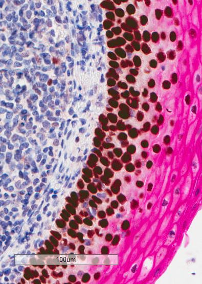
bioRxiv preprint first posted online Mar. 14, 2018; doi: http://dx.doi.org/10.1101/276501. The copyright holder for this preprint
        (which was not peer-reviewed) is the author/funder, who has granted bioRxiv a license to display the preprint in perpetuity.
                                         It is made available under a CC-BY 4.0 International license.

3751 2018MAR09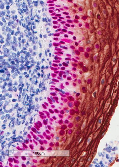
You can also read