BRACTIVATE: DENDRITIC BRANCHING IN MEDI- CAL IMAGE SEGMENTATION NEURAL ARCHITECTURE SEARCH
←
→
Page content transcription
If your browser does not render page correctly, please read the page content below
Under review as a conference paper at ICLR 2021

B RACTIVATE : D ENDRITIC B RANCHING IN M EDI -
CAL I MAGE S EGMENTATION N EURAL A RCHITECTURE
S EARCH
 Anonymous authors
 Paper under double-blind review

                                           A BSTRACT

         Researchers manually compose most neural networks through painstaking exper-
         imentation. This process is taxing and explores only a limited subset of possible
         architecture. Researchers design architectures to address objectives ranging from
         low space complexity to high accuracy through hours of experimentation. Neural
         architecture search (NAS) is a thriving field for automatically discovering archi-
         tectures achieving these same objectives. Addressing these ever-increasing chal-
         lenges in computing, we take inspiration from the brain because it has the most
         efficient neuronal wiring of any complex structure; its physiology inspires us to
         propose Bractivate, a NAS algorithm inspired by neural dendritic branching. An
         evolutionary algorithm that adds new skip connection combinations to the most
         active blocks in the network, propagating salient information through the network.
         We apply our methods to lung x-ray, cell nuclei microscopy, and electron mi-
         croscopy segmentation tasks to highlight Bractivate’s robustness. Moreover, our
         ablation studies emphasize dendritic branching’s necessity: ablating these con-
         nections leads to significantly lower model performance. We finally compare
         our discovered architecture with other state-of-the-art UNet models, highlight-
         ing how efficient skip connections allow Bractivate to achieve comparable results
         with substantially lower space and time complexity, proving how Bractivate bal-
         ances efficiency with performance. We invite you to work with our code here:
         https://tinyurl.com/bractivate.

1   I NTRODUCTION

Researchers manually composing
neural networks must juggle multiple
goals for their architectures. Archi-
tectures must make good decisions;
they must be fast, and they should
work even with limited computa-
tional resources. These goals are
challenging to achieve manually,
and researchers often spend months
attempting to discover the perfect
architecture.    To overcome these
challenges, we turn to the human Figure 1: Through Bractivate, we discover UNet architecture with
brain’s efficient neural wiring for high spatio-temporal efficiency by mimicking the brain’s dendritic
automated architecture discovery. branching.
Neuroscience already underlies core
neural network concepts: The perceptron (Rosenblatt, 1958) is directly analogous to a human
neuron. One of the brain’s fundamental learning mechanisms is dendritic branching (Greenough
& Volkmar, 1973) whereby active neurons send out signals for other neurons to form connections,
strengthening signals through that neural pathway. This neuroscience concept inspires us to
devise Bractivate, a Neural Architecture Search (NAS) algorithm for learning new efficient UNet
architectures networks, capable of being trained twice as fast as the traditional UNet, and often one

                                                  1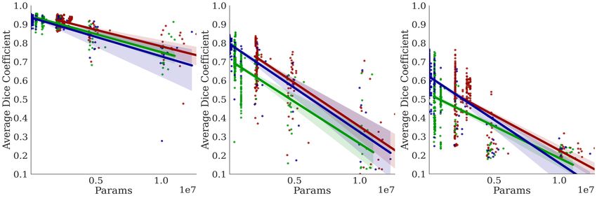
Under review as a conference paper at ICLR 2021

to two orders of magnitude lighter in terms of trainable parameters. We apply Bractivate on three
medical imaging segmentation problems: cell nuclei, electron microscopy, and chest X-ray lung
segmentation.
Medical image segmentation is a growing field in Deep Learning Computer Assisted Detection
(CAD): it is a powerful component in clinical decision support tools and has applications in retinal
fundus image, lung scan, and mammography analysis. Most papers now approach medical image
segmentation with the UNet (Ronneberger et al., 2015); the model architecture is straightforward:
it has symmetric, hierarchical convolutional blocks, which are components of an initial contracting
path and a final expanding path, with an apex bottleneck layer. Between parallel contracting and
expanding blocks, the traditional UNet contains skip connections that pass information through
concatenation (Ronneberger et al., 2015). Traditional UNet skip connections involve feature map
aggregation with same-scale convolutional blocks, but recent advances have yielded more complex
connections ranging from the UNet++ (Zhou et al., 2018) to the NasUNet (Weng et al., 2019). While
the UNet is a powerful tool, it does have many limitations:

      1. The depth necessary for many segmentation tasks is initially unknown, and traditional neu-
         ral architecture search (NAS) struggles to identify the optimal UNet depth.
      2. Researchers often manually choose skip connection locations, leading to potentially missed
         optimal connections.
      3. Scientists need a NAS algorithm addressing many implementation objectives, including
         computational time, number of model parameters, and robust segmentation performance.

On a broader level, discovering efficient UNet architectures is crucial because it can generate simpler
models for applications on mobile devices, which need low latency for online learning. In the
Telemedicine age, many medical applications rely on mobile Deep Learning to segment medical
images and process raw patient data (Xu et al., 2017; Vaze et al., 2020). We address the Medical
and Engineering fields’ need for efficiency with Bractivate, a NAS algorithm to discover lightweight
UNet architectures for medical image segmentation tasks. We present the following three primary
contributions:

      1. An evolutionary algorithm that non-randomly samples from a distribution of various UNet
         Model depths and skip connection configurations, with both tensor concatenation and ad-
         dition operators.
      2. ”Dendritic Branching”-inspired mutations that, just as in the brain, cause salient UNet
         blocks to branch to other blocks in the network through dendritic skip connections, cre-
         ating efficient networks that preserve information signals through the network.
      3. Bractivate generates high-performing models with lower space complexity than the current
         state-of-the-art.

The remainder of the paper is structured as follows: In Section 2, we discuss prior works, and what
gaps in the literature inspire us to propose Bractivate. Then, in Section 3, we discuss the search
algorithm and the dendritic branching mutation. Later, in Section 4, we implement our algorithm
with various experiments ranging from changing the search space depth to an ablation study. We
report our quantitative and qualitative results, along with baseline comparisons in Section 5 before
concluding in Section 6.

2   R ELATED W ORKS

Deep learning algorithms are often restricted to manual model design (Simonyan & Zisserman,
2014; He et al., 2016; Oktay et al., 2018; Ronneberger et al., 2015). To automate model schemes,
NAS is the process of selecting candidate architectures through various search strategies to achieve
optimal performance (Elsken et al., 2019). Advances in NAS have branched into different areas,
including evolutionary algorithms (Miller et al., 1989; de Garis, 1990; Yao, 1993; Fogel et al., 1990;
Angeline et al., 1994; Real et al., 2018; Yao, 1999) and automatic pattern recognition (Cai et al.,
2018; Radosavovic et al., 2020). While both approaches are merited, the tasks address image clas-
sification problems, and although some focus on skip connections, they lack deeper investigation

                                                  2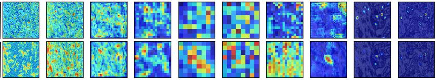
Under review as a conference paper at ICLR 2021

of their optimal configurations. Recent advances in the UNet have led to alternative skip connec-
tion implementations, including addition (Ghamdi et al., 2020), max out operations (Estrada et al.,
2019; Goodfellow et al., 2013) and multiplication by a gating function (Oktay et al., 2018). Ghamdi
et al. (2020) reports these connections’ improved efficacy over traditional concatenation, as they
overcome vanishing gradients and preserve salient features.
Auto-DeepLab, which Liu et al. (2019) present for semantic segmentation, is a graph-based NAS
algorithm that addresses changing model depth and connection locations in hierarchical models.
Building off this work, Zhou et al. (2020) propose a similar graph-search algorithm, termed UNet++,
for improved NAS; the final model incorporates dense skip connections to achieve multi-scale fea-
ture aggregation. Although UNet++ successfully addresses the model depth problem, it ignores
choosing the skip connection operator and relies on pretraining and pruning to generate skip con-
nection configurations.
The Differential Architecture Search (DARTs) algorithm by Liu et al. (2018) continuously relaxes
the architecture representation to enable gradient-based optimization. Advancing this algorithm,
Chen et al. (2019) proposes the Progressive Differentiable Architecture Search Algorithm (PDARTs)
to allow the searched model’s depth to grow during the search; when applied to ImageNet (Deng
et al., 2009), CIFAR-10 (Krizhevsky et al., 2009), or CIFAR-100 (Krizhevsky et al., 2009), the total
training time is approximately seven hours.
Although the DARTS and PDARTs algorithms are specific to image classification and sequential
model architecture, they lack applications for segmentation models. Weng et al. (2019) suggest a
NASUNet method with modified DARTs search for medical imaging segmentation; their approach
addresses searching for model parameters in the convolutional blocks to reduce the space complex-
ity found in attention-based (Oktay et al., 2018; Hy, 2018) and recurrent (Alom et al., 2018; Hy,
2018) UNets, yet NASUNet still preserves same-scale concatenation skip connections, overlooking
alternate skip connection possibilities across network blocks.
Many existing NAS algorithms use modified objective functions for evaluating the searched model
performance e.g. NAS-Bench-101 (Ying et al., 2019) uses the cross-entropy loss, Stochastic Neural
Architecture Search (SNAS) (Xie et al., 2019) devises a cost function deemed Memory Access
Cost (MAC) that incorporates the floating-point operations (FLOPs) and number of parameters,
and PDARTs (Chen et al., 2019) employs auxiliary loss (Szegedy et al., 2014). To target gaps in
the literature related to skip connection search for efficient models, we propose Bractivate, a NAS
algorithm inspired by the brain’s dendritic branching to facilitate optimal architecture discovery.

3     T HE B RACTIVATE NAS A LGORITHM

3.1   D ENDRITIC A RBORIZATION

Table 1 translates neuroscience into computational
terms we use throughout the paper. In the neuro- Table 1: Defining terms from neuroscience we
science field, dendritic branching occurs when stim- use and their parallels in Deep Learning.
ulating environments cause neurons to form new
connections (Greenough & Volkmar, 1973; Gree-             Neuroscience          Deep Learning
nough et al., 1985). These neural connections are
                                                          Soma                  Layer in a block
associated with learning, and even learning-impaired
                                                          Dendrite              Input Connection
children with fetal alcohol syndrome display lower        Axon                  Sender output Connec-
dendritic branching levels (Hamilton et al., 2010)                              tion
compared to their healthy peers. This branching phe-      Dendritic Branch      Connection to first
nomenon parallels Deep Neural Networks: in the                                  layer in an active
brain, dendrites form new connections to the hyper-                             receiving block
active soma: the perceptron’s activation function is
to the biological soma as the incoming connections are to dendrites. Perceptrons can be stacked
together to form multi-layer perceptrons (Rumelhart et al., 1986), with parallel architecture similar
to the brain, and this structure underlies convolutional neural networks (LeCun et al., 1995).
For the UNet, if we consider layer in the network’s blocks to be a neural soma, then we can think
about a block’s ”activity” as the mean-absolute value of its layers’ activations, as shown by Equa-

                                                 3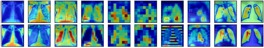
Under review as a conference paper at ICLR 2021
A) Evaluate Performance B) Identify Activity C) Branching
Conv-Transpose (M , M , F)
Conv-Transpose (M , M , F)
Conv-Transpose (M , M , F)
Batch Norm-1 (M , M , F)
Batch Norm-2 (M , M , F)
Batch Norm-1 (M , M , F)
Batch Norm-2 (M , M , F)
Batch Norm-1 (M , M , F)
Batch Norm-2 (M , M , F)
DropOut (M , M , F)
DropOut (M , M , F)
DropOut (M , M , F)
Conv-1-(M , M , F)
Conv-1-(M , M , F)
Conv-1-(M , M , F)
Conv-2 (M , M , F)
Conv-2 (M , M , F)
Conv-2 (M , M , F)
Addition Concatenation
M = Layer Dimension F = # of Filters
Low Activation High Activation
Figure 2: During each search iteration, Bractivate chooses a model from the randomly initialized search do-
main. After we train on the dataset, we evaluate its performance. To mutate, we identify the most active block
in the ”best model” architecture, as per Equation 1; to that active block, we initialize new skip connections
(branches) pointing from other blocks in the network to the highest-activation block, propagating their signals
through randomly chosen branches.
tion 1.
L
1X
Ab = |Al | (1)
L
l=0
where Ab represents block activation, b ∈ B, Al is the weight of each layer in the block, l, and L is
the total number of layers in the block. Knowing the block’s location, b, with max(Ab ), surrounding
blocks then form skip connections around this active node, a process analogous to dendritic branch-
ing. We apply this method to target conv and deconv layers, excluding batch normalization layers
as they contain static weights and high values that overwhelm the mutation’s layer selection. When
new connections are formed, across blocks with various tensor dimensions, we overcome spatial di-
mensional mismatch by resizing the incoming connection tensors to the receiving tensor by bilinear
interpolation.
3.2 NAS WITH D ENDRITIC B RANCHING M UTATIONS
We implement our dendritic
Efficiency
branching mutations to our evolu-
Evaluator tionary algorithm based on adding
dendritic branching mutations to
Best Model
a randomized search domain, as
Chosen Model
Genome displayed in Figure 3. This muta-
Sample Dendritic
tion applies a mutation operator to
Randomly
Branching
Mutation
form new skip connection combi-
Initialized Domain nations to the most active block in
the network. This NAS algorithm
Figure 3: Bractivate samples from a randomly initialized domain, initializes a random domain with
constrained by the model depth, D. The efficiency evaluator compares n model genotype. Each genotype
a selected model with the current ”best model” genome. If the ”best codes for the model’s depth, D.
model” outperforms the current model, we mutate the ”best model” It also encodes the number of
(Figure 2) and replace the chosen model with the mutated version in filters per conv and deconv layer,
the search space.
the degree of skip connections
for each block, and the skip connection operator type (concatenation or addition). A detailed
discussion on the genotype initialization and its micro-architecture is found in Appendix A.1. When
4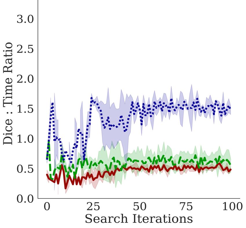
Under review as a conference paper at ICLR 2021

initializing the genotype, we constrain the number of feature maps to grow by 1.5 in the encoder
and decrease it by the same factor for each block in the decoder.

3.3   E FFICIENT L OSS

NAS methods often focus on accuracy as the main performance metric (Real et al., 2018; Ra-
dosavovic et al., 2020), but often lack consideration for discovered model space and time complexity.
To address this, we propose an efficient loss function for the NAS evaluation step. Traditional binary
cross-entropy is given by Equation 2. During the NAS mutation and selections, the search process
accelerates as ”better” models have faster training steps.

                                    m
                                1 X
                     BCL = −         (y × log(ŷi ) + (1 − yi ) × log(1 − ŷi ))                   (2)
                                m i=1 i

where m is the number of samples, yi is the sample image’s true segmentation mask tensor, and ŷi
is the model’s prediction tensor. We propose an alternate efficient loss function equation, Efficient
Loss Scaling (ELS ). It uses the number of model parameters, P , and the training time per epoch,
T.

E FFICIENCY L OSS S CALING
We also explore Efficiency penalty scaling where log(P ) and log(T ) scale the overall loss function
through multiplication, hence:

                               ELS = γ × log(P ) × log(T ) × BCL                                   (3)

In our experiments we set γ = 0.01. We use Equation 3 in Section 4.4 during model search.
A detailed ablation study on how this equation favors efficient networks and outperforms standard
BCL can be found in Appendix A.3.

4     E XPERIMENTS

4.1   I MPLEMENTATION

For the following experiments, we execute our programs using TensorFlow 2.3.0 and Keras 2.4.3,
using Python 3.6.9. For training the models and inference, we use a virtual machine with 25 GB of
RAM, 150 GB of disk space, and one Tesla v-100 GPU.

4.2   T HE DATA

To validate Bractivate NAS, we work with three datasets:

      1. The Montgomery Pulmonary Chest X-Ray (Jaeger et al., 2014) dataset (N = 138)
      2. The EPFL CVlab Electron Microscopy (Lucchi et al., 2011) dataset (N=330)
      3. The Kaggle Data Science Bowl Cell Nuclei Segmentation Challenge (Caicedo et al., 2019)
         dataset (N=670)

A detailed discussion of the dataset follows in Section 4.3.

4.3   P REPROCESSING

We resize all images in the three datasets to be 128×128 pixels with bilinear interpolation, and later
min-max normalize the pixels, so that pixel intensities are ∈ [0, 1]. We apply the same preprocessing
to the image masks. We use a 0.1 validation split percentage on all three datasets to evaluate the
optimized model loss function and the Dice coefficient during the search.

                                                  5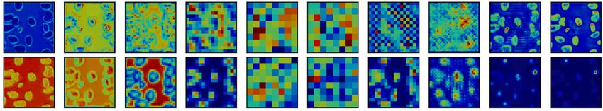
Under review as a conference paper at ICLR 2021

4.4   P ENALIZING T IME AND S PACE C OMPLEXITY

We experiment with using the standard binary BCL loss function in Equation 2, and the ELS
function in Equation 3. We record our results both with the Dice Score, representing the precision
and recall harmonic mean. We note that these loss functions are used only during the search step.
During full model training, we use the BCL loss on the ”best model” and use that for our later
experiments in Sections 4.7 and 4.8.

4.5   D ISCOVERING A RCHITECTURE

For all three datasets, we use the same Bractivate search algorithm based on dendritic branching. We
initialize our search domain as a queue with 20 randomly generate model genotypes and initialized
generated models from the genotypes; we then train the models for 50 epochs on the datasets with
Early Stopping. The Early Stopping has patience of 10 and min ∆ of 0.05. In a first-in-first-out
(FIFO) fashion, we evaluate each genotype in the queue: if the generated model has ELmin , then
it becomes the ”Best Model,” and we place this genotype back in the queue. Suppose it has a
EL > ELmin . In that case, the search algorithm mutates the ”Best Model” genotype with the
dendritic branching method described in Figure 2 before replacing the mutated genotype in the
search queue.

4.6   G OING D EEPER

We notice that the original Bractivate search algorithm with a minimum depth of two yields mainly
shallow networks, usually with two or three expanding and contracting blocks. To explore how
model depth affects the search algorithm’s results, we constrain the search space such that depth is
∈ [5, 10] and later ∈ [7, 10], and observe how the Dice coefficient and Dice:time ratio change while
using the ELS (Equation 3).

4.7   A BLATING B RANCHING C ONNECTIONS

We hypothesize that active block branching increases signal propagation, reducing time complexity,
and improving overall model performance. Thus, we must prove these new branches are necessary
for the model’s success by ablating them and measuring how their absence affects the model perfor-
mance. We perform these experiments by training the selected model architecture for 200 epochs
on the dataset, and use the Keract (Remy, 2018) library to measure layer activations on the most
active layer (the second convolutional layer in the decoder of a D = 5 deep UNet. For each layer,
we calculate the layer’s average activation from Equation 1 and then ablate all dendritic (input) con-
nections of the most active block. We record the results quantitatively with the Dice coefficient and
visualize them by analyzing changes in the activation heat maps.

4.8   BASELINE C OMPARISON

We compare Bractivate models to other state-of-the-art methods, including the standard UNet model
(Ronneberger et al., 2015), Wide-UNet (Zhou et al., 2018), UNet++ (Zhou et al., 2020), and
attention-based models . We obtain all model architectures from GitHub repositories made by these
models’ authors or by contributors who create implementations in Keras and use them with Xavier
weight initialization on the three datasets for comparison.

5     R ESULTS

We observe clear patterns pointing to how dendritic branching allows for efficient neural network
selection through our experimentation. Figure 4 presents an example of the discovered architecture
with the ELS function.

5.1   D ISCOVERED A RCHITECTURES

                                                  6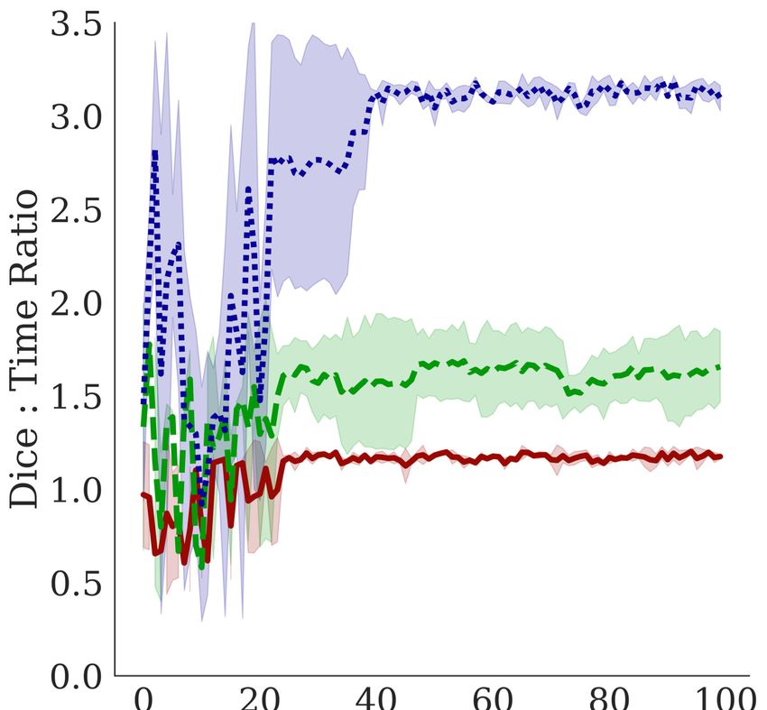
Under review as a conference paper at ICLR 2021
Because the mutations add more con-
nections to salient blocks, the most ac-
tive node has the highest input connec-
tions. Bractivate randomly assigns addi-
tion or concatenation connections to the
most active block, optimizing the best
connecting operator combination.
Figure 4 shows the discovered archi-
tecture when the search space is con-
strained such that D ∈ [5, 10]. Al Addition Concatenation
Ghamdi et al. (Ghamdi et al., 2020) re-
port that the addition operator can per- Figure 4: A sample of the discovered architecture through the
form as well or better than the concate- efficiency loss scaling in Equation 3. The green block is the
nation operator, so we incorporate both most active block, having the highest number of incoming den-
operators as modules into the NAS, with dritic connections.
each mutation yielding a new random
connecting operator combination to the
salient block.
5.2 D EPTH AND T IME -S PACE C OMPLEXITY
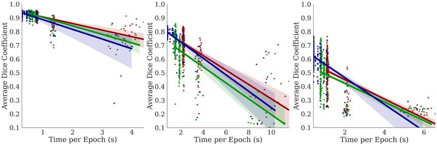
In Figure 5, we observe a negative correla-
Lungs EM Cell Nuclei tion between the training time per epoch
and the Dice coefficient, indicating that
Bractivate favors shallower over deeper
networks, as the skip connections branch-
ing towards active blocks carry the nec-
essary information for the segmentation
task. This slope varies between datasets;
a simple problem like segmenting two-
component lungs confers an overall larger
Dice: time ratio than the cell nuclei and
electron microscopy tasks. These prob-
lems are challenging because they have
more than two connected components,
yielding lower Dice: time ratios. More
importantly, our methods determine that
given the early stopping constraint, shal-
lower models (D = 2) with fewer pa-
rameters have comparable performance to
deeper models. When UNet models are
too deep (D ∈ [7, 10]), the input signal
may be lost in transformations, leading to
lower performance; shallower models pre-
D= 7 5 2
serve this signal.
Figure 5: Comparing the effects of model depth on the corre-
lations between the Dice coefficient performance metric and 5.3 S KIP
different depth constraint D over three trials. Top: Dice vs C ONNECTION A BLATION S TUDY
time per epoch; Middle: Dice vs. number of parameters
(spatial complexity) Bottom: Dice vs the number of model
params.
Our ablation study most strongly confirms
our hypothesis that dendritic branching to
active blocks significantly improves segmentation performance: Figure 6 examines the saliency
maps produced by the model architecture in Figure 4 before and after ablating connections to the
most active block. Before ablation, the most active block’s saliency maps show encoded information
that significantly influences the decoded feature maps during deconvolution.
After ablation, the saliency maps for the EM and nuclei segmentation tasks lack accurate late-stage
saliency maps. When the salient block has configured dendritic branches from neighboring blocks,
7
Under review as a conference paper at ICLR 2021

                    Enc1 Conv2    Enc2 Conv2   Enc3 Conv2   Enc4 Conv2   Enc5 Conv2       Dec1 Conv2   Dec2 Conv2   Dec3 Conv2   Dec4 Conv2   Dec5 Conv2   Dice

         Searched
                                                                                                                                                           0.956
Lungs
         Ablated

                                                                                                                                                           0.914
         Searched

                                                                                                                                                           0.900
EM
         Ablated

                                                                                                                                                           0.798
         Searched

                                                                                                                                                           0.889
Nuclei
         Ablated

                                                                                                                                                           0.800

Figure 6: Activation map comparison for the second deconv block’s second convolutional layer for the archi-
tecture described in Figure 4. Blue represents low saliency, and red represents high saliency. We ablate the most
salient block (The green block containing Decode2 Conv2) in the network and display activation differences
after ablation.

the output signal is highly accurate. However, when these vital encodings in the Decode 2 block
lack input from neighboring blocks, the output signal is degraded. This degradation is especially
true for the EM and nuclei segmentation tasks.
The EM and nuclei segmentation tasks contain more than two connected components; removing
dendrites to salient blocks prevents valuable information from neighboring blocks to travel to the
most salient block, degrading the signal through the last four blocks in the network. The model’s
Dice score is significantly lower in the ablated architecture than in the intact Bractivate-selected
model. The added information from these dendritic skip connections, explicitly targeting a salient
block in the model, generates more accurate saliency maps, helping the model learn faster. Before
ablation, activations are more salient during the decoding phase than post-ablation, where saliency
concentrates in the encoder. This observation may be because removing connections towards an
active block forces surrounding layers to compensate by increasing their activations.

5.4                 BASELINE C OMPARISON

Table 2: Comparing the spacial complexity of Bractivate with various state-of-the-art UNet architectures. The
top group represents manually-designed models. The middle row comprises differentiable search. The bottom
is ours, based on dendritic branching. We report the results as ”Dice (# Params).”

                                               Model                              Lung                      EM                   Nuc
                             UNet (Ronneberger et al., 2015)                0.925 (7.7e6)              0.811 (7.7e6)        0.833 (7.7e6)
                              R2-UNet (Alom et al., 2018)                   0.596 (9.5e7)              0.464 (9.5e7)        0.049 (9.5e7)
                             Attn-UNet (Oktay et al., 2018)                 0.954 (3.2e7)              0.937 (3.2e7)        0.721 (3.2e7)
                               UNet++ (Zhou et al., 2018)                   0.903 (9.0e6)              0.846 (9.0e6)        0.841 (9.0e6)
                              WideUNet (Zhou et al., 2018)                  0.888 (9.3e6)              0.811 (9.3e6)        0.828 (9.3e6)
                                 NasUNet (Weng et al., 2019)                0.934 (1.2e5)              0.729 (4.8e5)        0.774 (1.2e5)
                                           Bractivate                       0.942 (3.1e4)              0.929 (4.8e5)        0.878 (4.8e5)

Figure 7 highlights how Bractivate achieves comparable performance to larger models when ini-
tialized with Xavier initialization (Glorot & Bengio, 2010). Table 2 highlights how Bractivate is
significantly smaller than many of the other state-of-the-art models: it exchanges high spatial com-

                                                                                      8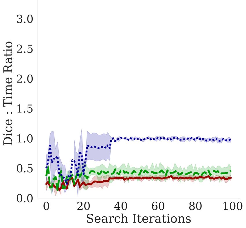
Under review as a conference paper at ICLR 2021

plexity for more skip connections, as these branches allow information to propagate through salient
blocks in the network. For domain-specific tasks, high parameters reduce the signal: noise ratio in
the network; simpler models like Bractivate rely on powerful skip connections, analogous to den-
drites, to carry most of the signal. Because these connections consist of simple concatenation or
addition operators, they greatly reduce the number of trainable parameters, preventing overfitting;
this speaks to Bractivate’s comparable–or better–Dice scores as compared to the baseline models.

              Lungs                           EM                            Nuclei

                                                                                                UNet
                                                                                               R2-UNet

                                                                                               AttnUNet

                                                                                               UNet++

                                                                                               WideUNet
                                                                                                NasUNet
                                                                                                Bractivate

Figure 7: Comparing model performance measured by the Dice score as a function of different model archi-
tectures. Note that these models are all Xavier initialized.

6    C ONCLUSION

Throughout this paper, we highlight how dendritic branching in the brain inspires efficient skip
connections in Deep Learning models. With our focus on segmentation, we present Bractivate as a
method for identifying skip connection configurations to elevate the traditional UNet. During the
search, Bractivate mutates the architecture so that the most salient blocks in the network branch out
their ”dendrites” to other network blocks. By replacing the oldest model in the search space with
the new mutated architecture, we accelerate the search rate.
The ablation study strongly supports our hypothesis that dendritic branching is necessary for efficient
model discovery; when we ablate dendritic connections to the most salient block, the Dice Score
decreases. Before and after ablation, the saliency maps reveal stark contrasts, with the ablated
activation maps lacking apparent features for the final segmentation layer in the UNet’s decoder.
We finally weigh our methods with other baselines, highlighting how smaller networks can perform
segmentation tasks well given limited pretraining data.
Overall, we present how optimally configured skip connections, inspired by the brain, yield robust
signal streaming paths through a lightweight network. Our algorithm is an asset to many mobile
medical computing technologies that rely on low latency and high computational efficiency.

R EFERENCES
Md Zahangir Alom, Mahmudul Hasan, Chris Yakopcic, Tarek M Taha, and Vijayan K Asari. Recurrent residual
 convolutional neural network based on u-net (r2u-net) for medical image segmentation. arXiv preprint
 arXiv:1802.06955, 2018.

Peter J Angeline, Gregory M Saunders, and Jordan B Pollack. An evolutionary algorithm that constructs
  recurrent neural networks. IEEE transactions on Neural Networks, 5(1):54–65, 1994.

Han Cai, Tianyao Chen, Weinan Zhang, Yong Yu, and Jun Wang. Efficient architecture search by network
  transformation. In AAAI, 2018.

Juan C Caicedo, Allen Goodman, Kyle W Karhohs, Beth A Cimini, Jeanelle Ackerman, Marzieh Haghighi,
   CherKeng Heng, Tim Becker, Minh Doan, Claire McQuin, et al. Nucleus segmentation across imaging
   experiments: the 2018 data science bowl. Nature methods, 16(12):1247–1253, 2019.

X. Chen, L. Xie, J. Wu, and Q. Tian. Progressive differentiable architecture search: Bridging the depth gap
  between search and evaluation. In 2019 IEEE/CVF International Conference on Computer Vision (ICCV),
  pp. 1294–1303, 2019.

                                                    9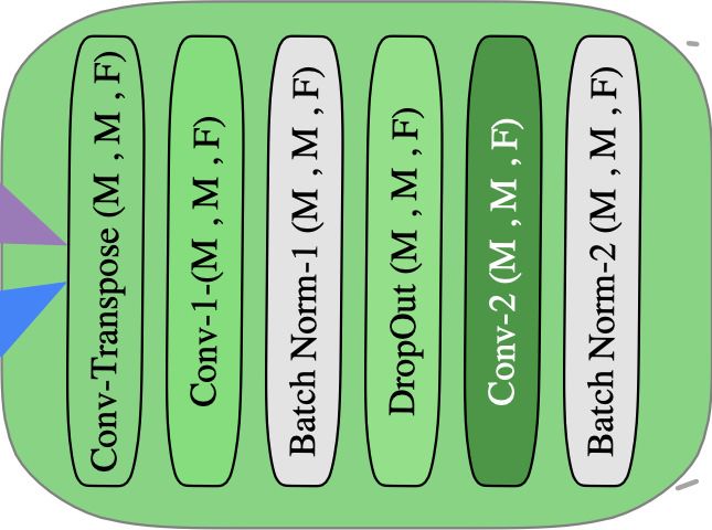
Under review as a conference paper at ICLR 2021

Hugo de Garis. Genetic programming: Modular evolution for darwin machines. In Proceedings of the 1990
  International Joint Conference on Neural Networks, pp. 194–197, 1990.

J. Deng, W. Dong, R. Socher, L. Li, Kai Li, and Li Fei-Fei. Imagenet: A large-scale hierarchical image
   database. In 2009 IEEE Conference on Computer Vision and Pattern Recognition, pp. 248–255, 2009.

Thomas Elsken, Jan Hendrik Metzen, and Frank Hutter. Neural architecture search: A survey. Journal of
  Machine Learning Research, 20(55):1–21, 2019.

Santiago Estrada, Ran Lu, Sailesh Conjeti, Ximena Orozco-Ruiz, Joana Panos-Willuhn, Monique M. B.
  Breteler, and Martin Reuter. Fatsegnet : A fully automated deep learning pipeline for adipose tissue seg-
  mentation on abdominal dixon MRI. CoRR, abs/1904.02082, 2019. URL http://arxiv.org/abs/
  1904.02082.

David B Fogel, Lawrence J Fogel, and VW Porto. Evolving neural networks. Biological cybernetics, 63(6):
  487–493, 1990.

M. A. Ghamdi, M. Abdel-Mottaleb, and F. Collado-Mesa. Du-net: Convolutional network for the detection of
  arterial calcifications in mammograms. IEEE Transactions on Medical Imaging, pp. 1–1, 2020.

Xavier Glorot and Yoshua Bengio. Understanding the difficulty of training deep feedforward neural networks.
  In Proceedings of the thirteenth international conference on artificial intelligence and statistics, pp. 249–
  256, 2010.

Ian Goodfellow, David Warde-Farley, Mehdi Mirza, Aaron Courville, and Y. Bengio. Maxout networks. 30th
   International Conference on Machine Learning, ICML 2013, 1302, 02 2013.

William T Greenough and Fred R Volkmar. Pattern of dendritic branching in occipital cortex of rats reared in
  complex environments. Experimental neurology, 40(2):491–504, 1973.

William T Greenough, John R Larson, and Ginger S Withers. Effects of unilateral and bilateral training in a
  reaching task on dendritic branching of neurons in the rat motor-sensory forelimb cortex. Behavioral and
  neural biology, 44(2):301–314, 1985.

Gillian F Hamilton, Lee T Whitcher, and Anna Y Klintsova. Postnatal binge-like alcohol exposure decreases
  dendritic complexity while increasing the density of mature spines in mpfc layer ii/iii pyramidal neurons.
  Synapse, 64(2):127–135, 2010.

K. He, X. Zhang, S. Ren, and J. Sun. Deep residual learning for image recognition. In 2016 IEEE Conference
   on Computer Vision and Pattern Recognition (CVPR), pp. 770–778, 2016.

Lee Jun Hy. Leejunhyun/image segmentation: Pytorch implementation of u-net, r2u-net, attention u-net, and
  attention r2u-net. https://github.com/LeeJunHyun/Image_Segmentation, 2018. (Accessed
  on 09/06/2020).

Stefan Jaeger, Sema Candemir, Sameer Antani, Yı̀-Xiáng J Wáng, Pu-Xuan Lu, and George Thoma. Two
   public chest x-ray datasets for computer-aided screening of pulmonary diseases. Quantitative imaging in
   medicine and surgery, 4(6):475, 2014.

Alex Krizhevsky, Vinod Nair, and Geoffrey Hinton. Cifar-10 and cifar-100 datasets. URl: https://www. cs.
  toronto. edu/kriz/cifar. html, 6:1, 2009.

Yann LeCun, Yoshua Bengio, et al. Convolutional networks for images, speech, and time series. The handbook
  of brain theory and neural networks, 3361(10):1995, 1995.

C. Liu, L. Chen, F. Schroff, H. Adam, W. Hua, A. L. Yuille, and L. Fei-Fei. Auto-deeplab: Hierarchical neural
   architecture search for semantic image segmentation. In 2019 IEEE/CVF Conference on Computer Vision
   and Pattern Recognition (CVPR), pp. 82–92, 2019.

Hanxiao Liu, Karen Simonyan, and Yiming Yang. DARTS: differentiable architecture search.                CoRR,
  abs/1806.09055, 2018. URL http://arxiv.org/abs/1806.09055.

Aurélien Lucchi, Kevin Smith, Radhakrishna Achanta, Graham Knott, and Pascal Fua. Supervoxel-based
  segmentation of mitochondria in em image stacks with learned shape features. IEEE transactions on medical
  imaging, 31(2):474–486, 2011.

Geoffrey F Miller, Peter M Todd, and Shailesh U Hegde. Designing neural networks using genetic algorithms.
  In ICGA, volume 89, pp. 379–384, 1989.

                                                      10
Under review as a conference paper at ICLR 2021
Ozan Oktay, Jo Schlemper, Loic Le Folgoc, Matthew Lee, Mattias Heinrich, Kazunari Misawa, Kensaku Mori,
Steven McDonagh, Nils Y Hammerla, Bernhard Kainz, et al. Attention u-net: Learning where to look for
the pancreas. arXiv preprint arXiv:1804.03999, 2018.
Ilija Radosavovic, Raj Prateek Kosaraju, Ross Girshick, Kaiming He, and Piotr Dollár. Designing network
design spaces, 2020.
Esteban Real, Alok Aggarwal, Yanping Huang, and Quoc V Le. Regularized evolution for image classifier
architecture search, 2018.
Philippe Remy. philipperemy/keract: Activation maps (layers outputs) and gradients in keras. https://
github.com/philipperemy/keract, 2018. (Accessed on 08/31/2020).
Olaf Ronneberger, Philipp Fischer, and Thomas Brox. U-net: Convolutional networks for biomedical image
segmentation. In Lecture Notes in Computer Science, pp. 234–241. Springer International Publishing, 2015.
Frank Rosenblatt. The perceptron: a probabilistic model for information storage and organization in the brain.
Psychological review, 65(6):386, 1958.
David E. Rumelhart, James L. McClelland, and CORPORATE PDP Research Group (eds.). Parallel Distributed
Processing: Explorations in the Microstructure of Cognition, Vol. 1: Foundations. MIT Press, Cambridge,
MA, USA, 1986. ISBN 026268053X.
Karen Simonyan and Andrew Zisserman. Very deep convolutional networks for large-scale image recognition,
2014.
Christian Szegedy, Wei Liu, Yangqing Jia, Pierre Sermanet, Scott Reed, Dragomir Anguelov, Dumitru Erhan,
Vincent Vanhoucke, and Andrew Rabinovich. Going deeper with convolutions, 2014.
Sagar Vaze, Weidi Xie, and Ana IL Namburete. Low-memory cnns enabling real-time ultrasound segmentation
towards mobile deployment. IEEE Journal of Biomedical and Health Informatics, 24(4):1059–1069, 2020.
Yu Weng, Tianbao Zhou, Yujie Li, and Xiaoyu Qiu. Nas-unet: Neural architecture search for medical image
segmentation. IEEE Access, 7:44247–44257, 2019.
Sirui Xie, Hehui Zheng, Chunxiao Liu, and Liang Lin. SNAS: stochastic neural architecture search. In Inter-
national Conference on Learning Representations, 2019.
Xiaowei Xu, Qing Lu, Tianchen Wang, Jinglan Liu, Cheng Zhuo, Xiaobo Sharon Hu, and Yiyu Shi. Edge seg-
mentation: Empowering mobile telemedicine with compressed cellular neural networks. In 2017 IEEE/ACM
International Conference on Computer-Aided Design (ICCAD), pp. 880–887. IEEE, 2017.
Xin Yao. Evolutionary artificial neural networks. International journal of neural systems, 4(03):203–222, 1993.
Xin Yao. Evolving artificial neural networks. Proceedings of the IEEE, 87(9):1423–1447, 1999.
Chris Ying, Aaron Klein, Eric Christiansen, Esteban Real, Kevin Murphy, and Frank Hutter. NAS-bench-
101: Towards reproducible neural architecture search. In Kamalika Chaudhuri and Ruslan Salakhutdinov
(eds.), Proceedings of the 36th International Conference on Machine Learning, volume 97 of Proceedings
of Machine Learning Research, pp. 7105–7114, Long Beach, California, USA, 09–15 Jun 2019. PMLR.
Z. Zhou, M. M. R. Siddiquee, N. Tajbakhsh, and J. Liang. Unet++: Redesigning skip connections to exploit
multiscale features in image segmentation. IEEE Transactions on Medical Imaging, 39(6):1856–1867, 2020.
Zongwei Zhou, Md Mahfuzur Rahman Siddiquee, Nima Tajbakhsh, and Jianming Liang. Unet++: A nested
u-net architecture for medical image segmentation. In Deep Learning in Medical Image Analysis and Mul-
timodal Learning for Clinical Decision Support, pp. 3–11. Springer, 2018.
A A PPENDIX
A.1 G ENOME M ICRO -A RCHITECTURE
When designing our search space, we formulate genotypes that code for model architectures. Fol-
lowing common patters in convolutional networks, and the UNet Ronneberger et al. (2015), we first
impose the following constraints on our search space:
11Under review as a conference paper at ICLR 2021
• The network blocks must be symmetrical. This means that the number of blocks both in the
network encoder and decoder are identical, with mirror internal layer configurations (types
of layers, numbers of filters, and number of layers in the block)
• The network must be hierarchical. When designing models for medical image segmenta-
tion, we rely on hierarchical backbones for both the encoder and decoder, as reflected in
Figure 4.
• We constrain skip connection directionality. In the network, skip connections only occur
from earlier to later layers in the background.
Figure 8 shows the standard micro-architecture for the contracting and expanding parts of the net-
work. We also note that while the layer arrangements are constant, the number of filters, n, for each
block is initially random. However, each integer value of filter numbers is scaled by a factor of 1.5
for each subsequent block, as Figure 8 highlights.
A)
Channels = n
Batc
Norm h
1 Con
v2D
1
B) Batc
Norm h
2
Dro
pou
t (10
%)
Con
v2D
Channels = n 2
Batc
Norm h
Con 3
v2D Max
Tran P oo
spo ling
se
Con
v2D
1
Batc
Norm h
1
Dro
pou
t (10
%)
Con
v2D
2
Batc
Norm h
2
Figure 8: Each prism represents a layer in the overall repeating block motifs in the network. A) The con-
tracting block micro-architecture for one bloc. Note that this motif repeats throughout the contracting phase
of the network. n, the number of channels, is factored by 1.5 for each subsequent contracting block. B) The
Expanding block of the micro-architecture. n, the number of channels, is factored by .75 for each subsequent
expanding block.
A.2 GPU RUN -T IME
Overall, the search algorithm had GPU run-times, as shown in Table 3. We note that these results
are reported after running the search algorithm on a Tesla-v100. The reported values are an average
of three independent trials. Oftentimes, the run time was dependent on the dataset size. Because the
Cell Nuclei dataset had the highest number of sample images, it took longer to train on as compared
to the smaller Lung dataset.
Table 3: GPU Run-time for all three datasets with time measured in hours averaged over three trials with image
dimensions of 128 × 128.
Dataset Run Time (hrs)
Lungs 0.483 ± 0.067
EM 0.878 ± 0.164
Cell Nuclei 1.31 ±. 0.170
12Under review as a conference paper at ICLR 2021
A.3 A BLATING E FFICIENT L OSS
BCL
ELS
Figure 9: The efficiency loss scaling (ELS) loss function selects a smaller model (orange) that can perform at
the level as one selected simply by BCL alone (blue).
We also examine the effect of ablating the efficiency loss’ parameter and time penalty terms on the
overall model selection. Through our investigation, we find that the efficiency loss does help the
model select smaller models, that can perform at the level of larger models selected by the BCL loss
function. Figure 9 highlights this trend for the Lung Dataset. The results are averaged over three
trial runs.
We see that removing the penalty terms for high space and time complexity still yields high-
performing models. However, these models are larger, and computationally costly.
13You can also read