Breast Cancer - Diagnosis, Evaluation and Treatment
←
→
Page content transcription
If your browser does not render page correctly, please read the page content below
Breast Cancer
         Breast cancer is a malignant tumor in or around breast tissue. It usually
         begins as a lump or calcium deposit that develops from abnormal cell
         growth. Most breast lumps are benign, but some can be pre-cancerous
         or cancerous. Breast cancer can be localized (initially appearing within
         the breast) or metastatic (spread to another part of the body).

         Your doctor will likely perform a physical exam to evaluate a breast
         lump. To determine whether that lump is benign, your doctor may order
         mammography, breast ultrasound, breast MRI, PET/CT or
         scintimammography. If the lump is indeed benign, no further action may
         be needed. However, your doctor may want to monitor it to see if it
         changes, grows, or disappears over time. If the tests are inconclusive,
         your doctor may perform a biopsy using ultrasound, x-ray, or magnetic resonance imaging guidance. Breast cancer treatment
         depends on the tumor's size, extent of disease spread, type, receptor status, tumor growth rate and the patient’s general health.
         Treatments include surgery, radiation therapy, chemotherapy, hormone therapy or a combination thereof.

         What is breast cancer?
         Breast cancer is a malignant tumor that grows in or around the breast tissue, mainly in the milk ducts and glands. A tumor usually
         starts as a lump or calcium deposit that develops as a result of abnormal cell growth. Most breast
         lumps (https://www.radiologyinfo.org/en/info/breastlumps) are benign but some can be premalignant (may become cancer) or
         malignant.

         Breast cancer is classified as either primary or metastatic. The initial malignant tumor that develops within the breast tissue is
         known as primary breast cancer. Sometimes, primary breast cancer can also be found when it is spread to lymph nodes that are
         close by in the armpit. Metastatic breast cancer, or advanced cancer, is formed when cancer cells located in the breast break away
         and travel to another organ or part of the body.

         The cause of breast cancer is still unknown. However, some risk factors include:
                Age
                Gender
                Race
                Family history and genetic factors
                History of previous cancers (such a colon or ovarian cancer)
                Dense breast (https://www.radiologyinfo.org/en/info/dense-breasts) (meaning there is a lot of ducts, glands, fibrous tissue
                and less fatty tissue)
                Body Mass Index
                Use of hormone replacement therapies
                Alcohol use
                Age of first period, number of viable pregnancies, and age of first childbirth
Breast Cancer                                                                                                                             Page 1 of 5
Copyright© 2021, RadiologyInfo.org                                                                                              Reviewed Jun-15-2020
Risk factors are not a sure indication that you will develop breast cancer. Some women with breast cancer have no risk factors. On
the other hand, many women with risk factors never develop the disease.
Breast cancer symptoms may not appear in the early stages of the disease. However, as the cancer develops, symptoms may
include:
A lump or thickening in or near the breast or underarm
Pain or tenderness in the breast or nipple
Clear or bloody nipple discharge
Change in the breast or nipple such as color, shape or size
Swelling in the armpit
Redness or flakiness around the nipple or skin of the breast
How is breast cancer diagnosed and evaluated?
Your primary doctor will begin by asking you about your medical history, risk factors and symptoms. You will also undergo a
physical exam.
In order to determine if a breast lump is malignant or benign, one or more of the following imaging tests may be performed:
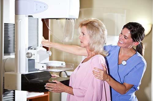
Mammogram (https://www.radiologyinfo.org/en/info/mammo) : Mammography is a type of x-ray examination used to
examine the breasts. This type of imaging involves exposing the breasts to a small amount of ionizing radiation to obtain
pictures of the inside of the breasts. See the Safety page (https://www.radiologyinfo.org/en/info/safety-xray) for more
information about x-rays.
Breast ultrasound (https://www.radiologyinfo.org/en/info/breastus) : Breast ultrasound uses sound waves to create pictures
of the inside of the breasts. Breast ultrasound can capture images of areas of the breast that may be difficult to see with
mammography. It can also help to determine whether a breast lump is a solid mass or a cyst.
Breast MRI: (https://www.radiologyinfo.org/en/info/breastmr) During breast MRI, a powerful magnetic field, radio
frequency pulses and a computer will be used to produce detailed pictures of the inside of the breasts. MRI is helpful in
evaluating breast lumps that are not visible with mammography or ultrasound, particularly in women with dense breast
tissue.
PET/CT (https://www.radiologyinfo.org/en/info/pet) : This type of nuclear imaging combines PET scans and CT scans to
provide images that pinpoint the anatomic location of abnormal metabolic activity within the breasts. It can detect breast
cancer, determine if it has spread, assess the effectiveness of a treatment plan and determine if the cancer has returned after
treatment.
Scintimammography (https://www.radiologyinfo.org/en/info/scintimammo) : This imaging test, also known as nuclear
medicine breast imaging, may be used to investigate a breast abnormality that has been discovered on mammography. The
procedure is noninvasive and involves the injection of a radiotracer, or drug that emits radioactivity, into the patient.
Because the radiotracer accumulates differently in different kinds of tissue, it can help physicians determine whether cancer
could be present, thus helping determine whether a biopsy or additional follow-up is necessary. While it is not a primary
screening tool and does not replace mammography, some physicians have used it as an additional tool in women who are at
elevated risk for breast cancer, but cannot undergo MRI screening.
If a lump is proven to be benign by these exams, no further steps may be needed. Your doctor may want to monitor the area at
future visits to check if the breast lump has changed, grown or gone away.
If these tests do not clearly show that the lump is benign, a biopsy may be necessary. If so, one of the following image-guided
procedures may be performed:
Breast Cancer Page 2 of 5
Copyright© 2021, RadiologyInfo.org Reviewed Jun-15-2020Ultrasound-guided biopsy (https://www.radiologyinfo.org/en/info/breastbius) : During this type of biopsy, ultrasound
imaging is used to visualize a breast lump. An interventional radiologist will advance a thin needle to the site of the lump
using the real-time ultrasound images and remove some tissue for evaluation under a microscope. The biopsy procedure is
usually quick, but it may take a few days before the final tissue analysis (pathology report) is ready.
Stereotactic (x-ray guided) biopsy (https://www.radiologyinfo.org/en/info/breastbixr) : During this type of biopsy, a digital
mammography x-ray machine is used to produce real-time pictures of the breast. A radiologist uses these live pictures to
guide placement of a needle to the site of the lump and remove tissue samples for further evaluation.
MRI-guided biopsy (https://www.radiologyinfo.org/en/info/breastbimr) : An MRI machine is used to produce pictures of the
breasts and help the radiologist guide a needle to the site of the lump to remove a tissue sample.
How is a breast cancer treated?
The type of treatment recommended depends on the size and type of the tumor, its growth rate, and the general health of the
patient. Treatment options include:
Surgery: Surgery can consist of mastectomy or breast conservation therapy (BCT).
Mastectomy is an operation to remove the entire breast, and usually the entire nipple. Often an axillary (armpit)
sampling is also done which removes the glands under the arm called axillary nodes. The surgeon may evaluate just
one or two nodes (sentinel node/s) or may perform a more extensive axillary dissection to check for disease spread.
Mastectomy sometimes requires a hospital stay. A drainage tube is sometimes temporarily left in the surgical cavity
after a mastectomy to help prevent fluid accumulation. Women who undergo a mastectomy have the option of breast
reconstruction.
BCT surgery (such as lumpectomy, partial mastectomy, segmental mastectomy or quadrantectomy) removes the
breast tumor and a margin of surrounding normal tissues. Radiation therapy usually follows lumpectomy to eliminate
any microscopic cancer cells in the remaining breast tissue. The purpose of BCT is to give women the same cure rate
they would have if they were treated with a mastectomy but to leave the breast intact, with an appearance and texture
as close as possible to what they had before treatment. The surgeon may remove some lymph nodes by performing a
sentinel lymph node procedure or axillary dissection at the same time as the lumpectomy procedure or later.
Radiation therapy (https://www.radiologyinfo.org/en/info/intro_onco) : Radiation therapy uses high-energy x-rays (photons)
or a stream of particles. When radiation is used at therapeutic doses (many times those used for x-ray imaging exams) it can
destroy abnormal cancer cells in the breasts. Your radiation therapy procedure might include:
External Beam Therapy (EBT) (https://www.radiologyinfo.org/en/info/ebt) : EBT, also called external radiation
therapy, delivers one or more beams of high-energy x-rays to a tumor in the breast. Beams are generated outside the
patient (usually by a linear accelerator) and are targeted at the tumor site. These high energy x-rays can deliver their
dose to the area of the tumor to destroy the cancer cells and, with careful treatment planning, spare the surrounding
normal tissues. No radioactive sources are placed inside the patient's body. See the
(http://www.radiologyinfo.org/en/info.cfm?pg=linac#part_four)
LINAC (https://www.radiologyinfo.org/en/info/linac) page for more information about linear accelerator.
Intensity-Modulated Radiation Therapy (IMRT) (https://www.radiologyinfo.org/en/info/imrt) : IMRT is an advanced
mode of high-precision radiotherapy that uses computer-controlled linear accelerators to deliver precise radiation
doses to a malignant tumor or specific areas within the tumor. IMRT allows for the radiation dose to conform more
precisely to the three-dimensional (3-D) shape of the tumor by modulating—or controlling—the intensity of the
radiation beam in multiple small volumes. IMRT also allows higher radiation doses to be focused on regions within
the tumor while minimizing the dose to surrounding normal critical structures. Treatment is carefully planned by using
3-D computed tomography (CT) or magnetic resonance (MRI) images of the patient in conjunction with
computerized dose calculations to determine the dose intensity pattern that will best conform to the tumor shape.
Typically, combinations of multiple intensity-modulated fields coming from different beam directions produce a
custom tailored radiation dose that maximizes tumor dose while also minimizing the dose to adjacent normal tissues.
Brachytherapy: (https://www.radiologyinfo.org/en/info/brachy) Brachytherapy, also called internal radiation therapy,
allows a physician to use a higher total dose of radiation to treat a smaller area and in a shorter time. In breast cancer,
brachytherapy is used as radiation therapy to treat the partial breast only. It may be either temporary or permanent,
Breast Cancer Page 3 of 5
Copyright© 2021, RadiologyInfo.org Reviewed Jun-15-2020although temporary brachytherapy is the most commonly used form of brachytherapy in breast cancer. Accelerated
partial breast irradiation with temporary brachytherapy places a highly radioactive material inside a device with one or
more catheters (slender tubes) in or near a tumor for a specific amount of time and then withdraws it. Temporary
brachytherapy is administered at a high-dose rate (HDR) for five days twice daily. This is a form of partial breast
radiation therapy.
Intraoperative Radiation Therapy (IORT): IORT is a type of radiation therapy that is delivered during the time of
surgery while the tumor cavity is exposed. Select women with early breast cancer may be offered IORT. High doses
of radiation therapy are delivered to the surgical bed only. This is a form of partial breast radiation.
There are certainly advantages and disadvantages with each type of radiation therapy that should be discussed with the
radiation oncologist.
Chemotherapy: Patients may also undergo chemotherapy if there is a risk that the cancer may have spread outside of the
breast to other body organs. Chemotherapy drugs may be taken by pill or by injection and are sometimes used in
combination with radiation therapy. In some cases, breast cancer will be treated with chemotherapy before it has been
removed by surgery. This is called neoadjuvant chemotherapy. When chemotherapy is used after surgery, it is called
adjuvant.
Sometimes, the tumor may be subjected to genomic testing to help determine how the cancer may behave and to analyze a
series of genes in the tumor. This information can help patients make a more informed decision about whether or not they
should undergo chemotherapy.
Hormone Therapy: Hormone therapy is sometimes offered for patients with ER (estrogen) or PR + (progesterone) disease.
Tamoxifen is usually offered for premenopausal women. For postmenopausal women, sometimes, an aromatase inhibitor is
offered as a hormone blockade.
Targeted Cancer Therapy: Targeted cancer treatments are drugs that are sometimes used in tumors that have genetic
changes that make them different from normal cells. Targeted drugs specifically attack cancer cells that have these genetic
changes. They may be offered in combination with chemotherapy.
A combination of any of these treatment options may be performed.
See the Breast Cancer Treatment (https://www.radiologyinfo.org/en/info/breast-cancer-therapy) page for more information.
Which test, procedure or treatment is best for me?
Breast Cancer Screening (https://www.radiologyinfo.org/en/info/article-appropriateness-
criteria#c9276d7c026c41649ed125328190de26)
Breast Imaging of Pregnant and Lactating Women (https://www.radiologyinfo.org/en/info/article-appropriateness-
criteria#6a2a97c01cff4f5bb62d85b025bb5968)
Monitoring Response to Neoadjuvant Systemic Therapy for Breast Cancer (https://www.radiologyinfo.org/en/info/article-
appropriateness-criteria#24412e38cc3c4d23b0ed436c0ce762a4)
Disclaimer
This information is copied from the RadiologyInfo Web site (http://www.radiologyinfo.org) which is dedicated to providing the highest quality
information. To ensure that, each section is reviewed by a physician with expertise in the area presented. All information contained in the
Web site is further reviewed by an ACR (American College of Radiology) - RSNA (Radiological Society of North America) committee,
comprising physicians with expertise in several radiologic areas.
However, it is not possible to assure that this Web site contains complete, up-to-date information on any particular subject. Therefore, ACR
and RSNA make no representations or warranties about the suitability of this information for use for any particular purpose. All information
is provided "as is" without express or implied warranty.
Please visit the RadiologyInfo Web site at http://www.radiologyinfo.org to view or download the latest information.
Note: Images may be shown for illustrative purposes. Do not attempt to draw conclusions or make diagnoses by comparing these images to
other medical images, particularly your own. Only qualified physicians should interpret images; the radiologist is the physician expert trained
in medical imaging.
Breast Cancer Page 4 of 5
Copyright© 2021, RadiologyInfo.org Reviewed Jun-15-2020Copyright
This material is copyrighted by either the Radiological Society of North America (RSNA), 820 Jorie Boulevard, Oak Brook, IL 60523-2251 or
the American College of Radiology (ACR), 1891 Preston White Drive, Reston, VA 20191-4397. Commercial reproduction or multiple
distribution by any traditional or electronically based reproduction/publication method is prohibited.
Copyright ® 2021 Radiological Society of North America, Inc.
Breast Cancer Page 5 of 5
Copyright© 2021, RadiologyInfo.org Reviewed Jun-15-2020You can also read