Case Report Atypical Nodular Pulmonary Kappa Light-Chain Deposition
←
→
Page content transcription
If your browser does not render page correctly, please read the page content below
Hindawi
Case Reports in Pathology
Volume 2021, Article ID 5578885, 4 pages
https://doi.org/10.1155/2021/5578885

Case Report
Atypical Nodular Pulmonary Kappa Light-Chain Deposition

 Jessy Nellipudi ,1,2 John Brealey ,3 Sonja Klebe ,2,3 and David Lance1,4
 1
 Department of Cardiothoracic Surgery, Flinders Medical Centre, Southern Adelaide Local Health Network,
 South Australia, Australia
 2
 College of Medicine and Public Health, Flinders University, South Australia, Australia
 3
 Department of Anatomical Pathology, Flinders Medical Centre, Southern Adelaide Local Health Network,
 South Australia, Australia
 4
 Fellow of the Royal Australian College of Surgeons, Australia

 Correspondence should be addressed to Jessy Nellipudi; jessy.nellipudi@gmail.com

 Received 5 January 2021; Revised 23 February 2021; Accepted 25 February 2021; Published 12 March 2021

 Academic Editor: Piero Tosi

 Copyright © 2021 Jessy Nellipudi et al. This is an open access article distributed under the Creative Commons Attribution License,
 which permits unrestricted use, distribution, and reproduction in any medium, provided the original work is properly cited.

 We report a case of an incidental positron emission tomography avid right middle lobe lesion which was increasing in size. Due to
 concerns regarding malignancy, the patient underwent right middle lobectomy. Microscopic examination showed a 12 × 10 × 10
 mm poorly circumscribed lesion composed of eosinophilic material. The material labelled strongly for kappa light chains;
 however, Congo red stain was only weakly positive and without “apple-green” positive birefringence under polarised light.
 Electron microscopy revealed fibrillar amyloid-like material. The features were those of kappa light-chain deposition.

1. Introduction monary disease and ischaemic heart disease. External
 physical examination was unremarkable. Chest computed
Amyloid is a common manifestation of tissue deposition of tomography (CT) showed a right middle lobe nodule which
abnormal proteins. It is defined as extracellular deposition of increased from 11 mm to 16 mm over six months. A positron
fibrillar proteinaceous material that stains positively with emission tomography (PET) scan showed low radiolabeled
Congo red stain and shows apple-green birefringence under [18F]-2-fluoro-2-deoxy-D-glucose (FDG) uptake with a
polarised light, with beta-pleated sheet structure by electron standardised uptake value (SUV) max of 2.0 and no evidence
microscopy (EM) [1]. Localised nodular amyloid deposition of metastatic disease (Figure 1). CT-guided biopsy identified
in the lung (pulmonary amyloidoma) is rare and usually found eosinophilic amorphic material, which was Congo red nega-
incidentally on chest radiography in older adults [2]. Kappa tive, with no definitive diagnosis reached. Due to the volume
light chains are a common cause of amyloid; in some increase of the suspicious lesion, a right middle lobe lobec-
instances, kappa light chains form deposits distinct from amy- tomy was performed.
loid. This is referred to as light-chain deposition disease [3],
very rarely manifesting within the lung, where it may be seen
in diffuse or nodular forms [4]. We report a case of pulmonary 3. Microscopy
kappa light-chain deposition in a 67-year-old Caucasian man.
 Microscopic examination showed a 12 × 10 × 10 mm poorly
 circumscribed lesion composed of dense eosinophilic material
2. Case Report with areas of calcification and ossification. Perivascular eosin-
 ophilic material was noted, especially at the periphery of the
A 67-year-old male ex-smoker (>20 years) with a 30 pack- mass-like deposit, and was not seen diffusely throughout the
year history was investigated for morning cough and clear lung (Figure 2(a)). Congo red stain was only weakly positive
sputum with no constitutional symptoms and an active life- and did not demonstrate classical “apple-green” positive bire-
style. Past medical history included chronic obstructive pul- fringence under polarised light (Figure 2(b)). The material
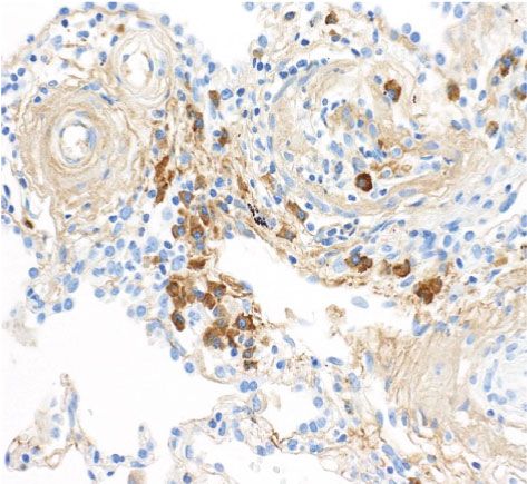
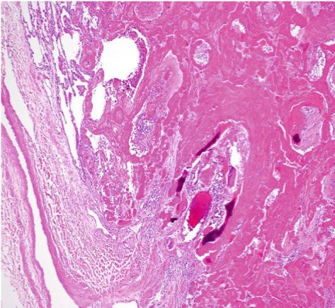
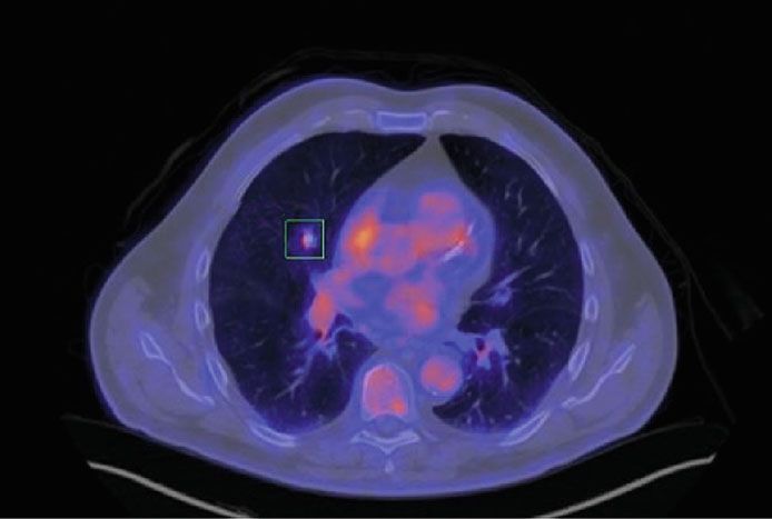
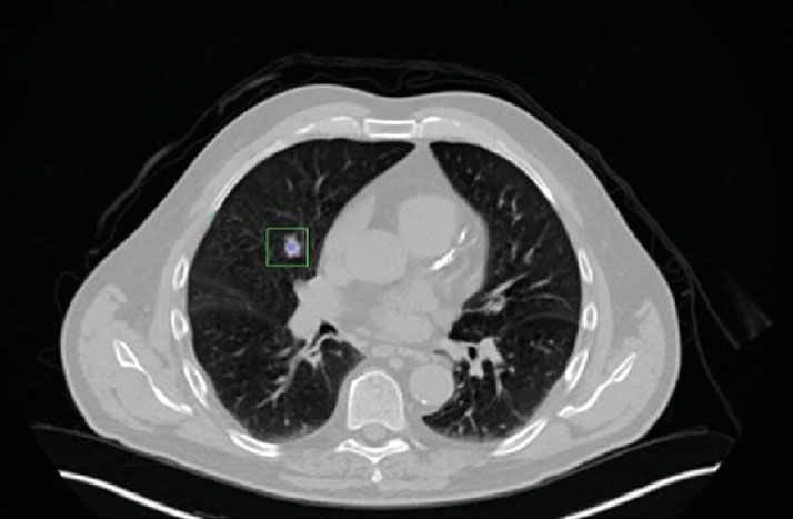
2 Case Reports in Pathology

 SUVm 2.0 SUVm 2.0

 (a) (b)

Figure 1: (a) Computed tomography image with the corresponding (b) positron emission tomography image showing 16 mm right middle
lobe lung lesion (FDG avid, SUV max 2.0).

 (a) (b)

 (c)

Figure 2: (a) Dense extracellular eosinophilic material, presenting as a mass-like deposit with areas of calcification and ossification (arrow),
perivascular eosinophilic deposits were noted, especially at the periphery of the mass-like deposit, and are seen better in (c). Perivascular
eosinophilic material was not seen diffusely throughout the lung (magnification ×400). (b) Congo red staining when viewed under
polarised light (apple-green birefringence under polarised light not visualised) (magnification ×400). (c) Kappa light-chain
immunochemistry shows positivity within the amorphous material and plasma cells (^) (magnification ×400).

labelled strongly for kappa (k), but not for lambda (λ) light area, fibrils were more dispersed and measured 4-7 nm in
chains, nor amyloid P. A moderate number of plasma cells diameter (Figure 3).
showed labelling exclusively for k light chains, raising the pos-
sibility of a monoclonal population (Figure 2(c)). The labelling 5. Haematology
for k light chains highlighted plasma cells. The surrounding
lung parenchyma showed no significant pathology. Haematology workup showed no serum paraproteins nor
 evidence of B cell monoclonality, no atypical expression of
4. Electron Microscopy T cell markers. The serum kappa/lambda light-chain ratio
 was 1.4 (reference range: 0.25–1.65). There were no urinary
The material in question was scattered throughout the lung Bence Jones proteins and no bony lytic lesions on whole-
parenchyma and occasionally was adjacent to plasma cells. body CT. Bone marrow biopsy was not performed.
At low magnification, the material had a worm-like appear-
ance measuring 10-20 microns in diameter. In cross-section, 6. Comment
the material was composed of a central core of electron-dense
material that ultrastructurally resembled elastin and an outer Immunoglobulin light-chain deposits in tissues can present
zone of amyloid-like fibrils (not shown in the figure). In one as primary amyloid light-chain (AL) amyloidosis (AL
Case Reports in Pathology 3

 5.35 nm
 6.82 nm

 4.36 nm

 1 m 50 nm

 (a) (b)

Figure 3: Electron microscopy: (a) electron-dense amyloid-like material (scale bar = 1 micron); (b) fibrils measuring 4-7 nm in diameter
observed at higher magnification (scale bar = 50 nanometer).

amyloidosis) or light-chain deposition disease (LCDD) [5]. did not reveal the classical appearance of “apple-green” bire-
AL amyloidosis is a monoclonal plasma cell or rarely lym- fringence and amyloid P was negative. The material labelled
phoplasmacytic proliferative disorder in which monoclonal strongly for k light chain but not for λ-light chain, and elec-
immunoglobulin light chains are deposited throughout tis- tron microscopy revealed fibrillar amyloid-like material mea-
sues. AL amyloid deposits are composed of protein fibrils suring 4-7 nm and larger worm-like formations 10-20
with a common core structure, consisting of anti-parallel β- microns in diameter. The material in question was unusual
strands (less commonly, parallel β-strands). This specific, in that it had an amyloid-like component. However, the
highly ordered ultrastructure of amyloid fibrils accounts for diameter of most of the fibrillar substructure is less than that
their characteristic property of binding Congo red dye when observed for amyloid (typically 7-12 nm).
viewed under cross-polarised light produces the classical Congo red staining is a screening test, but mass spectros-
“apple-green” birefringence [1, 5]. Similar to AL amyloidosis, copy in conjunction with immunohistochemistry is now
LCDD may be related to monoclonal plasma cell proliferative regarded as the “gold standard” for the diagnosis of amyloid
disorder, or it may rarely be idiopathic (ICD-O-coded since heterogeneity in Congo red staining has been reported
9769/1) [3] and can involve the lung as a diffuse or a nodular [8]. Electron microscopy is more objective as fibril diameter
form. Pulmonary light-chain deposition (PLCDD) is rare, can be measured and in this case was clearly less than that
especially in the localised form. Similar to AL amyloidosis, typical of amyloid, further supported by the lack of Congo
PLCDD also demonstrates positive k light-chain immuno- red staining and labelling for P-protein. The difficulty in this
chemistry within amorphous material [6]. However, in case, which showed focal amyloid-like features on EM, is how
PLCDD, light chains failed to assume the β-pleated sheet to best designate the lesion. Kappa light-chain deposition
configuration and result in granular deposits seen by electron with focal amyloidoma-like features, as an intermediate
microscopy (EM) and lack the disordered meshwork of 8- between focal amyloid and PLCDD, may be deemed the best
10 nm fibrils, characteristics of amyloid [5–7]. Consequently, designation.
PLCDD can be differentiated from amyloid by the absence of PET scan is an important diagnostic and staging modal-
Congo red staining and distinct EM appearances [6, 7]. ity for the evaluation of pulmonary nodules. Concerning
 In the World Health Organisation (WHO) 2017 revised PET scan in the assessment of pulmonary amyloid lesions,
classification of tumours of haematopoietic and lymphoid there is varying evidence of pulmonary amyloid lesions
tissue, LCDD is classified under the monoclonal immuno- showing intense to no FDG uptake [9]. Light-chain deposi-
globulin disposition diseases. Whilst LCDD frequently occur tion disease has also been reported to show varying degree
in association with plasma cell myeloma or in patients with of FDG uptake [7]. Whilst PET scans are important in eval-
M protein and marrow plasma cells as monoclonal gammo- uating lung lesions, their use in the detection of pulmonary
pathy of undetermined significance, some cases are idio- amyloid or light-chain deposition is not well established.
pathic or occur in association with another Furthermore, pulmonary light-chain deposition may
lymphoproliferative disorder [3]. Given the morphological also be associated with underlying lymphoproliferative or
and immunochemical characteristics of the lesion in ques- immunological disorders. In a case series of 46 patients
tion, in this case, a firm diagnosis is unable to be established, conducted by Milani et al. [10], 11 patients had urinary
and morphological may represent an intermediate between or serum monoclonal proteins detected, 13 patients had
AL amyloidosis and PLCDD. abnormal free chain ratio, Sjogren’s disease was diagnosed
 Our patient presented with a solitary FDG avid (SUV in three patients, and MALT cell lymphoma was diag-
max 2.0) nodule, which was concerning for cancer. Hence, nosed in two patients [10]. Therefore, further haematolog-
the decision to proceed with lobectomy was made with diag- ical workup is essential. This case did not reveal any
nostic and curative intent. Microscopically, the material was suggestion of systemic amyloidosis, multiple myeloma, or
densely eosinophilic and on Hematoxylin and Eosin (H&E) evidence of other lymphoproliferative disorders, therefore
staining suggestive of amyloid. However, Congo red staining necessitating clinical surveillance.
4 Case Reports in Pathology

 In conclusion, we describe a case of k light-chain deposi-
tion whose morphological, histochemical, and electron
microscopy features do not allow classification within a spe-
cific category recognised by the current WHO classification,
thus expanding the spectrum of monoclonal immunoglobu-
lin deposition disease lesions. Regardless of the diagnosis,
haematological review to identify any underlying lympho-
proliferative diseases is essential since they may present with
hematolymphoid malignancies after prolonged follow-up.

Consent
Written informed consent has been obtained from the
patient.

Conflicts of Interest
The authors declare that they have no conflicts of interest.

References
 [1] A. D. Wechalekar, J. D. Gillmore, and P. N. Hawkins, “Sys-
 temic amyloidosis,” The Lancet, vol. 387, no. 10038,
 pp. 2641–2654, 2016.
 [2] K. Matsumoto, M. Ueno, Y. Matsuo, S. Kudo, K. Horita, and
 Y. Sakao, “Primary solitary amyloidoma of the lung: findings
 on CT and MRI,” European Radiology, vol. 7, no. 4, pp. 586–
 588, 1997.
 [3] S. S. H. CE, N. L. Harris, E. S. Jaffe, S. A. Pileri, H. Stein, and
 J. Thiele, “WHO Classification of Tumours of Haematopoietic
 and Lymphoid Tissues,” Revised 4th ed editionRevised 4th ed
 edition, , 2017.
 [4] M. Yee, B. Delahunt, and P. A. Russell, “Nodular pulmonary
 light chain deposition disease,” Pathology, vol. 48, no. 5,
 pp. 515–518, 2016.
 [5] P. Bhargava, J. M. Rushin, E. J. Rusnock et al., “Pulmonary
 light chain deposition disease: report of five cases and review
 of the literature,” American Journal of Surgical Pathology,
 vol. 31, no. 2, pp. 267–276, 2007.
 [6] P. Wei, R. Tao, Y. Liu et al., “Pulmonary light chain deposition
 disease: a case series and literature review,” Annals of transla-
 tional medicine, vol. 8, no. 9, 2020.
 [7] M. Baqir, T. Moua, D. White, E. S. Yi, and J. H. Ryu, “Pulmo-
 nary nodular and cystic light chain deposition disease: a retro-
 spective review of 10 cases,” Respiratory Medicine, vol. 164,
 article 105896, 2020.
 [8] K. Bowen, N. Shah, and M. Lewin, “AL-amyloidosis presenting
 with negative Congo red staining in the setting of high clinical
 suspicion: a case report,” Case reports in nephrology, vol. 2012,
 Article ID 593460, 4 pages, 2012.
 [9] M.-J. Dong, K. Zhao, Z.-F. Liu, G.-L. Wang, and J. Yang, “Pri-
 mary pulmonary amyloidosis misdiagnosed as malignancy on
 dual-time-point fluoro-deoxyglucose positron emission tomo-
 graphy/computed tomography: a case report and review of the
 literature,” Oncology Letters, vol. 9, no. 2, pp. 591–594, 2015.
[10] P. Milani, M. Basset, F. Russo, A. Foli, G. Palladini, and
 G. Merlini, “The lung in amyloidosis,” European Respiratory
 Review, vol. 26, no. 145, article 170046, 2017.
You can also read


