Case Report Concurrent Central Diabetes Insipidus and Acute Myeloid Leukemia - Hindawi.com
←
→
Page content transcription
If your browser does not render page correctly, please read the page content below
Hindawi
Case Reports in Hematology
Volume 2021, Article ID 8898671, 5 pages
https://doi.org/10.1155/2021/8898671

Case Report
Concurrent Central Diabetes Insipidus and Acute
Myeloid Leukemia

          Stephanie L. Pritzl , Daniel R. Matson , Mark B. Juckett , and David J. Ciske
          University of Wisconsin School of Medicine and Public Health, Madison, Wisconsin, USA

          Correspondence should be addressed to David J. Ciske; dciske@dshealthcare.com

          Received 18 September 2020; Revised 2 February 2021; Accepted 8 February 2021; Published 16 February 2021

          Academic Editor: Håkon Reikvam

          Copyright © 2021 Stephanie L. Pritzl et al. This is an open access article distributed under the Creative Commons Attribution
          License, which permits unrestricted use, distribution, and reproduction in any medium, provided the original work is
          properly cited.
          Central diabetes insipidus (CDI) is a rare reported complication of acute myeloid leukemia (AML). The onset of AML-associated
          CDI often precedes the diagnosis of AML by weeks or months and is considered an adverse prognostic indicator in this setting.
          The mechanism of AML-associated CDI is not known; however, it is often reported in the setting of cytogenetic events resulting in
          MDS1 and EVI1 complex locus protein (MECOM) gene overexpression. Here, we describe a case of new onset CDI which
          preceded a diagnosis of AML by 1 month. We detail the clinical and laboratory evaluation of the patient’s CDI, and we describe the
          pathological and laboratory workup of their AML, which ultimately yielded a diagnosis of AML with myelodysplasia-related
          changes. Additional cytogenetic findings included the identification of the t (2;3)(p23;q27), which leads to MECOM gene
          overexpression and which to our knowledge has not previously been reported in the setting of AML-associated CDI. The patient
          received induction chemotherapy followed by allogeneic hematopoietic stem cell transplantation but experienced disease relapse
          and passed away nine months after initial diagnosis. We emphasize that new onset CDI can occur as a rare complication of AML
          where it portends a poor prognosis and may be related to AML subtypes displaying MECOM gene dysregulation.

1. Introduction                                                         polydipsia. Physical exam was notable for intact visual fields
                                                                        and no evidence of hypovolemia. His medications included
Central diabetes insipidus (CDI) as a complication of acute             only aspirin (81 mg daily) and simvastatin (40 mg daily).
myeloid leukemia (AML) is rare, and the underlying                      Laboratory testing revealed leukopenia and macrocytic
mechanism(s) of AML-associated CDI remains incom-                       anemia (Table 1), in the setting of a previously normal
pletely understood. When it occurs, the onset of CDI typ-               complete blood count (CBC) one year prior. Comprehensive
ically precedes the diagnosis of AML by 1-2 months [1].                 metabolic panel, hemoglobin A1c, and urinalysis were un-
However, CDI may also occur at the time of AML diagnosis                remarkable. However, urine osmolality was inappropriately
or as the initial manifestation of AML relapse [1, 2]. AML-             low in conjunction with an elevated serum osmolality
associated CDI is hypothesized to represent an adverse                  (Table 1). Polyuria was confirmed with a 24-hour urine
prognostic indicator of AML, even when CDI symptoms are                 collection. An 8-hour water deprivation test was completed
ameliorated by administration of desmopressin (DDAVP)                   and demonstrated worsening hypernatremia with a sub-
[1–3]. Here, we present a case of CDI preceding an initial              optimal response in urinary concentration (Table 1). A
diagnosis of AML.                                                       subsequent desmopressin challenge was notable for de-
                                                                        creased urine output and increased urine osmolality (Ta-
                                                                        ble 1). Levels of plasma vasopressin and copeptin, which
2. Case Presentation
                                                                        comprises a portion of the vasopressin precursor molecule,
A previously healthy 71-year-old man presented to his                   were not measured. Other hormones dependent on pituitary
primary care physician with abrupt onset of polyuria and                function including serum prolactin, free T4, cortisol (AM),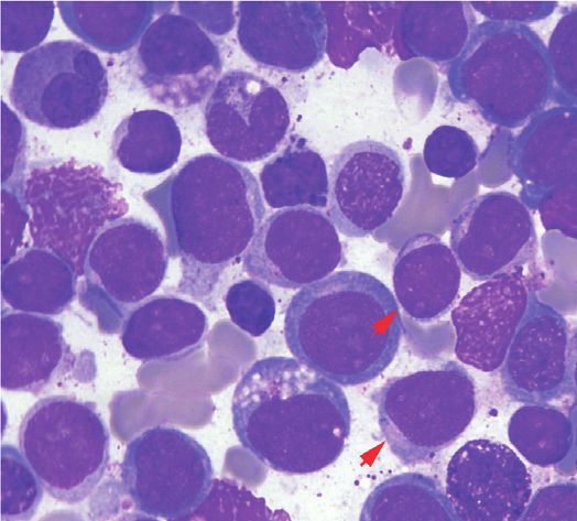
2 Case Reports in Hematology
Table 1: Laboratory data.
Laboratory testing at diagnosis
Assay Result Normal values
WBC 2.3 K/μL 3.8–10.5 K/μL
Hb 13.5 g/dL 13.6–17.2 g/dL
MCV 101 fL 80–97 fL
24-hour urine collection 12.5 L urine output 0.8–2.0 L
Prolactin 8.9 ng/mL 3.5–19.4 ng/mL
Cortisol (AM) 18.6 μg/dL 10–20 μg/dL
Free T4 0.81 ng/L 0.70–1.48 ng/L
Total testosterone 338 ng/dL 300–720 ng/dL
8-hour water deprivation test
Assay 0 hours 8 hours Normal values
Serum sodium 144 mmol/L 149 mmol/L 136–145 mmol/L
Serum osmolality 302 mOsm/kg 308 mOsm/kg 278–298 mOsm/kg
Urine osmolality 123 mOsm/kg 235 mOsm/kg 50–1200 mOsm/kg
Desmopressin challenge
Time Urine output (mL) Urine osmolality (mOsm/kg)
12-1 PM 725 87
1 PM 2 mcg DDAVP SubQ
1-2 PM 160 341
2-3 PM 80 467
and free testosterone were all within normal limits. MRI syndrome, display significant morphologic dysplasia in the
head demonstrated lack of the normal posterior pituitary bone marrow at leukemic presentation, or when their AML
bright spot. The laboratory and imaging findings were harbors characteristic cytogenetic findings that are associ-
consistent with CDI, and the, patient began treatment with ated with myelodysplasia [5]. These characteristic cytoge-
DDAVP nasal spray which led to rapid improvement in his netic findings include deletions in chromosome 5, as seen in
symptoms. our patient.
During this 21-day period, serial CBCs revealed a per- The patient received induction chemotherapy with li-
sistent leukopenia (WBC 3.4 K/μL), worsening macrocytic posomal daunorubicin and cytarabine which failed to reduce
anemia (Hb 11.7 g/Dl and MCV 107 fL), and a progressive his bone marrow blast count. This was followed by two cycles
thrombocytosis (PLT 500 K/μL and normal 160–370 K/μL). of azacytidine and lenalidomide and reinduction with flu-
A manual differential performed on peripheral blood darabine, cytarabine, and granulocyte colony-stimulating
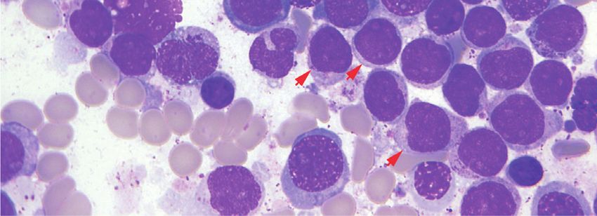
revealed 17% circulating blasts, and bone marrow biopsy factor. Three weeks following his second induction, the bone
demonstrated a hypercellular marrow virtually effaced by marrow blast count percentage was 95%. He then received
leukemic blasts (Figure 1(a)). Flow cytometry revealed a an allogeneic stem cell transplantation (alloSCT) following
myeloid immunophenotype confirming a diagnosis of AML, pretreatment with an experimental radiolabeled anti-CD45
without features worrisome for acute promyelocytic leu- monoclonal antibody (IOMAB-01). Unfortunately, he ex-
kemia. Routine ancillary testing for newly diagnosed AML perienced disease relapse 1 month after transplant and ul-
includes (minimally) the following studies performed on the timately passed away 9 months after his initial diagnosis.
leukemic blast population: karyotyping by classical cyto- Interestingly, prior to transplant, he required 40 mcg/day of
genetics, evaluation for core binding factor rearrangements DDAVP 0.01% nasal spray (20 mcg twice daily) to control
by either fluorescence in situ hybridization (FISH) or his CDI symptoms, but following his transplant, he was able
classical cytogenetics, and mutational analysis of the fms-like to space out the dosing of his DDAVP to every 4-5 days.
tyrosine kinase 3 (FLT3) gene [4].
In this case, cytogenetic analyses revealed the t (2;3)(p23;
q27), which is a chromosomal translocation often associated 3. Discussion
with MDS1 and EVI1 complex locus (MECOM) gene
overexpression, as well as the del (5) q31;q33) (Figure 1(b)). AML-associated CDI is a rare but increasingly recognized
FISH was negative for core binding factor rearrangements, complication of AML. In perhaps the largest study of AML-
and molecular analyses revealed no mutation in FLT3. Based associated CDI to date, Ladigan et al. analyzed 51 reports of
on the presence of the del (5)(q31;q33), a final diagnosis of adults with myeloid malignancies and associated CDI [1].
AML with myelodysplasia-related changes (AML-MRC) was They found a median age of diagnosis of 48 years, which is
rendered. AML-MRCs are a subtype of AML that are as- younger than the median age of 65 years reported across all
sociated with a poor prognosis. They are diagnosed when new AML diagnoses [6]. At least 9 cases have also been
AML patients have a known history of myelodysplastic reported in the pediatric population [7–13]. In addition,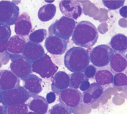
Case Reports in Hematology                                                                                                                        3

                                                                      1                  2                    3                 4             5

                                                                      6              7             8              9         10           11

                                                                     12         13            14         15            16           17    18

                                                                           19            20        21             22        X        Y
                                    (a)                                                                 (b)

Figure 1: Bone marrow aspirate and AML cytogenetic karyotype: (a) wright-stained bone marrow aspirate showing increased blasts
(examples highlighted by red arrowhead; 1000x original magnification); (b) AML karyotype demonstrating the t (2;3)(p23;q27) (black
arrows) and del (5q)(q31;q33) (red arrow).

patients with AML-associated CDI show a female pre-                However, it should be noted that leukemic infiltrates are not
dominance (59% female), compared to a mild male pre-               uncommonly seen at autopsy in AML patients who never
dominance across all AMLs. The majority (45/51) of these           developed CDI [2, 17, 18]. More recently, multiple case
cases occur in the setting of de novo AMLs, wherein the            series have revealed a strong association between AML-
patient has no known prior diagnosis of a primary bone             associated CDI and cytogenetic abnormalities in the con-
marrow neoplasm. The remaining cases are comprised of              current AML, chiefly inversions involving chromosome 3
either myelodysplastic syndrome (MDS), which is a com-             (inv (3)) and monosomies involving chromosome 7
mon precursor to AML, or AML transformed from aplastic             (monosomy 7) [1, 3, 19, 20]. Interestingly, at least a subset of
anemia, MDS, or chronic myelomonocytic leukemia                    cases with inv (3) harbor fusion events leading to MECOM
(CMML).                                                            gene overexpression, which has been shown to drive both
    The diagnosis of AML-associated CDI is essentially             leukemogenesis and dysmegakaryopoiesis [3, 21, 22]. It is
identical to the diagnosis of CDI in any other setting [14, 15].   theorized that the resulting abnormal platelets could ad-
Most patients (75%) will be diagnosed with CDI concur-             versely impact circulating vasopressin function, as 90% of
rently with their AML diagnosis, with most remaining cases         peripheral vasopressin is platelet-bound [1–3]. Cytogenetic
presenting no more than 2 months prior to or following their       studies in our patient revealed the t (2;3)(p23;q27), a
AML diagnosis [1]. While the majority of patients with             translocation that also typically results in MECOM over-
AML-associated CDI (61%) will have no associated brain             expression and which to our knowledge has not been pre-
imaging abnormalities, some may have pathologic findings            viously reported in the setting of AML-associated CDI [23].
including loss of the posterior pituitary bright spot or           Interestingly, our patient also showed a progressive
nodular thickening/attenuation of the pituitary stalk [1].         thrombocytosis, an uncommon event in AML that has
MRI of our patient revealed absence of the posterior pitu-         nonetheless been reported in the setting of the t (2;3) and
itary bright spot which can be indicative of hypothalamic          MECOM overexpression [24]. Finally, our patient showed a
pituitary dysfunction. The normal pituitary bright spot seen       greatly decreased requirement for DDAVP following
on T1-weighted MRI is thought to result from the T1-               alloSCT, after which his circulating platelets would have
shortening effect of stored vasopressin in the posterior pi-        been derived from donor marrow. This decreased require-
tuitary [16].                                                      ment for DDAVP and even complete resolution of CDI
    The pathogenesis of AML-associated CDI remains un-             following AML treatment has been previously reported [1]
clear although there are hints that dysmegakaryopoiesis and        and argues against pituitary destruction as the cause of
resulting platelet dysfunction could lie at the heart of this      AML-associated CDI in these cases.
disorder. Previously, clinicians hypothesized that CDI oc-             Clinicians treating a patient with AML-associated CDI
curred secondary to pituitary damage as a consequence of           must correct the patient’s CDI while also identifying an
tumor-mediated destruction or infection. Early cohort              optimal treatment strategy for their AML. 90% of reported
studies of deceased patients with leukemia-associated CDI          AML-associated CDI cases have a documented response to a
noted two types of lesions that could be routinely identified       vasopressin analog (mainly DDAVP), with the majority of
at autopsy in the supraopticohypophyseal system: (1) leu-          patients demonstrating a marked improvement in CDI
kemic infiltrates and/or (2) thrombosis of small vessels [16].      symptoms upon treatment [1]. Our patient’s CDI symptoms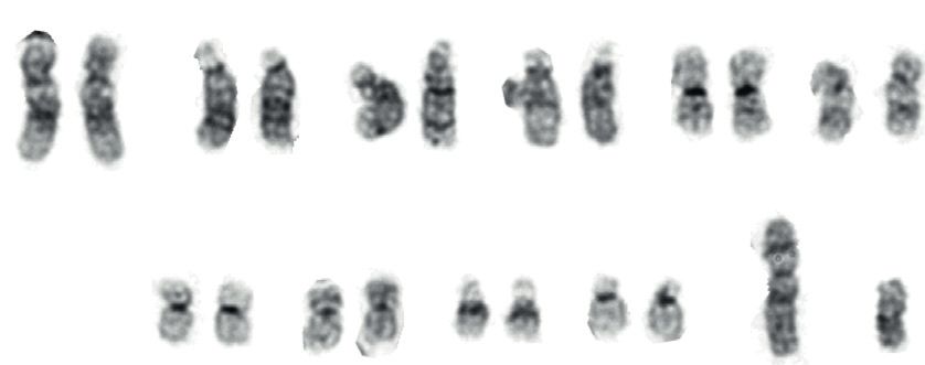
4                                                                                                      Case Reports in Hematology

are rapidly improved with DDAVP nasal spray. In com-                 findings in this case. DRM is further supported by Grant
parison, treatment of the underlying AML depends on a                number T32-HL007899 from the NHLBI.
number of factors, including the presence or absence of
prognostically important cytogenetic and molecular findings           References
and the patient’s ability to tolerate therapy. In this respect, it
is important to note that both monosomy 7 and inv (3)                 [1] S. Ladigan, T. Mika, A. Figge et al., “Acute myeloid leukemia
constitute unfavorable-risk cytogenetic findings in AML and                with central diabetes insipidus,” Blood Cells, Molecules, and
are independently associated with worse rates of complete                 Diseases, vol. 76, pp. 45–52, 2019.
remission and overall survival [25–27]. Thus, independent of          [2] P. Dy, P. Chua, J. Kelly, and S. Liebman, “Central diabetes
CDI, most AML-associated CDI patients would already be                    insipidus in the setting of acute myelogenous leukemia,”
expected to have a 5-year AML-related survival of only 5-                 American Journal of Kidney Diseases, vol. 60, no. 6,
                                                                          pp. 998–1001, 2012.
10%, compared to a 5-year survival of 29% across all AMLs
                                                                      [3] E. H. Cull, J. M. Watts, M. S. Tallman et al., “Acute myeloid
[6]. In general, there is also a paucity of data regarding                leukemia presenting with panhypopituitarism or diabetes
optimal induction regimens for AML patients with associ-                  insipidus: a case series with molecular genetic analysis and
ated CDI. In 39 patients who received treatment with either               review of the literature,” Leukemia & Lymphoma, vol. 55,
standard-dose cytarabine and daunorubicin (often referred                 no. 9, pp. 2125–2129, 2014.
to as 7 + 3) or high-dose cytarabine, the rate of complete            [4] D. A. Arber, M. J. Borowitz, M. Cessna et al., “Initial diag-
remission (CR) was only 15% [28–33]. Sufficient reports of                  nostic workup of acute leukemia: guideline from the college of
other treatment regimens are not readily available. Perhaps               American pathologists and the American society of hema-
not surprisingly given these data, AMLs in patients with                  tology,” Archives of Pathology & Laboratory Medicine,
AML-associated CDI confer a dismal prognosis, with an                     vol. 141, no. 10, pp. 1342–1393, 2017.
                                                                      [5] D. A. Arber and H. P. Erba, “Diagnosis and treatment of
overall 1-year survival as low as 20% and a median overall
                                                                          patients with acute myeloid leukemia with myelodysplasia-
survival time of only 6 months [1, 3]. However, CDI-as-                   related changes (AML-MRC),” American Journal of Clinical
sociated AML patients who underwent alloSCT showed                        Pathology, vol. 154, no. 6, pp. 731–741, 2020.
markedly better outcomes, with 40% surviving to 12 months.            [6] NIH, Acute Myeloid Leukemia-Cancer Stat Facts, NIH,
This is consistent with the survival benefit seen with alloSCT             Bethesda, MD, USA, 2018, https://seer.cancer.gov/statfacts/
following first CR in AML patients with unfavorable risk                   html/amyl.html.
cytogenetics [34]. Thus, the best available evidence suggests         [7] M. C. Joseph and S. E. Levin, “Leukaemia and diabetes
that patients with AML-associated CDI typically have un-                  insipidus,” BMJ, vol. 1, no. 4979, pp. 1328–1322, 1956.
favorable-risk cytogenetics and stand to benefit from in-              [8] G. E. Bergman, H. J. Baluarte, and J. L. Naiman, “Diabetes
tensive induction chemotherapy regimens followed (when                    insipidus as a presenting manifestation of acute myelogenous
possible) by an alloSCT in the first CR. Unfortunately, our                leukemia,” The Journal of Pediatrics, vol. 88, no. 2, p. 355,
                                                                          1976.
patient never achieved a CR despite receiving multiple in-
                                                                      [9] D. J. Kanabar, D. R. Betts, B. Gibbons, J. E. Kingston, and
duction regimens, and although he underwent an alloSCT                    O. B. Eden, “Monosomy 7, diabetes insipidus and acute
facilitated by pretreatment with an experimental therapy, he              myeloid leukemia in childhood,” Pediatric Hematology and
rapidly relapsed.                                                         Oncology, vol. 11, no. 1, pp. 111–114, 1994.
    We reiterate that CDI is a rare but recognized com-              [10] S. Roy and W. W. Johnson, “Diabetes insipidus in a child with
plication of AML which is associated with a poor prognosis.               erythromyelocytic leukemia,” Archives of Pediatrics & Ado-
We support future studies examining the consequences of                   lescent Medicine, vol. 119, no. 1, pp. 82–85, 1970.
MECOM gene dysregulation in AML and vasopressin                      [11] U. Betkerur, A. Shende, and P. Lanzkowsky, “Acute myelo-
function in both the pituitary and peripheral circulation.                blastic leukemia presenting with diabetes insipidus,” The
                                                                          American Journal of the Medical Sciences, vol. 273, no. 3,
                                                                          pp. 325–328, 1977.
Data Availability                                                    [12] H. A. Frangoul, D. W. W. Shaw, D. Hawkins, and J. Park,
No data sets were generated in the preparation of this report.            “Diabetes insipidus as a presenting symptom of acute mye-
                                                                          logenous leukemia,” Journal of Pediatric Hematology/Oncol-
                                                                          ogy, vol. 22, no. 5, pp. 457–459, 2000.
Disclosure                                                           [13] W. Wössmann, A. Borkhardt, R. Gossen, F.-J. Göbel, and
                                                                          A. Reiter, “Acute myeloid leukemia presenting with diabetes
This case report was prepared as part of the employment of
                                                                          insipidus,” European Journal of Pediatrics, vol. 161, no. 3,
the authors at the University of Wisconsin Hospitals and                  pp. 161-162, 2002.
Clinics.                                                             [14] A. Garrahy, C. Moran, and C. J. Thompson, “Diagnosis and
                                                                          management of central diabetes insipidus in adults,” Clinical
Conflicts of Interest                                                     Endocrinology, vol. 90, no. 1, pp. 23–30, 2019.
                                                                     [15] W. Fenske and B. Allolio, “Current state and future per-
The authors declare that there are no conflicts of interest.               spectives in the diagnosis of diabetes insipidus: a clinical
                                                                          review,” The Journal of Clinical Endocrinology & Metabolism,
Acknowledgments                                                           vol. 97, no. 10, pp. 3426–3437, 2012.
                                                                     [16] V. I. Miller and W. G. Campbell, “Diabetes insipidus as a
The authors would like to acknowledge the Wisconsin State                 complication of leukemia: a case report with a literature re-
Lab of Hygiene for providing images of the cytogenetic                    view,” Cancer, vol. 28, no. 3, pp. 666–673, 1971.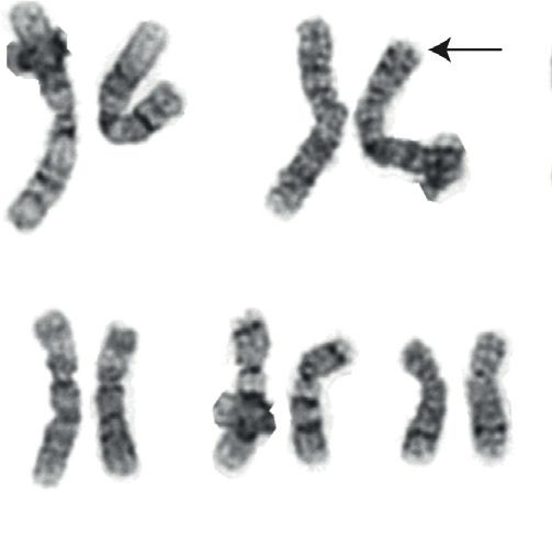
Case Reports in Hematology                                                                                                                  5

[17] K. Yamagami and K. Yoshioka, “Central diabetes insipidus                  Netherlands Journal of Medicine, vol. 56, no. 2, pp. 45–47,
     presenting as an early sign of acute myeloid leukemia,”                   2000.
     Clinical Advances in Hematology & Oncology: H&O, vol. 10,          [31]   L. Pagano, M. T. Voso, S. Sica, and G. Leone, “Recovery from
     no. 6, pp. 400-401, 2012.                                                 diabetes insipidus associated with AML after a BMT condi-
[18] K. Loukidis, E. Papadakis, N. Anagnostou et al., “Polyuria due            tioning regimen including busulfan,” Bone Marrow Trans-
     to central diabetes insipidus presenting as an early mani-                plantation, vol. 11, no. 2, pp. 175-176, 1993.
     festation of acute myeloid leukemia,” Clinical Advances in         [32]   C. C. Yen, C. H. Tzeng, J. H. Liu et al., “Acute myelomonocytic
     Hematology & Oncology: H&O, vol. 10, no. 6, pp. 399-400,                  leukemia preceded by secondary amenorrhea and presenting
     2012.                                                                     with central diabetes insipidus: a case report,” Zhonghua Yi
[19] C. Castagnola, M. Morra, B. Bemasconi, C. Astori,                         Xue Za Zhi, vol. 60, no. 4, pp. 213–218, 1997.
     S. Santagostino, and C. Bemasconi, “Acute myeloid leukemia         [33]   A. Lawrie, D. A. J. Stevenson, T. N. Doig, M. A. Vickers, and
     and diabetes insipidus: results in 5 patients,” Acta Haema-               D. J. Culligan, “Acute myeloid leukemia presenting in a
     tologica, vol. 93, no. 1, pp. 1–4, 1995.                                  mother and daughter pair with the identical acquired kar-
[20] A. De La Chapelle and R. Lahtinen, “Monosomy 7 predisposes                yotypic abnormality consisting of inversion 3q21q26 and
     to diabetes insipidus in leukaemia and myelodysplastic syn-               monosomy 7: a review of possible mechanisms,” Cancer
     drome,” European Journal of Haematology, vol. 39, no. 5,                  Genetics, vol. 205, no. 11, pp. 599–602, 2012.
     pp. 404–411, 1987.                                                 [34]   W.-J. Hong and B. C. Medeiros, “Unfavorable-risk cytoge-
[21] M. Sonmez, N. Erkut, T. S. Tat, F. Celep, U. Cobanoglu, and               netics in acute myeloid leukemia,” Expert Review of Hema-
     H. O. Ersoz, “Can a high platelet count be responsible for                tology, vol. 4, no. 2, pp. 173–184, 2011.
     diabetes insipidus in acute myelogenous leukemia with
     monosomy 7 and inversion 3 (q21q26)?” International Journal
     of Hematology, vol. 90, no. 2, pp. 273-274, 2009.
[22] K. Morishita, E. Parganas, C. L. William et al., “Activation of
     EVI1 gene expression in human acute myelogenous leukemias
     by translocations spanning 300-400 kilobases on chromosome
     band 3q26,” Proceedings of the National Academy of Sciences,
     vol. 89, no. 9, pp. 3937–3941, 1992.
[23] J. L. Huret, M. Ahmad, M Arsaban et al., “Atlas of genetics and
     cytogenetics in oncology and haematology in 2013,” Nucleic
     Acids Research, vol. 41, no. Database issue, pp. D920–D924,
     2013.
[24] K. Yamamoto, A. Okamura, Y. Sanada, K. Yakushijin,
     H. Matsuoka, and H. Minami, “Marked thrombocytosis and
     dysmegakaryopoiesis in acute myeloid leukemia with t (2;3)
     (p22; q26.2) and EVI1 rearrangement,” Annals of Hematology,
     vol. 92, no. 12, pp. 1713–1715, 2013.
[25] D. Grimwade, H. Walker, F. Oliver et al., “The importance of
     diagnostic cytogenetics on outcome in AML: analysis of 1,612
     patients entered into the MRC AML 10 trial,” Blood, vol. 92,
     no. 7, pp. 2322–2333, 1998.
[26] M. L. Slovak, K. J. Kopecky, P. A. Cassileth et al., “Karyotypic
     analysis predicts outcome of preremission and postremission
     therapy in adult acute myeloid leukemia: a Southwest on-
     cology group/eastern cooperative oncology group study,”
     Blood, vol. 96, no. 13, pp. 4075–4083, 2000.
[27] J. C. Byrd, K. Mrózek, R. K. Dodge et al., “Pretreatment
     cytogenetic abnormalities are predictive of induction success,
     cumulative incidence of relapse, and overall survival in adult
     patients with de novo acute myeloid leukemia: results from
     cancer and leukemia group B (CALGB 8461),” Blood, vol. 100,
     no. 13, pp. 4325–4336, 2002.
[28] F. Nakamura, Y. Kishimoto, T. Handa, Y. Arai, and K. Mitani,
     “Myelodysplastic syndrome with central diabetes insipidus
     manifesting hypodipsic hypernatremia and dehydration,”
     American Journal of Hematology, vol. 75, no. 4, pp. 213–216,
     2004.
[29] R. P. Mozersky, V. K. Bahl, D. Meisner, and H. Patel, “Di-
     abetes insipidus, acute myelogenous leukemia, and mono-
     somy 7,” The Journal of the American Osteopathic Association,
     vol. 96, no. 2, pp. 116–118, 1996.
[30] P.     Nieboer,     E.    Vellenga,    R.   Adriaanse,      and
     A. A. van de Loosdrecht, “Central diabetes insipidus pre-
     ceding acute myeloid leukemia with t (3;12) (q26;p12),” The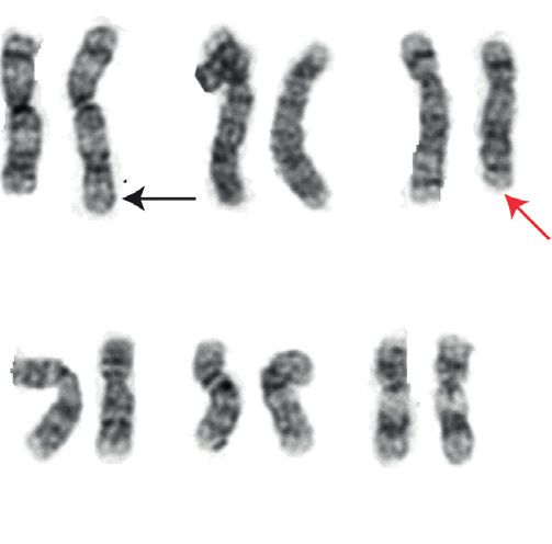
You can also read