Journal of Orthopaedics Study and Sports Medicine
←
→
Page content transcription
If your browser does not render page correctly, please read the page content below
1

Journal of Orthopaedics Study and Sports
Medicine
                                                                                                     Genesis-JOSSM-1(1)-02
                                                                                                         Volume 1 | Issue 1
                                                                                                               Open Access

Current Thinking in Pin-Site Management in
External Hexagonal Frames
                 1*                      2
Gordon Slater and Luke Mathen
1
  MBBS (UNSW) FRACS FA Ortho A Private Practice Double Bay Sydney
2
  Lead research assistant, Department of biomedical engineering, Sydney University

*
    Corresponding author: Gordon Slater, MBBS (UNSW) FRACS FA Ortho A Private Practice Double Bay Sydney

                                                                                   ©
Citation: Slater G, Mathen L. (2021) Current Thinking                    Copyright 2021 by Slater G. All rights reserved. This
in Pin-Site Management in External Hexagonal                             is an open access article distributed under the terms
Frames. J Orthop Study Sports Med. 1(1):1-9.                             of the Creative Commons Attribution License, which
                                                                         permits unrestricted use, distribution, and
Received: September 01, 2021 | Published:                                reproduction in any medium, provided the original
September 20, 2021                                                       author and source are credited.

     Abstract
     In this article, the authors review the literature for the current thinking in management of pin sites after external
     framing. Pin site management remains a challenge after external framing and is perhaps the most significant barrier
     to the use of this technology. Infection after framing ranges from 1% to 100% depending on the criteria used to
     diagnose. Clarity will be sought with regards to defining and identifying infection, as well as the appropriate
     treatment to infection. The paper looks at current methods of managing pin sites, and their advantages and
     shortcomings. The authors will also investigate the future of pin-site management, and where it is headed.
     Literature has differing opinions on a gold standard for managing pin sites. This paper will aim to provide the best
     practice to lower and control the incidence of infection for external fixation during post-operative care. This paper
     will aim to provide a consensus that will facilitate specialists, nurses, and patients in their respective management of
     infection.

     Keywords
     Percutaneous pins; Staphylococcus aureus; Escherichia coli; Skin redness

Review Article | Slater G, et al. J Orthop Study Sports Med 2021, 1(1)-02.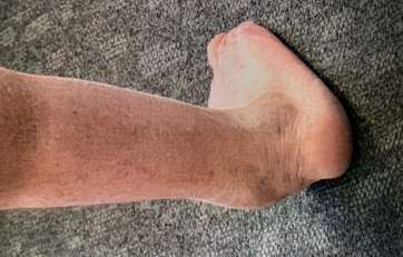
2
Introduction
External fixation is a popular technique that attaches percutaneous pins to an external frame to provide
support for the joint in case of injury or limb reconstruction (Fragomen, 2007). Despite the effectiveness
of external fixation, it is often associated with the troubling complication of pin site infection [1].
Infections vary in degree of severity for each patient, and it is important the proper pin site
management is followed to minimise the detrimental impacts of infection. The potential outcomes of
infection are damaging both commercially and in terms of patient health. Such outcomes include
increased clinic visits, additional treatment such as antibiotics or surgery, osteomyelitis, instability due
to pin loosening and frame removal [2], not to mention the potential psychological implications for
prolonged treatment. Literature has differing opinions on a gold standard for managing pin sites. This
paper will aim to provide the best practice to lower and control the incidence of infection for external
fixation during post-operative care.
Background
What is external fixation and why is it important? Why is infection associated commonly with
external fixation and what are the challenges? What is infection and how is it measured?
External fixation
External fixation frames are used to provide limb and joint support. Typically applications include limb
reconstructions following trauma or due to congenital factors, arthrodesis, distraction, management of
osteomyelitis, limb salvage in charcot and deformity correction [3]. External fixators have gained some
popularity due to their numerous benefits. Compared with internal plates and nails, external frames are
cause less trauma to the adjacent soft tissues, blood supply to the area and the periosteum. They are
indicated for patients with open wounds, skin contusions, or chronic trauma where skin is populated
with grafts and muscle flaps. External fixator devices are also adaptable to the circumstances of
individual patients, and can be attached in multiple planes [3]. This allows a frame to correct multiple
different deformities in the same limb. It can even be used in competing strategies of fusion and
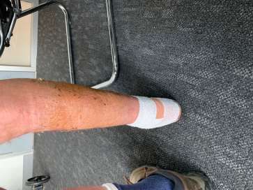
distraction arthroplasty in adjacent joints. One of the most popular configuration is demonstrated below
were the struts are placed to allow for three dimensional manipulation of the limb (Figure 1).
Figure 1: External fixator frame.
Review Article | Slater G, et al. J Orthop Study Sports Med 2021, 1(1)-02.3
Idiopathic fixed pes planovalgus deformity treated with key hole osteotomies of calcaneus and M1 to
M5 than manipulated by slow correction over 4 weeks.(M=metatarsal).Portals have been left open and
good healing is demonstrated (Figure 2).
Figure 2: Pre-operative and post-operative external fixation.
Infection
There are two main classification systems commonly found in literature to grade infections occurring at
the pin site. One popular system defines three different categories of infection following external fixator
frame attachment: Reaction, Colonisation, and Infection [4]. These three categories are useful as they
denote qualitative data that can be observed by patients. Reaction represents normal changes that
occur post-surgery, which are expected to resolve within 72 hours, including skin redness, pin site heat
and minor levels or serous fluid or blood discharge. Colonisation involves further redness and warmth
post 72 hours and accommodates for pain and exudate occurring at one or more of the pin sites.
Infection involves purulent discharge and potential loosening of the pin. This review also includes
quantitative measures of bacterial counts at each stage. Whilst this classification system is useful for
patients to identify the severity of their infection, it provides no solution to the infection, and fails to
account for infection that may be deeper inside the tissue. A typical six-point grading system is
expressed in (Table 1).
Review Article | Slater G, et al. J Orthop Study Sports Med 2021, 1(1)-02.4
Level of Infection Symptoms Treatment Method
Grade 1 Slight redness around the pin. Minimal Increase level of cleaning and
discharge. Should improve with proper perform massaging to prevent pin
care and hygiene. Can progress into adherence. Ensure wound is
serious infection. appropriately draining and remove
crusts.
Grade 2 Skin redness, discharge, and pain in soft Administer oral antibiotics. Oral
tissues. antibiotic depends on the bacteria,
which is usually Staphylococcus
aureus.
Grade 3 Grade 2 infections that continue despite Administer intravenous antibiotics.
improved care and oral antibiotic. Pins potentially re-sited. External
fixation continues.
Grade 4 Severe infection that involves multiple The pin is removed. External fixation
pins. Pins may be loosened. is discontinued.
Grade 5 Tissue is infected and bone may be Infected tissue is removed surgically
involved. Osteolysis and sequestrum to prevent the spread of infection.
formation is present from radiographs. Will resolve after fixator removal.
Grade 6 Chronic osteomyelitis (infection of the Typical treatment will involve
bone). debridement or treatment through
antibiotic agents. Can lead to long-
term disability and discomfort.
Table 1: Symptoms and treatment methods for different grades of pin-site infection.
Grade 1-3 infections are classified as minor, and can be managed with the external fixator still in place.
Grade 4-6 infections are major, and required abandonment of external fixation [1]. Grade 1 and 2 are
useful for patients in their management of pin sites, however this framework is excessively detailed and
may be intimidating for patients. For managing pin sites at home, Lee-Smith’s “Reaction, Colonisation
and Infection” framework is the recommended guideline due to its simplicity and clarity.
Bacteria typically found
It is important to be aware of the type of bacteria present in a pin-site infection in order to properly
administer antibiotics. Staphylococcus Aureus is the most common type of bacteria found [6]. They exist
frequently on skin surfaces and mucus, and can thus easily become implicated when a medical device
penetrates the skin. Staphylococcus Epidermis and Escherichia coli (E. coli) are other bacteria that may
cause pin site infections [7].
Review Article | Slater G, et al. J Orthop Study Sports Med 2021, 1(1)-02.5 Management General principles There are a few general principles for management in which there is consensus in the literature.. First, hand hygiene must be performed at all times when taking care of pin sites, particularly in removing dressings, washing the sites, showering and massaging [1]. Additionally, cross-contamination between pin sites should always be avoided [4]. Using something like a toothbrush for cleaning encourages cross- contamination, so should not be used. Pin site management should also aim to minimise the use of antibiotics, as a result of the increasing presence of drug-resistant micro-organisms. Antibiotics should only be taken if necessary and in consultation with the doctor [4]. Crusts The overall consensus regarding crusts based on research and practical experience would recommend that crusts should be removed. Crust removal reduces the infection rate as it allows fluid to be moved away from the wound site, and prevents it from being held up in the wound area [8]. It also allows the wound site to be visualised which is paramount in the prevention of infection. [4]. The method of crust removal remains uncertain. A gentle scab peeling approach is the most logical method, and persistent scabs can be dry rubbed gently with gauze [4]. An alternate perspective highlights the nature of crusts to act as a biological dressing, and postulates they should be kept. The study revealed that patients where crust was retained had lower infection rates. However, where infection did occur, the group that retained crusts had more severe infection and required a more urgent administration of antibiotics. It was concluded that crusts should be retained so long as the site was uninfected [9]. Despite this, the clear advantages of crust removal through visualisation and drainage is preferable than the chance of a severe infection occurring from crust retention. Regular crust removal is something every patient should engage in to minimise the risk of infection. Cleaning Various studies using different cleaning solutions have failed to demonstrate a statistical significance to recommend one cleaning solution over another.. Many sterile and non-sterile techniques exist in literature, and typical solutions studied include sterile chlorohexidine, betadine, alcohol and hydrogen peroxide. Non-sterile solutions include saline, soap and warm sterile water. A few studies suggest there is no significant benefit between different cleaning solutions [10]. Despite this, chlorhexidine alone has been shown to less effective than when it is combined with alcohol. The study recommends avoiding this solution closer to the wound, and instead using saline to encourage granulation and remove exudate or blood [11]. Another study has found chlorhexidine to be superior to saline in its cleaning capability, with a high presence of S. aureus in saline treatment sites. Whilst chlorhexidine didn’t lead to lower infection rates, it resulted in lower bacterial colonisation and decreased use of antibiotics when compared to saline [2]. Alcohol is damaging to the skin and can cause pain at the pin sites, so should be avoided. Iodine-based products should also be avoided due to metal corrosion, staining and high costs [4]. Hydrogen peroxide represents a source of contention. It has damaging properties towards healthy skin tissue, however it is effective in killing bacteria and wound debridement [11]. It is recommended that hydrogen peroxide is used scarcely and only in event of grade 3 infections. Some studies indicate Review Article | Slater G, et al. J Orthop Study Sports Med 2021, 1(1)-02.
6
saline as something that has higher infection rates compared to control groups, however it is still
recommended as a washing agent [6].
Dressing
Studies have once again failed to demonstrate superiority in terms of type of dressing used, however it
is clear that some form of dressing is essential for prevention of bacterial infection by absorbing blood
and exudate. An important role of the dressing is also to prevent tenting, which occurs where the skin
climbs up along the wire. Tenting can lead to skin tenderness and may need to be addressed surgically.
Prevention can be managed through pressure when applying a dressing [11]. It is also paramount that
the dressing does not leak fibres into the skin. For example, gauze rubbing against the wound area may
result in detachment of fibres that can enter the wound site [11].
Managing patients from a psychological perspective
The importance of physical care is paramount and must be adequate to minimise infection, however the
important of emotional and psychological care for the patient is often overlooked. We identify sufficient
education and guidance to be an appropriate measure to deal with the intimidating nature of having an
external fixator frame. Written and verbal information can reduce anxiety for patients. Lee-Smith
clarifies how providing psychosocial support can help patients deal with issues pertaining to self-image,
which will in turn result in greater compliance with cleaning protocol, and thus improve pin site
management [4]. External fixation devices have been shown to contribute to patient anxiety and
depression, with more than half of patients experiencing some form of anxiety or depression, due to
uncertainty, fearful reaction, post-removal re-injury. Results suggests use of support groups and
education [12].
Help patients deal with the operation – provide information, education, contact
How can patient handouts be more effective?
o Visual information
o More education
o More clarity on the process, e.g. clarity with infection definitions.
Future Directions
What will be potential best practices going forward?
There are several areas of interest for pin site management that would increase the utility of external
fixation as common practice for joint salvage. One study demonstrated the effectiveness of a lipid
stabilized hydroxyapatite / chlorhexidine coating. The study used goats and demonstrated a decreased
infection rate and improved fixation of the frame [13]. A more recent study postulated the use of
antimicrobial gauze as a dressing to reduce the incidence of infection. Using polyhexamethylene
biguanide-impregnated gauze reduced the risk of infection in a level 1 therapeutic study.
Lee-Smith 2001: Pins impregnated with anti-bacterial agents [4].
Review Article | Slater G, et al. J Orthop Study Sports Med 2021, 1(1)-02.7
Lee 2012: Antimicrobial gauze as a dressing reduces pin site infection. PHMB polyheamethylene
biguanide. Effective against range of bacteria including s. Aureus which causes most infections in
pin sites [14].
Our experience
We reviewed our last 50 consecutive distraction frames. No patients were excluded. In that time there
were 720 separate pin sites that were monitored. Our resume was:
Antibiotics used with induction. Usually Keflex 1gm or floucloxacillin 1gm.
Chlormycetin or betadine ointment applied to pin sites intra-operatively.
Proximal tibial pins were placed with diamond tipped predrilling.
All pins fixations were smooth interference fit 2.7 mm hard wire system . No use of Steinman
pins or tensioned wires.
Wires were inserted without tourniquet. Minimise the chance of tissue necrosis.
The patient was administered antibiotics for a four week period.Discharge from hospital pre
planned and within 24 to 48 hrs.
pin sites were lavaged with hydrogen peroxide or betadine twice daily for the first 4 weeks.
This was continued if issues. Note that use of swabs and gauze not used unless a crust was to form.
Simplification of the cleaning process is recommended as patients manage the pin sites themselves and
cross contamination is a very high risk.
Almost all wire points experienced redness at some point. These largely resolved with the standard
protocol.
1 patient had a grade 3 infection requiring 3 weeks of intravenous antibiotics. This involved two
calcaneal pin sites. 2 sites. Staphylococcus aureus.
1 patient had a grade 4 infection requiring removal and replacement of a wire. This was a proximal pin
site. Staphyloccus aureus
Discussion
There an increasing desire in the community to salvage limbs and joints and employ minimally invasive
technologies to achieve this. In the foot and ankle external framing is a technique that can be used to
achieve these objectives in increasing indications. The scourge on the technology has always been the
possibility of infection at the pin-sites. This frontier represents a defect in the normal barrier of the body
to the external environment this is compounded by there being a foreign body at this surface.
The definition of what represents an infection is also contentious. Most likely redness after surgery of 72
hours is a normal and transitory state. Even so it should not be ignored as some of these will lead to a
bone fide infection. If we define infection as only occurring if redness is persisting after 72 hr, spreading
erythema, temperature increase, pain (nb beware charcot patients) and or purulent discharge than
infection with an appropriate resume should fall well below 5%. Infection should rarely deteriorate to
the point that wire removal is required.
Review Article | Slater G, et al. J Orthop Study Sports Med 2021, 1(1)-02.8
Conclusion
External fixation using ringed hexagonal external framing systems is complex surgery requiring intricate
pre-operative planning and effective post-operative management to maximise the chance of success.
Hexagonal framing systems allow combinations of minimally invasive surgical techniques that can be
combine minimally invasive image guided corrections, fusion and distraction technologies. It is likely
that with increasing advances in biologics that joint salvage procedures and minimally invasive
techniques will increase the usage of frames.
There is no consensus how to minimise the chance of pin site infection. The definition of what
substantiates an infection also confuses the data. Depending on how sensitive the definition of infection
of the pin sites is infection can be deemed to occur from 1 to 100%. There is consensus that infection of
pin sites in external framing remains one of the most serious and frequent complications that occur with
external framing. Our approach is to begin with optimising the health of the patient. Carefully plan the
pin positions, minimise thermal necrosis and engage aggressive measure to pre-empt potential
infection.
What are key takeout’s from this?
Place wires without tourniquet to decrease the possibility of thermal necrosis.
Tibial wires more likely to generate heat and experience necrosis and secondary infection. direct
wires away from the thick anterior cortical bone.
Cleaning in particularly of crust is very important
Minimise the chance of cross contamination by the patient or health worker in your cleaning
resume.
Virtually all patients will get erythema around the pin sites this is likely not infected but a normal
reaction.
The most likely bacteria accounted is Staphylococcus Aureus
References
1. RG Checketts, AG MacEachem, M. Otterbum. (2000) "Pin Track Infection and the Principles of Pin Site Care," in
Orthofix External Fixation in Trauma and Orthopaedics, G. De Bastiani, A. G. Apley, and A. Goldberg Eds. London:
Springer London,pp:97-103.
2. NH Kazmers, AT Fragomen, SR Rozbruch. (2016) "Prevention of pin site infection in external fixation: a review of
the literature," (in eng), Strategies Trauma Limb Reconstr. 11(2):75-85.
3. AT Fragomen, SR Rozbruch. (2007) The Mechanics of External Fixation. HSS J. 3(1):13-29.
4. LS Jenny, S Julie, D Peter, J Rebecca, K Julia. (2001) Pin site management. Towards a consensus: part 1. J Orthop
Nurs.5(1):37-42.
5. S Melanie, S Mike. (2000) External fixation-the incidence of pin site infection: a prospective audit. J Orthop
Nurs. 4(2):59-63.
6. C Henry. (1996) Pin sites: Do we need to clean them?. Practice Nurs. 7(4):12-17.
7. D Ceroni, C Grumetz, O Desvachez, S Pusateri, P Dunand, et al. (2016) From prevention of pin-tract infection to
treatment of osteomyelitis during paediatric external fixation," (in eng). J Child Orthop. 10(6):605-12.
8. M Trigueiro. (1983) Pin site care protocol," (in eng). Can Nurse. 79(8):24-6.
Review Article | Slater G, et al. J Orthop Study Sports Med 2021, 1(1)-02.9 9. S Britten, A Ghoz, B Duffield, PV Giannoudis. (2013) Ilizarov fixator pin site care: The role of crusts in the prevention of infection. Injury. 44(10):1275-78. 10. A Lethaby, J Temple, J Santy‐Tomlinson. (2013) Pin site care for preventing infections associated with external bone fixators and pins. Cochrane Database Syst Rev. 3(12):CD004551. 11. A Timms, H Pugh. (2013) Pin site care: guidance and key recommendations," (in English), Nursing Standard. 27(1):50-5. 12. S Dheensa, S Thomas. (2012) Investigating the relationship between coping, quality of life and depression/anxiety in patients with external fixation devices. Int J Orthop Trauma Nurs. 16(1):30-8. 13. E DeJong. (2001) Antimicrobial Efficacy of External Fixator Pins Coated with a Lipid Stabilized Hydroxyapatite/Chlorhexidine Complex to Prevent Pin Tract Infection in a Goat Model. J trauma. 50(8):1008-14. 14. CK Lee, YP Chua, A Saw. (2012) Antimicrobial gauze as a dressing reduces pin site infection: a randomized controlled trial," (in eng), Clin Orthop Relat Res. 470(2):610-5. Review Article | Slater G, et al. J Orthop Study Sports Med 2021, 1(1)-02.
You can also read