Case Report Middle Ear Ceruminous Gland Adenoma Obstructing the Eustachian Tube Orifice
←
→
Page content transcription
If your browser does not render page correctly, please read the page content below
Hindawi
Case Reports in Otolaryngology
Volume 2021, Article ID 5987353, 3 pages
https://doi.org/10.1155/2021/5987353

Case Report
Middle Ear Ceruminous Gland Adenoma Obstructing the
Eustachian Tube Orifice

          Hamin Jeong, Haemin Noh, and Chang-Hee Kim
          Department of Otorhinolaryngology-Head and Neck Surgery, Konkuk University Medical Center,
          Research Institute of Medical Science, Konkuk University School of Medicine, Seoul, Republic of Korea

          Correspondence should be addressed to Chang-Hee Kim; ryomachang@gmail.com

          Received 3 May 2021; Accepted 7 July 2021; Published 14 July 2021

          Academic Editor: Akinobu Kakigi

          Copyright © 2021 Hamin Jeong et al. This is an open access article distributed under the Creative Commons Attribution License,
          which permits unrestricted use, distribution, and reproduction in any medium, provided the original work is properly cited.
          Ceruminous glands are located in the skin of the cartilaginous portion of the external auditory canal, and ceruminous gland
          adenoma originating from the middle ear mucosa is extremely rare. We report a case of middle ear ceruminous gland adenoma
          which caused long-standing otomastoiditis and mixed hearing loss with a large air-bone gap by obstructing the bony Eustachian
          tube. We discuss the clinical characteristics and histologic features of the present case.

1. Introduction                                                       anterosuperior quadrant of the tympanic membrane without
                                                                      discharge (Figure 1(a)). A nonenhanced temporal bone
Cerumen, which plays an important role in protecting the              computed tomography (TBCT) demonstrated soft tissue
ear from infection and mechanical damage, is produced by              density obstructing the bony portion of the Eustachian tube
the ceruminous gland and sebaceous gland. The human                   and opacification in the middle ear and mastoid cavity with
ceruminous glands are modified apocrine glands and located             an intact bony labyrinth (Figure 1(b)). A pure tone audi-
in the skin of the cartilaginous portion of the external au-          ometry (PTA) revealed the left-side mixed hearing loss with
ditory canal [1, 2]. The ceruminous gland neoplasms in the            a large air-bone gap (Figure 1(c)). A retroauricular canal
middle ear cavity are extremely rare because ceruminous               wall-up tympanomastoidectomy was performed, and a
glands are not distributed in the middle ear cavity, and only         polypoid mass filling the anterior part of the tympanic cavity
few cases have been reported in the English literature [3–10].        was revealed (Figure 2(a)) and completely removed. His-
In the present study, we report a case of the middle ear              tological examination revealed a nonencapsulated mass
ceruminous gland adenoma which caused long-standing                   composed of glandular structures lined by two layers of
otomastoiditis and conductive hearing loss by obstructing             epithelium originating from the ceruminous gland, which
the Eustachian tube orifice. This work has been reported in            was consistent with ceruminous gland adenoma
line with the SCARE guidelines [11].                                  (Figure 2(b)). Luminal cells were positive for cytokeratin 7
                                                                      (Figure 2(c)). One year after surgery, the left tympanic
2. Case Presentation                                                  membrane appeared normal (Figure 1(d)), TBCT revealed
                                                                      no residual mass in the tympanic cavity (Figure 1(e)), and
A previously healthy 56-year-old woman complained of left-            conductive hearing loss was much improved (Figure 1(f )).
side hearing loss over a 10-month period. She had been
receiving treatment at another hospital and undergone                 3. Discussion
ventilation tube insertion surgery in the left ear 4 month
prior. The patient reported that the left-side hearing loss was       Although ceruminous gland neoplasms are relatively
not relieved after the surgery. On the otoendoscopic ex-              common in other mammals [12], they are highly uncommon
amination, a pinkish mass was seen through the                        in humans [2]. Ceruminous glands are primarily located in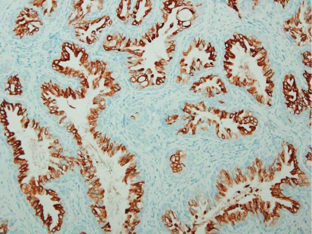
2 Case Reports in Otolaryngology
–10
0
10
20
30
40
50
60
70
80
90
100
110
120
125 250 500 1k 2k 4k 8k
(a) (b) (c)
–10
0
10
20
30
40
50
60
70
80
90
100
110
120
125 250 500 1k 2k 4k 8k
(d) (e) (f )
Figure 1: Preoperative (a–c) and postoperative (d–f ) clinical findings. (a) Preoperative otoendoscopic examination revealed a pinkish mass-
like lesion (black arrow) behind the anterior portion of the tympanic membrane. Middle ear effusion is observed (white arrow) despite
previous ventilation tube insertion (black arrow head). (b) Axial view of temporal bone computed tomography (TBCT) demonstrated soft
tissue density obstructing the bony portion of the Eustachian tube (black arrow) and mastoiditis (white arrows). (c) Pure tone audiometry
showed conductive hearing loss with an air-bone gap of 37 dB in the left side. (d) Postoperative otoendoscopic examination revealed normal
tympanic membrane. (e) Axial view of TBCTdemonstrated a clean mastoid cavity (white arrow) and no residual mass in the bony portion of
the Eustachian tube (black arrow). (f ) Pure tone audiometry showed that the air-bone gap was reduced to 5 dB in the left side.
(a) (b) (c)
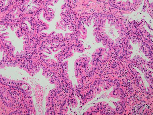
Figure 2: (a) A nonencapsulated, polypoid, pinkish mass (black arrow) and gray-colored mass with fibrotic consistency (white arrow) were
seen in the anterior part of the tympanic cavity. Black arrow heads, malleus handle; blue arrow, tympanomeatal flap. (b) Tubular tumor cell
structures with intervening bands of tumor stroma are observed, hematoxylin-eosin X100. (c) Immunohistochemical staining for cyto-
keratin 7 was positive in luminal cells, X100.
the skin lining cartilaginous portion of the external auditory pleomorphic adenoma, and ceruminous syringocystade-
canal, and most tumors originating from the ceruminous noma papilliferum, and malignant tumors, which consist of
glands are found in the external auditory canal in humans ceruminous adenocarcinoma, ceruminous adenoid cystic
[2, 13]. Ceruminous gland neoplasms in the middle ear carcinoma, and ceruminous mucoepidermoid carcinoma
cavity are extremely rare, and it has been suggested that these [2]. Among these, ceruminous gland adenoma is histolog-
tumors originate from ectopic ceruminous glands or arise ically characterized by cuboid and cylindrical cells with an
from the mucosal lining of the middle ear [8, 9]. Ceruminous eosinophilic cytoplasm and hyperchromatic round nuclei,
gland neoplasms can be categorized into benign tumors, without mitotic figures [2]. Macroscopically, the tumor is
which consist of ceruminous adenoma, ceruminous fibrotic, gray or pink colored, and poorly vascularized.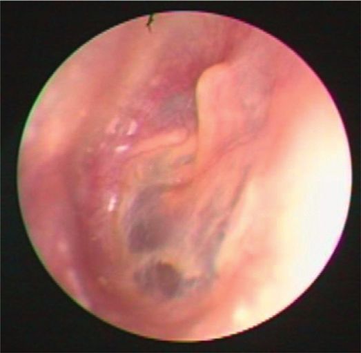
Case Reports in Otolaryngology                                                                                                     3

Although ceruminous gland adenomas in the middle ear             References
cavity are usually confined in the middle ear without erosion
of the ossicles or bony labyrinth [10], the mastoid cavity can    [1] M. Stoeckelhuber, C. Matthias, M. Andratschke et al., “Hu-
also be involved [4]. Involvement of the Eustachian tube, as          man ceruminous gland: ultrastructure and histochemical
                                                                      analysis of antimicrobial and cytoskeletal components,” The
observed in our patient, may occur infrequently [4].
                                                                      Anatomical Record Part A: Discoveries in Molecular, Cellular,
    Progressive aural fullness, hearing loss, otalgia, and            and Evolutionary Biology, vol. 288A, no. 8, pp. 877–884, 2006.
otorrhea are known as typical symptoms of middle ear              [2] P. Nagarajan, “Ceruminous neoplasms of the ear,” Head and
neoplasm [4–10]. Our patient received medical treatment               Neck Pathology, vol. 12, no. 3, pp. 350–361, 2018.
including oral antibiotics for several months under the           [3] P. Schenk, A. Handisurya, and M. Steurer, “Ultrastructural
diagnosis of otitis media and underwent ventilation tube              morphology of a middle ear ceruminoma,” Orl, vol. 64, no. 5,
insertion before visiting our clinic. However, despite pro-           pp. 358–363, 2002.
longed treatment, hearing loss was progressively aggravated.      [4] K. Orendorz-Fraczkowska, M. Jaworska, W. Gawron, and
The large air-bone gap, which was observed in the affected             R. Badowski, “Middle-ear ceruminous adenoma as a rare
ear of our patient, may be attributed to middle ear effusion           cause of hearing loss and vertigo: case reports,” Auris Nasus
and attachment of tumor to the malleus as observed in                 Larynx, vol. 32, pp. 393–397, 2005.
                                                                  [5] A. Grossman, W. H. Mathews, and M. B. Gravanis, “Cer-
Figure 1. Pure tone audiometry showed mild high-frequency
                                                                      uminal adenoma of the middle ear and external ear canal,” The
sensorineural hearing loss on both ears, which may be                 Laryngoscope, vol. 74, no. 2, pp. 241–244, 1964.
explained as age-related hearing loss. In our patient, tym-       [6] D. A. Gillanders, A. J. Worth, and L. H. Honore, “Ceruminous
panomastoidectomy was performed after TBCT evaluation,                adenoma of the middle ear,” Canadian Journal of Otolaryn-
and ceruminous gland adenoma was histologically                       gology, vol. 3, pp. 194–201, 1974.
diagnosed.                                                        [7] F. Peytz and A. S. Ohlsen, “Ceruminoma in the tympanic
                                                                      cavity,” Acta Oto-Laryngologica, vol. 53, no. 2-3, pp. 391–396,
4. Conclusions                                                        1961.
                                                                  [8] J. F. Pallanch, L. H. Weiland, T. J. McDonald, G. W. Facer, and
The present study demonstrated that ceruminous gland                  S. G. Harner, “Adenocarcinoma and adenoma of the middle
adenoma, which originated from the middle ear, can ob-                ear,” The Laryngoscope, vol. 92, no. 1, pp. 47–54, 1982.
struct the bony portion of the Eustachian tube and cause          [9] V. J. Hyams and L. Michaels, “Benign adenomatous neoplasm
                                                                      (adenoma) of the middle ear,” Clinical Otolaryngology, vol. 1,
progressive conductive hearing loss and otomastoiditis.
                                                                      no. 1, pp. 17–26, 1976.
Thus, though extremely rare, the middle ear ceruminous           [10] B. Arnold, C. Zietz, J. Muller-Hocker, and T. P. Wustrow,
gland adenoma should be taken into consideration in the               “Adenoma of the middle ear mucosa,” European Archives of
differential diagnosis when conductive hearing loss and                Oto-Rhino-Laryngology: Official Journal of the European
otitis media are progressively worsening and do not respond           Federation of Oto-Rhino-Laryngological Societies (EUFOS):
to long-term conventional treatment.                                  Affiliated with the German Society for Oto-Rhino-Laryngology
                                                                      - Head and Neck Surgery, vol. 253, pp. 65–68, 1996.
Ethical Approval                                                 [11] R. A. Agha, M. R. Borrelli, R. Farwana et al., “The SCARE 2018
                                                                      statement: updating consensus surgical case report (SCARE)
This study was approved by the Institutional Review Board             guidelines,” International Journal of Surgery, vol. 60,
(No. 2021-03-072).                                                    pp. 132–136, 2018.
                                                                 [12] P. Moisan and G. Watson, “Ceruminous gland tumors in dogs
                                                                      and cats: a review of 124 cases,” Journal of the American
Consent                                                               Animal Hospital Association, vol. 32, no. 5, pp. 448–452, 1996.
                                                                 [13] A. A. Lott Limbach, A. P. Hoschar, L. D. R. Thompson,
Written informed consent was obtained from the patient for
                                                                      E. B. Stelow, and D. J. Chute, “Middle ear adenomas stain for
publication of this case report and accompanying images.              two cell populations and lack myoepithelial cell differentia-
                                                                      tion,” Head and Neck Pathology, vol. 6, no. 3, pp. 345–353,
Conflicts of Interest                                                 2012.

The authors declare that there are no conflicts of interest
with respect to the research, authorship, and/or publication
of this article.

Authors’ Contributions
Hamin Jeong wrote the draft of the manuscript and inter-
preted the results. Haemin Noh analyzed and interpreted the
data. Chang-Hee Kim conceived, designed, and supervised
the study and critically revised the manuscript.

Acknowledgments
This work was supported by Konkuk University in 2020.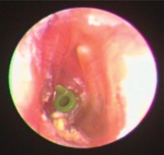
You can also read