Milodowski, E., Amengual-Batle, P., Beltran, E., Gutierrez-Quintana, R., & De Decker, S. (2019). Clinical findings and outcome of dogs with ...
←
→
Page content transcription
If your browser does not render page correctly, please read the page content below
Milodowski, E., Amengual-Batle, P., Beltran, E., Gutierrez-Quintana, R., &
De Decker, S. (2019). Clinical findings and outcome of dogs with unilateral
masticatory muscle atrophy. Journal of Veterinary Internal Medicine, 33(2),
735-742. https://doi.org/10.1111/jvim.15373

Publisher's PDF, also known as Version of record
License (if available):
CC BY-NC
Link to published version (if available):
10.1111/jvim.15373

Link to publication record in Explore Bristol Research
PDF-document

This is the final published version of the article (version of record). It first appeared online via Wiley at
https://doi.org/10.1111/jvim.15373 . Please refer to any applicable terms of use of the publisher.

University of Bristol - Explore Bristol Research
General rights

This document is made available in accordance with publisher policies. Please cite only the published
version using the reference above. Full terms of use are available:
http://www.bristol.ac.uk/pure/about/ebr-terms
Received: 27 July 2018       Accepted: 6 November 2018
DOI: 10.1111/jvim.15373

STANDARD ARTICLE

Clinical findings and outcome of dogs with unilateral
masticatory muscle atrophy
Emily Jayne Milodowski1 | Pablo Amengual-Batle2 | Elsa Beltran1 |
Rodrigo Gutierrez-Quintana2                             | Steven De Decker1

1
  Department of Veterinary Clinical Science
and Services, Royal Veterinary College,               Background: Little is known about the spectrum of underlying disorders in dogs with unilateral
University of London, Hatfield, England,              masticatory muscle (MM) atrophy.
United Kingdom
                                                      Objectives: To evaluate the clinical presentation, magnetic resonance imaging (MRI) findings,
2
 School of Veterinary Medicine, College of
                                                      and outcome of dogs with unilateral MM atrophy.
Medical, Veterinary and Life Sciences,
University of Glasgow, Glasgow, Scotland,             Animals: Sixty-three client-owned dogs.
United Kingdom                                        Methods: The medical database was retrospectively reviewed for dogs that underwent MRI for
Correspondence                                        evaluation of unilateral MM atrophy. Imaging studies were reviewed and follow-up information
Steven De Decker, Department of Veterinary            was obtained from telephone interviews.
Clinical Science and Services, Royal Veterinary
College, University of London, Hawkshead
                                                      Results: Presumptive trigeminal nerve sheath tumor (pTNST) was diagnosed in 30 dogs (47.6%);
Lane, North Mymms, Hatfield, AL9 7TA                  survival time varied from 1 day to 21 months (median, 5 months). Other extra-axial mass lesions
England, United Kingdom.                              were observed in 13 dogs (20.6%); survival time varied from 6 days to 25 months (median,
Email: sdedecker@rvc.ac.uk
                                                      2.5 months). In 18 dogs (28.6%), no abnormalities were observed on MRI; neurological signs
                                                      only progressed in 1 dog. Diagnosis had a significant influence on the type of neurological
                                                      abnormalities, with additional neurological deficits observed in most dogs with pTNST and in all
                                                      dogs with other extra-axial mass lesions. Diagnosis had a significant effect on euthanasia at the
                                                      time of diagnosis and likelihood of neurological deterioration. Dogs with mass lesions were more
                                                      likely to be euthanized or experience neurological deterioration, whereas these outcomes
                                                      occurred less often in dogs in which no causative lesion could be identified.
                                                      Conclusions and Clinical Importance: Trigeminal nerve sheath tumors should not be considered
                                                      the only cause of unilateral MM atrophy. Our results illustrate the importance of performing a
                                                      neurological examination and MRI when evaluating dogs with unilateral MM atrophy.

                                                      KEYWORDS

                                                      cranial nerves, facial asymmetry, neuritis, trigeminal nerve sheath tumor

1 | I N T R O D U C TI O N                                                          which receives its innervation from the facial nerve.1 Bilateral mastica-
                                                                                    tory muscle (MM) atrophy occurs relatively commonly in dogs and can
The muscles of mastication are responsible for movement of the man-                 be caused by several underlying disease processes. Comparatively,
dible during prehension and mastication of food. They include the                   facial asymmetry caused by unilateral MM atrophy is observed less
temporalis, masseter, pterygoideus, mylohyoideus, and digastricus                   frequently and is associated with a limited number of differential diag-
muscles. The mandibular branch of the trigeminal nerve innervates                   noses.2 Neoplasia of the trigeminal nerve is thought to be the most
each of these with the exception of the caudal belly of the digastricus,            common cause of unilateral MM atrophy.2,3 By far the most common
                                                                                    type of tumor affecting the trigeminal nerve is malignant nerve sheath
                                                                                    tumor with other tumors, such as lymphoma, occurring less com-
Abbreviations: CSF, cerebrospinal fluid; MM, masticatory muscle; MRI, mag-
netic resonance imaging; PLR, pupillary light reflex; pTNST, presumptive trigem-    monly.4 Trigeminal nerve sheath tumors (TNST) are an uncommon
inal nerve sheath tumor; TNST, trigeminal nerve sheath tumor.                       intracranial neoplasm in dogs, for which unilateral MM atrophy

This is an open access article under the terms of the Creative Commons Attribution-NonCommercial License, which permits use, distribution and reproduction in any
medium, provided the original work is properly cited and is not used for commercial purposes.
© 2018 The Authors. Journal of Veterinary Internal Medicine published by Wiley Periodicals, Inc. on behalf of the American College of Veterinary Internal Medicine.
J Vet Intern Med. 2019;33:735–742.                                                                                    wileyonlinelibrary.com/journal/jvim       735
736 MILODOWSKI ET AL.
appears to be a consistent clinical feature.3 The nature of additional the clinical records included signalment, clinical signs, neurological
clinical signs seen is reflected by the local effects of trigeminal nerve examination findings, results of diagnostic investigations, and any treat-
dysfunction and those secondary to potential brainstem compression. ment received. Dogs were excluded if clinical records or imaging studies
Reported findings in animals with TNST therefore include decreased were unavailable for review or if unilateral MM atrophy was incidentally
facial sensation, facial rubbing, absent palpebral reflex, decreased cor- observed or not the predominant clinical indication for referral. All medi-
neal sensation, ocular pathology, decreased menace response, torti- cal files and imaging studies were reviewed by a board-certified neurol-
collis, ipsilateral Horner's syndrome, enophthalmia, and paradoxical ogist (Steven De Decker) to evaluate if potential cases could be
3,5–8 included or should be excluded. After reviewing the MRI studies,
vestibular syndrome. The auditory tube is controlled by the tensor
tympani and tensor veli palatini muscles innervated by the trigeminal included dogs were divided into 4 diagnostic categories: (1) presumptive
nerve and the levator veli palatini and salpingopharyngeus muscles trigeminal nerve sheath tumor (pTNST); (2) other extra-axial mass
innervated by the vagus nerve. Trigeminal nerve lesions therefore also lesions affecting the cerebellopontine angle or petrosal part of the tem-
have been associated with auditory tube dysfunction and middle ear poral bone; (3) unilateral MM atrophy with no causative lesion identified
effusion in dogs.9–11
These neoplasms typically affect older dogs and on MRI; and (4) dogs that could not be classified into any of the above
are considered progressive.3,7,9 Other differential diagnoses for unilat- categories (Figure 1).
6
eral MM atrophy include neuropathies such as trigeminal neuritis, idi- For the purpose of the study, pTNST was defined as a unilateral,
5
opathic trigeminal neuropathy, and also could include myopathies, well-circumscribed, extra-axial, homogenously contrast-enhancing
traumatic injury, or extension of other extra-axial mass lesions.12 To mass at the level of the pons, or associated with 1 or more branches
date, little is known about the spectrum, distribution, progression, and of the trigeminal nerve within the caudal cranial fossa, middle cranial
outcome of underlying causes for unilateral MM atrophy. Our aims fossa, or in the extracranial peripheral division of its branches or
therefore were to investigate the various etiologies, presenting signs, both.3,6,9,11 Other extra-axial mass lesions affecting the cerebellopon-
magnetic resonance imaging (MRI) findings, and outcome of dogs pre- tine angle or petrosal part of the temporal bone, but not arising from
senting with unilateral MM atrophy. The results of our study may help the trigeminal nerve or ganglion, were classified in a separate group.
guide clinical investigations, as well as improve understanding of the Magnetic resonance imaging of the head of each dog was per-
disease progression both for accurate prognostication, treatment formed under general anesthesia using a permanent 1.5T Magnet
selection, and management of owner expectations. (Intera, Philips Medical Systems, Eindhoven, the Netherlands, or Sie-
mens Magnetom Essenza, Frimley, United Kingdom). The MRI
sequences included a minimum of T2-weighted (TW2; repetition time
2 | MATERIALS AND METHODS [TR] [ms], echo time [TE] [ms] 3333/110) sagittal and transverse images
and transverse fluid attenuation inversion recovery images (FLAIR;
The study was approved by the local clinical research ethical review TR/TE, 6000/120). Transverse plane T1-weighted (T1W) images
board (URN: M2016 0088). The electronic medical databases of 2 aca- (TR/TE, 515/15) were acquired before and after IV injection of gadolin-
demic referral hospitals were searched from June 2003 to October ium contrast material (0.1 mL/kg gadoterate meglumine, Dotarem,
2016 for dogs presented with unilateral MM atrophy as their predomi- Guerbet, Milton Keynes, England). Slice thickness was 3.5 mm in all
nant clinical sign. Dogs were included in the study if: (1) they were planes with an interslice gap of 0.9 mm in the sagittal and 1 mm in the
referred for further investigation of unilateral MM atrophy as the pre- transverse planes. Images of dogs with pTNST were reviewed by a
dominant clinical sign, (2) complete medical records were available for board-certified neurologist (Elsa Beltran), who did not perform the initial
review, (3) facial asymmetry with unilateral MM atrophy was confirmed evaluation of the imaging studies, using Osirix DICOM viewer (Osirix
on clinical examination, and (4) MRI of the head was performed and Foundation, V.5.5.2 Geneva, Switzerland). This reviewer was aware of
images were available for review. Histopathological confirmation was the suspected diagnosis of pTNST in each case, but was blinded to the
not necessary to be included in this study. Information retrieved from signalment, and general physical and neurological examination findings.
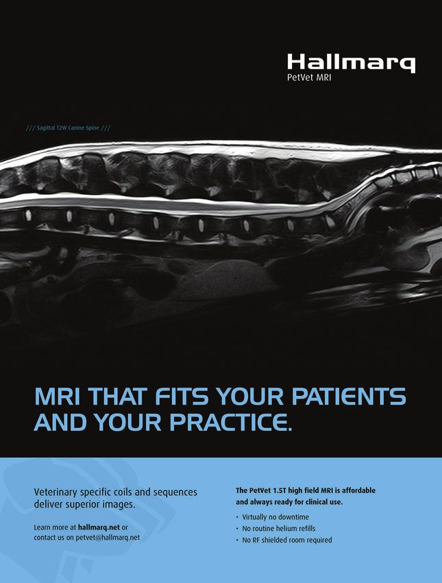
FIGURE 1 T1-weighted transverse images after IV administration of gadolinium contrast material in 3 dogs presenting for further evaluation of
right-sided unilateral masticatory muscle atrophy. Atrophied muscles are indicated with asterisk. A, Presumptive trigeminal nerve sheath tumor
(arrow). B, An extra-axial mass affecting the cerebellopontine angle (arrow). C, No abnormalities were detected on magnetic resonance imaging
with the exception of unilateral masticatory muscle atrophyMILODOWSKI ET AL. 737
The trigeminal nerve was evaluated using 5 variables as previously 9 years 3 months (range 1 year 6 months to 13 years). Breeds
13
reported for oculomotor nerve dysfunction: (1) enlargement (mild or included Labrador Retriever (n = 5), Boxer, English Bull Terrier, West
marked); (2) intensity on T2W and T1W images; (3) degree of contrast Highland White Terrier, Staffordshire Bull Terrier (n = 2 for each);
enhancement (none, mild, or marked); (4) pattern of contrast enhance- 11 breeds were represented by 1 dog and 6 dogs were crossbreeds.
ment (focal or diffuse and homogenous or heterogeneous enhance- Left-sided unilateral MM atrophy was seen in 17 dogs and right-
ment); and (5) anatomical region of the lesion. The presence of middle sided atrophy in 13 dogs. Duration of clinical signs varied from 10 days
ear effusion was recorded for all included dogs. to 2 years before presentation (median, 2.75 months). Masticatory
Follow-up to discharge was collated from both referral centers, and muscle atrophy was the only clinical sign in 5 dogs. Neurological
subsequent follow-up from the referring veterinarian also was obtained. examination identified additional abnormalities in the remaining
Initially, the referring veterinary surgeon of each animal was contacted 25 dogs (83%) and included enophthalmos (n = 12 dogs), facial hyper-
by telephone. For dogs that were deceased, the date, cause of death, esthesia (n = 2), decreased or absent ipsilateral palpebral reflex
and last recorded neurological status at the time of death were (n = 10), head tilt (n = 6) opposite to the side of MM atrophy in
recorded. In accordance with local ethical and welfare committee guide- 2 dogs, generalized ataxia (n = 6), proprioceptive deficits (n = 6), cer-
lines, owners of deceased dogs were not contacted further. For all dogs vical hyperesthesia (n = 6), obtundation (n = 5), decreased ipsilateral
that were presumed to be still alive at the time of study enrollment, a menace response (n = 5), positional strabismus (n = 5), ipsilateral Hor-
letter was sent to the owner outlining the study aims along with a stan- ner's syndrome (n = 4), decreased gag reflex (n = 4), decreased pupil-
dardized questionnaire before being contacted for a telephone inter- lary light reflex (PLR; n = 1), nystagmus (n = 1), and tetraparesis
view. Owners were asked to comment on neurological progression and (n = 1). A Schirmer’s tear test identified decreased tear production in
improvement of clinical signs. Telephone interviews were performed by 2 dogs. Magnetic resonance imaging disclosed middle ear effusion
1 of 2 investigators (Emily J. Milodowski for cases from the Royal veter- in 18 dogs (60%). The effusion was ipsilateral to the MM atrophy in
inary College and PA-B for cases from Glasgow University). The ques- 16 dogs, whereas bilateral effusion was observed in 2 dogs. The MRI
tionnaire was approved by the local ethics and welfare committee appearance of the pTNSTs was recorded (Table 1). In 9 of 30 dogs,
(Supporting Information Questionnaire). cerebrospinal fluid (CSF) analysis using CSF from cisternal puncture
Statistical analysis was performed by commercial statistical soft- was performed; total nucleated cell count was normal (738 MILODOWSKI ET AL.
lomustine (n = 1). Outcome data were available for 22 of the 27 dogs. Six dogs were euthanized at the time of diagnosis without treat-
Twenty-one of these 22 dogs were dead at the time of study enroll- ment because of the nature of the diagnosis and severity of clinical
ment, whereas 1 dog was still alive despite progression of vestibular signs. Follow-up data were available for 6 of the remaining 7 cases.
signs 7 months after diagnosis. Two dogs were euthanized in the first One dog did not receive any specific type of treatment and was
7 days after treatment was initiated. They were euthanized because euthanized 6 days after diagnosis because of progression of clinical
of progression of clinical signs, and both had received prednisolone signs. Three dogs were treated with prednisolone alone; 1 was eutha-
after diagnosis. Seventeen other dogs were euthanized between nized after 7 days, 1 survived for 2 months, and the remaining dog
9 days and 24 months (median, 8 months) after diagnosis of pTNST survived for 6 months after diagnosis. All 3 dogs experienced progres-
because of continuous progression of neurological signs. Progression sion of neurological signs before euthanasia, including deterioration in
of neurological signs included development of vestibular signs in mentation and vestibular signs. One dog was treated with a combina-
13 dogs, loss of facial sensation (n = 5), loss of corneal reflex (n = 3), tion of prednisolone, hydroxyurea, and lomustine. This dog was eutha-
collapse episodes (n = 2), seizures (n = 2), Horner's syndrome (n = 2), nized 3 months later because of progression of tetraparesis and
decreased gag reflex and dysphagia (n = 2), obtundation (n = 1), ton- generalized ataxia. The final dog was treated with a combination of
gue deviation (n = 1), and cervical hyperesthesia (n = 1). Two dogs prednisolone, hydroxyurea, and radiotherapy. This dog experienced
were euthanized for unrelated causes, 1 because of developing persis- neurological deterioration beginning 6 months after diagnosis. The
tent blindness after cataract surgery 5 months after diagnosis, the dog developed Horner's syndrome, decreased facial sensation, and
other because of severe osteoarthritis 21 months after diagnosis. head tilt, and was euthanized 25 months after diagnosis. Overall sur-
Neither dog was reported to have experienced neurological deteriora- vival time for this group varied therefore from 6 days to 25 months
tion after diagnosis. For the purpose of the study, these 2 dogs were after diagnosis (median, 2.5 months).
included in the survival analysis. Overall survival time for this group
varied from 1 day to 1 year and 9 months after diagnosis (median 3.1.2 | Dogs without an underlying lesion detected
5 months). on MRI
In 18 of the 63 dogs, no identifiable lesion was detected on MRI of
3.1.1 | Dogs with other extra-axial mass lesions affecting the head except for unilateral MM atrophy. This group consisted of
the cerebellopontine angle or petrosal part of the 14 males (4 neutered) and 4 females (2 neutered). Median age at diag-
temporal bone nosis was 7 years 10 months (range, 2 years 10 months to 11 years
Of the 13 dogs with other extra-axial mass lesions, 6 were females 1 month). Breeds included Staffordshire Bull Terrier (n = 5 dogs), Lab-
(3 neutered) and 7 were neutered males. Median age at diagnosis was rador Retriever (n = 4), 7 breeds were represented by 1 dog, and
7 years 5 months (range 5 years 1 month to 11 years 5 months). This 2 dogs were cross breeds. Twelve dogs had left-sided and 6 had right-
group included 2 Labrador Retrievers, 2 Boxers, 5 breeds were repre- sided MM atrophy. Duration of clinical signs before presentation
sented by 1 dog, and 4 dogs were cross breeds. The duration of clini- varied from 1 day to 1 year (median, 2 months). In 9 dogs, facial asym-
cal signs varied from 2 weeks to 1 year (median 1 month). Six dogs metry as a result of MM atrophy was the only clinical sign detected.
had left-sided and 7 dogs had right-sided MM atrophy. All dogs Neurological examination disclosed additional abnormalities in the
(100%) had additional abnormalities on neurological examination remaining 9 dogs (50%) and included Horner’ syndrome (n = 5 dogs),
including decreased palpebral reflex (n = 5 dogs), head tilt (n = 5), decreased or absent facial sensation (n = 3), decreased corneal sensa-
which was toward the affected side in 4 dogs, generalized tion (n = 2), decreased palpebral reflex (n = 2), and decreased menace
ataxia (n = 5), decreased mentation (n = 4), decreased tear production response (n = 1). A Schirmer’s tear test identified decreased tear pro-
(n = 4), proprioceptive deficits (n = 4), decreased menace response duction in 1 dog.
(n = 3), Horner's syndrome (n = 3), decreased corneal reflex (n = 2), Assessment of MRI findings indicated middle ear effusion in
strabismus (n = 2), cervical hyperesthesia (n = 2), nystagmus (n = 2), 3 dogs; 1 dog had bilateral effusion, 1 dog had ipsilateral effusion,
ipsilateral facial hyperesthesia (n = 1), and abnormal PLR (n = 1). Two whereas the effusion was contralateral to the MM atrophy in the 3rd
dogs had corneal ulceration. dog. In 12 dogs, CSF analysis using CSF obtained by cisternal puncture
Assessment of MRI indicated extra-axial mass lesions ipsilateral to was performed; total nucleated cell count was mildly increased in
the MM atrophy, which did not arise from the trigeminal nerve or gan- 1 case (8 cells/μl; reference interval,MILODOWSKI ET AL. 739
(n = 4 dogs), prednisolone in combination with cyclophosphamide (dogs with other extra-axial mass lesions and dogs in which no abnor-
(n = 1), gabapentin (n = 1), gabapentin and radiotherapy (n = 1), or malities were detected) and dogs with pTNST (P > .05). Diagnosis had
hydroxyurea alone (n = 1). One dog received a combination of neomy- a significant influence on the occurrence of middle ear effusion
cin, polymyxin, and dexamethasone ophthalmic ointment and cyclospor- (P = .008). Dogs with pTNST were more likely to have middle ear
ine A ointment for ocular pathology and decreased tear production, but effusion compared to dogs with no causative lesion observed on MRI
no further treatment specifically related to MM atrophy. Four dogs (P = .003). No significant differences were found between dogs with
were lost to follow-up and of the remaining 13 dogs, 8 were dead other extra-axial mass lesions and dogs with pTNST or between dogs
(median survival time, 25 months; range, 7 days to 65 months), and with other extra-axial mass lesions and dogs in which no causative
5 remained alive at the time of study enrollment (at 18, 33, 35, 36, and lesion was observed on MRI (P > .05). Diagnosis significantly influ-
54 months after diagnosis). Of the dogs that were dead, only 1 was enced whether or not treatment was attempted (P = .004). Dogs diag-
euthanized because of neurological disease. This dog did not experience nosed with other extra-axial mass lesions were more likely to be
progression of clinical signs after diagnosis until developing progres- euthanized at the time of diagnosis compared to either the pTNST
sively decreased mentation and nonambulatory tetraparesis 38 months group (P = .01) or those with no causative lesion identified on MRI
after diagnosis. No further diagnostic tests were performed before (P = .01). No significant difference was found between dogs with
euthanasia and this dog was treated with prednisolone, gabapentin, and pTNST and those in which no underlying cause could be identified
radiotherapy after diagnostic tests for unilateral MM atrophy were per- (P > .05). Diagnosis significantly influenced the likelihood of neurolog-
formed. It is therefore unclear if this dog was euthanized for reasons ical deterioration after diagnosis (P = .0007). Neurological deteriora-
related to the initial presentation of unilateral MM atrophy. The remain- tion occurred significantly less often in dogs in which no causative
ing 7 dogs died or were euthanized for unrelated reasons without fur- lesion could be identified on MRI compared to dogs with pTNST
ther progression of neurological signs. (P = .006) and other extra-axial mass lesions (P = .003). No significant
Of the 5 dogs still alive at the time of data collection, none had difference was found for the likelihood of neurological deterioration
experienced progression of neurological signs. In 2 dogs, the MM between dogs with pTNST and other extra-axial mass lesions. Diagno-
atrophy resolved without treatment. Another dog retained persistent
sis did not have a significant influence on any of the other evaluated
stable facial asymmetry after discharge. In the 2 remaining dogs, uni-
variables (P > .02; Bonferroni correction for multiple comparisons).
lateral MM muscle atrophy progressed after initial clinical presenta-
tion. One of these dogs did not receive any treatment, whereas the
other dog developed bilateral MM atrophy while being treated with 4 | DI SCU SSION
prednisolone.
We evaluated the spectrum of clinical characteristics, progression, and
outcome of different underlying causes of unilateral MM atrophy in
3.2 | Dogs that could not be classified into groups
dogs. Our results indicate that pTNST should be considered the most
Two dogs were presented with unilateral MM atrophy and concurrent
common cause of unilateral MM atrophy, representing almost half of
sciatic neuropathy of the contralateral limb. No abnormalities of the
the dogs included in this study, but other diagnoses constituted over
lumbosacral area, trigeminal nerves, cerebellopontine area, or petrosal
half of the cases. These included other extra-axial masses not arising
part of the temporal bone were identified on MRI. The 1st dog was a
from the trigeminal nerve, whereas in 28.6% of cases no causative
7-year 1-month-old male neutered cross breed, and improved with
lesion was observed on MRI studies of the head. This finding suggests
treatment using gabapentin and meloxicam. The dog was alive at the
that, although an important differential diagnosis, TNST should not be
time of data collection, 47 months after diagnosis. The 2nd dog was
considered the only cause of unilateral MM atrophy. Our results fur-
an 8 year 2-months-old male intact Labrador Retriever. This dog
ther suggest that unilateral MM atrophy can be caused by a variety of
received multiple treatments with vincristine, cyclophosphamide,
underlying benign and malignant conditions. This finding emphasizes
prednisolone, and cytosine arabinoside because of suspected lym-
the importance of making as accurate as possible a diagnosis by per-
phoma. Resolution of unilateral MM atrophy was recorded over
forming a complete neurological examination in combination with
12 months, but sciatic deficits were reported persistently over the
advanced diagnostic imaging in dogs presenting with unilateral MM
next 3 years. This dog was euthanized 50 months after diagnosis.
atrophy. Although all included dogs were presented with unilateral
Necropsy identified generalized peripheral neuropathy, with the right
MM as their predominant clinical sign, neurological examination iden-
sciatic nerve most affected. No inflammatory or neoplastic cells were
tified additional abnormalities in most dogs with pTNST and in all dogs
detected in the trigeminal or sciatic nerve.
with other extra-axial mass lesions. Signs of intracranial involvement
including head tilt, ataxia, proprioceptive deficits, and obtundation
3.3 | Comparison between groups commonly were reported in these groups of dogs. Alternatively, in half
Diagnosis had a significant influence on the type of abnormalities of dogs with no causative lesion observed on MRI, neurological exami-
detected on neurological examination. Dogs with other extra-axial nation did not identify any additional abnormalities, and neurological
mass lesions had head tilt and ataxia significantly more often (P = .02 deficits always were limited to the trigeminal nerve or neural struc-
for both) compared to dogs without an underlying cause identified on tures in close proximity of the trigeminal nerve, such as the sympa-
MRI. No significant differences were observed between both groups thetic or facial nerve. Intracranial signs such as head tilt, ataxia,740 MILODOWSKI ET AL.
proprioceptive deficits, or obtundation were not observed in this as masticatory myositis.2,15 In half of affected cases, neurological exami-
group of dogs. nation did however identify abnormalities suggestive of dysfunction of
As expected, pTNST was the most common cause of unilateral other branches of the trigeminal nerve or dysfunction of neural struc-
MM atrophy in our study population. Observed clinical signs, neuro- tures in close proximity to the trigeminal nerve. Additionally, testing for
logical deficits, and MRI findings were similar to those previously type 2M antibodies indicative of masticatory myositis was negative in
3,7,9
reported. Although recent studies have focused on clinical out- all tested cases. These findings could suggest that unilateral MM atro-
comes after advanced treatment strategies, including stereotactic phy without abnormalities observed on MRI could represent a neuropa-
radiotherapy7,9 and volumetric-modulated arc radiotherapy,14 the nat- thy instead of a myopathy in some cases. Unilateral MM atrophy as the
ural progression and survival of dogs with TNST after conservative sole clinical sign in combination with normal MRI findings is a rare clini-
treatment has rarely been reported. Although 2 of 27 dogs in which cal presentation in humans.16–18 This presentation is considered benign
treatment was attempted underwent radiotherapy, the remaining and is referred to as pure trigeminal motor neuropathy. It is character-
25 dogs included in our study received conservative medical treat- ized by trigeminal motor paralysis without sensory trigeminal distur-
ment or no specific treatment at all. The median survival time of bances or involvement of other cranial nerves.16–18 Although this
4 dogs treated conservatively was 12 months in 1 study (range, clinical presentation is similar to that observed in the dogs of our study,
5-21 months),3 whereas the median survival time of 10 dogs was only several animals had clinical signs suggestive of trigeminal sensory neu-
12 days in a more recent study (range, 1-577 days), with 40% of con- ropathy and involvement of cranial nerves in close proximity to the tri-
servatively treated dogs being alive at 1 year after diagnosis.9 In geminal nerve. It is possible that MRI was not sensitive enough to
agreement with these findings, the median survival time in our study detect small lesions along the anatomical pathway of the trigeminal
was 5 months and ranged from 1 day to 21 months. In agreement nerve. Thus, it remains uncertain if unilateral MM atrophy in dogs with-
with previous reports, reliable assessment of outcome was compli- out abnormalities on MRI represents a benign or malignant condition.
cated by variation in clinical presentation, MRI findings, and type of This uncertainty is reflected by the fact that 1 dog was euthanized at
medical management, and also because several dogs were euthanized the time of diagnosis, approximately half of the dogs did not receive
at the time of or soon after diagnosis.3,9 Although the prognosis of any specific type of treatment, and a variety of treatments were recom-
dogs with pTNST is poor, it is difficult to accurately predict the out- mended in the remaining dogs. With the exception of 1 dog, neurologi-
come and survival time of dogs treated conservatively for pTNST. In cal deterioration was not observed in any of the dogs in which MRI
7,9
agreement with previous studies, progression of clinical signs was failed to identify an underlying cause for unilateral MM atrophy. The
characterized by progressive brainstem compression or other central dog that was euthanized for neurological deterioration had received
nervous system signs, such as vestibular signs, multiple cranial nerve radiotherapy and developed decreased mentation and nonambulatory
deficits, seizures, or obtundation. tetraparesis 38 months after MRI was performed. Unfortunately, no
Our results suggest that other extra-axial mass lesions affecting further diagnostic tests were performed at the time of euthanasia. This
the cerebellopontine angle or petrosal portion of the temporal bone dog may have developed neurological signs because of trigeminal nerve
should be considered as important differential diagnoses for unilateral pathology, but it also is possible that this dog could have developed an
MM atrophy in dogs. Lesions producing trigeminal motor nerve dys- unrelated neurological disorder or suffered from late effect radiation
function and subsequent MM atrophy may occur anywhere along the toxicity, which is characterized by persistent progressive neurologic
course of the trigeminal nerve from its motor nucleus in the pons to signs >6 months after radiation treatment without imaging evidence of
its distal peripheral nerve endings. The motor nucleus of the trigeminal tumor progression.19 It remains unclear if this dog was euthanized for
nerve is found at the level of the middle and rostral cerebellar pedun- reasons related or unrelated to its clinical presentation for unilateral
cles just rostral to where the cerebellar peduncles merge with the cer- MM atrophy. A study evaluating the outcome of dogs with suspected
ebellum. After the trigeminal motor neurons exit the pons, they pass TNSTs after stereotactic radiotherapy suggested that late radiation
into the canal of the trigeminal nerve in the petrosal portion of the effects cannot always be distinguished from tumor progression.7 Most
temporal bone where the trigeminal ganglion also is located.1 Thus, of the remaining treated dogs received prednisolone with or without
lesions affecting the cerebellopontine angle or petrosal portion of the additional chemotherapy. It can be questioned however if: (1) medical
temporal bone can cause MM atrophy. treatment is necessary in these dogs, (2) benefits of empirical medical
Although unilateral MM atrophy often is caused by conditions treatment outweigh costs and potential adverse effects; and (3) if treat-
associated with a poor prognosis, MRI did not identify a lesion in >25% ment with prednisolone complicates assessment of clinical progression.
of our cases. Magnetic resonance imaging did not show any abnormali- The adverse effects of prednisolone are well characterized and include
ties in the pons or along the anatomical pathway of the trigeminal muscle atrophy, which often predominantly affects the temporalis mus-
nerve. It is unclear whether or not dogs in this group all were affected cles.20,21 For these reasons and because none of the dogs that did not
by the same underlying pathology or a more heterogeneous group of receive medical treatment experienced progression of neurological defi-
disorders. Potential underlying conditions could be inflammatory, trau- cits, we currently do not recommend starting empirical treatment when
matic, neoplastic, degenerative, or idiopathic in nature. Although MRI does not identify an underlying cause for unilateral MM atrophy.
Staffordshire Bull Terriers and Labrador Retrievers most often were Our study had several limitations. Although previously published
affected by this clinical presentation, it is unclear if a true breed predis- imaging criteria were used, diagnoses were not confirmed by histopa-
position exists. Unilateral MM atrophy theoretically could be caused by thology. It has been determined previously that histopathology is
disorders affecting the trigeminal nerve or the MMs themselves, such required for accurate diagnosis because the MRI appearance of TNSTMILODOWSKI ET AL. 741
can be mimicked by nonneoplastic conditions such as trigeminal neuri- ORCID
22
tis. Assessment of outcome was complicated by the fact that some Rodrigo Gutierrez-Quintana https://orcid.org/0000-0002-3570-
clients elected euthanasia without attempting further treatment, by 2542
variation in treatment protocols for the remaining dogs, by variable Steven De Decker https://orcid.org/0000-0002-2505-2152
clinical presentations, and by different imaging findings among dogs
within the same diagnosis category. Assessment of outcome was fur-
ther complicated by the retrospective nature of the study. Only dogs RE FE RE NC ES
assessed for further evaluation of facial asymmetry caused by unilat- 1. de Lahunta A, Glass E. Lower motor neuron: general somatic efferent,
eral MM atrophy were included. Dogs that had unilateral MM atrophy cranial nerve. In: Veterinary Neuroanatomy and Clinical Neurology 3rd
Ed. St. Louis, MO: Elsevier; 2009:135-167.
as part of a more complex clinical presentation were not included. Our
2. Jeffery N, Granger N. Neurological abnormalities of the head and face.
study therefore does not include all possible underlying causes for In: Platt S, Gloucester ON, eds. BSAVA Manual of Canine and Feline
unilateral MM atrophy in dogs, but rather those disorders associated Neurology. 4th ed. Gloucester, UK: British Small Animal Veterinary
with unilateral MM atrophy as a predominant clinical sign. Association; 2013:213-231.
3. Bagley RS, Wheeler SJ, Klopp L, et al. Clinical features of trigeminal
Despite these limitations, our study had several new clinically
nerve sheath tumors in 10 dogs. J Am Anim Hosp Assoc. 1998;34:
relevant findings. Unilateral MM atrophy is an uncommon clinical 19-25.
presentation associated with facial asymmetry, altered facial aes- 4. Mayhew PD, Bush WW, Glass E. Trigeminal neuropathy in dogs: a
thetics, and a variable prevalence of additional neurological deficits. retrospective study of 29 cases (1991-2000). J Am Anim Hosp Assoc.
2002;38:262-270.
Historically, unilateral MM atrophy has been associated with a poor
5. Saunders JH, Poncelet L, Clercx C, et al. Probable trigeminal nerve
prognosis and has been assumed to be most commonly caused by schwannoma in a dog. Vet Radiol Ultrasound. 1998;39:539-542.
trigeminal nerve neoplasia. Although our study confirmed that 6. Schultz RM, Tucker RL, Gavin PR, et al. Magnetic resonance imaging
pTNST was the most common cause, unilateral MM atrophy is not of acquired trigeminal nerve disorders in six dogs. Vet Radiol Ultra-
sound. 2007;48:101-104.
pathognomonic for PNST and alternative etiologies are implicated in
7. Hansen KS, Zwingenberger AL, Theon AP, Pfeiffer I, Kent MS. Treat-
>50% of cases. More than 25% of dogs with unilateral MM atrophy ment of MRI diagnosed trigeminal peripheral nerve sheath tumors by
have no lesions detected on MRI and generally do not experience stereotactic radiotherapy in dogs. J Vet Intern Med. 2016;30:1112-
further disease progression. Although additional studies are neces- 1120.
8. Cizinauskas S, Lang J, Maier R, Fatzer R, Jaggy A. Paradoxical vestibu-
sary to determine the underlying pathology in this last group of
lar disease with trigeminal nerve-sheath tumor in a dog. Schweiz Arch
dogs, this finding illustrates that unilateral MM atrophy can be Tierheilkd. 2001;143:419-425.
caused by a variety of malignant and benign conditions. This obser- 9. Swift KE, McGrath S, Nolan MW, et al. Clinical and imaging findings,
treatments, and outcomes in 27 dogs with imaging diagnosed trigemi-
vation emphasizes the importance of performing a complete neuro-
nal nerve sheath tumors: a multi-center study. Vet Radiol Ultrasound.
logical examination and advanced diagnostic tests in dogs 2017;58:679-689.
presenting with unilateral MM atrophy. 10. Kent M, Glass EN, de Lahunta A, Platt SR, Haley A. Prevalence of
effusion in the tympanic cavity in dogs with dysfunction of the
trigeminal nerve: 18 cases (2004-2013). J Vet Intern Med. 2013;27:
ACKNOWLEDGMENTS 1153-1158.
11. Wessmann A, Hennessey A, Goncalves R, Benigni L, Hammond G,
The results of this study were presented in abstract form at the 30th Volk HA. The association of middle ear effusion with trigeminal nerve
Annual Symposium of the European Society of Veterinary Neurology, mass lesions in dogs. Vet Rec. 2013;173:449.
12. Schellhas KP. MR imaging of muscles of mastication. Am J Roentgenol.
September 21-23, 2017, Helsinki, Finland.
1989;153:847-855.
13. Tetas Pont R, Freeman C, Dennis R, Hartley C, Beltran E. Clinical and
magnetic resonance imaging features of idiopathic oculomotor neu-
CONF LICT OF IN TE RE ST DEC LARAT ION ropathy in 14 dogs. Vet Radiol Ultrasound. 2017;58:334-343.
14. Dolera M, Malfassi L, Marcarini S, et al. High dose hypofractioned fra-
Authors declare no conflict of interest.
meless volumetric modulated arc radiotherapy is a feasible method for
treating canine trigeminal nerve sheath tumors. Vet Radiol Ultrasound.
2018;59:624-631. https://doi.org/10.1111/vru.12637 [Epub ahead
OFF- LABE L ANT IMICR OBIAL DE CLARAT ION of print].
15. Evans J, Levesque D, Shelton GD. Canine inflammatory myopathies: a
Authors declare no off-label use of antimicrobials.
clinicopathologic review of 200 cases. J Vet Intern Med. 2004;18:
679-691.
16. Chiba M, Echigo S. Unilateral atrophy of the masticatory muscles
INS TITUTIONAL ANIMAL CARE AND U SE C OMMITTEE and mandibular ramus due to pure trigeminal motor neuropathy: a
(IACUC) OR OTHER APPROVAL DECLARAT ION case report. Oral Surg Oral Med Oral Pathol Oral Radiol. 2012;113:
e30-e34.
This study was approved by the Royal Veterinary College clinical 17. Wilson MH, Hodgson EJ, Felstead AM. Focal atrophy of the mastica-
research ethical review board (URN: M2016 0088). tory muscles caused by pure trigeminal motor neuropathy: case
report. Br J Oral Maxillofac Surg. 2016;54:e13-e14.
18. Kämppi A, Kämppi L, Kemppainen P, Kanerva M, Toppila J,
Auranen M. Focal atrophy of the unilateral masticatory muscles
HUMAN ETHICS APPROVAL DECLARATION
caused by pure trigeminal motor neuropathy: case report. Clin Case
Authors declare human ethics approval was not needed for this study. Rep. 2018;6:939-943.742 MILODOWSKI ET AL.
19. Greene-Schloesser D, Robbins ME, Pfeiffer AM, Shaw EG, Wheeler SUPPOR TI NG I NFORMATION
KT, Chan MD. Radiation-induced brain injury: a review. Front Oncol.
2012;2:1-18. Additional supporting information may be found online in the Sup-
20. De Decker S, Gielen IM, Duchateau L, et al. Evolution of clinical signs porting Information section at the end of the article.
and predictors of outcome after conservative medical treatment for
disk-associated cervical spondylomyelopathy in dogs. J Am Vet Med
Assoc. 2012;240:848-857.
21. Jeffery ND. Corticosteroid use in small animal neurology. Vet Clin How to cite this article: Milodowski EJ, Amengual-Batle P,
North Am Small Anim Pract. 2014;44:1059-1074. Beltran E, Gutierrez-Quintana R, De Decker S. Clinical findings
22. Beltran E, Grundon R, Stewart J, Biggi M, Holloway A, Freeman C. and outcome of dogs with unilateral masticatory muscle atrophy.
Imaging diagnosis — unilateral trigeminal neuritis mimicking periph-
eral nerve sheath tumor in a horse. Vet Radiol Ultrasound. 2016;57: J Vet Intern Med. 2019;33:735–742. https://doi.org/10.1111/
E1-E4. jvim.15373You can also read