Cervical chondroid chordoma in a standard dachshund: a case report
←
→
Page content transcription
If your browser does not render page correctly, please read the page content below
Stigen et al. Acta Veterinaria Scandinavica 2011, 53:55
http://www.actavetscand.com/content/53/1/55

 CASE REPORT                                                                                                                                Open Access

Cervical chondroid chordoma in a standard
dachshund: a case report
Øyvind Stigen1*, Nina Ottesen1, Hans Gamlem2 and Caroline P Åkesson3

 Abstract
 A ten-year-old male standard dachshund was presented with a history of neck pain and progressive gait
 disturbances. Following a neurological examination and diagnostic imaging, including CT, a neoplastic lesion
 involving the third and fourth cervical vertebrae was suspected. The lesion included an extradural mass on the
 right side of the spinal canal causing a local compression of the cervical cord. Surgery, using a modified dorsal
 laminectomy procedure, was performed in order to decompress the cervical spinal cord. Histopathological
 examination of the extradural mass indicated that the tumour was a chondroid chordoma. Following discharge,
 the quality of life for the dog was very good for a sustained period, but clinical signs recurred at 22 months. The
 dog was euthanased 25 months post-surgery. On post-mortem examination, a regrowth of neoplastic tissue was
 found to have infiltrated the bone and spinal cord at C3-C4. This is the first report to show that palliative surgery
 can offer successful long-lasting treatment of chondroid chordoma of the cervical spine in the dog.

Background                                                                           This is the first report describing surgical treatment
Chordomas are slow-growing, locally destructive neo-                               and follow-up of a chordoma in the cervical spine of a
plasms arising in the cerebrospinal axis from remnants                             dog.
or derivatives of the notochord. These tumours are
uncommon in humans and in animals, and only a few                                  Case presentation
cases have been reported in dogs [1,2], cats [3,4], rats                           A ten-year-old, male, longhaired standard dachshund of
[5] and mink [6]. There are several reports of chordo-                             11 kg, presenting with lethargy and ataxia was referred
mas in ferrets where the tumour is typically located on                            to the Norwegian School of Veterinary Science for eva-
the tail [7-9].                                                                    luation and treatment. It had a three month history of
  By clinicopathologic, immunohistochemical and DNA                                gradual behavioural change, reluctance to exercise and
flow cytometric studies, three subtypes of chordomas                               increasing stumbling on the right thoracic limb. A week
are recognized in humans: 1) conventional chordoma, 2)                             before admission, the referring veterinarian also identi-
chondroid chordoma and 3) chordoma with a malignant                                fied neck pain, a slight degree of general muscular atro-
spindle cell component [10-12]. Distinct subtypes of                               phy and tetraparesis with an absence of the
chordomas have not been described in other species.                                proprioceptive positioning response in all four limbs.
  To the authors’ knowledge, eight cases of chordomas                              The referring veterinarian then prescribed 10 mg pre-
or probable chordomas in dogs have been reported pre-                              dnisolone orally once daily for seven days.
viously, and the locations of the tumours varied. Two
were located in the cranial region [13,14], two within                             Clinical findings
the cervical vertebral canal [15,16], two in the sacro-coc-                        On referral, the dog was bright and alert. Clinical exam-
cygeal area [13,17], one in the spinal cord at the level of                        ination revealed a reduced body condition including
L5-6 [18] and one was located between the trachea and                              general muscular atrophy. The claws of the third and
ventral muscles of the first cervical vertebrae [19].                              fourth digits were worn on all four limbs, especially on
                                                                                   the right thoracic limb.
* Correspondence: oyvind.stigen@nvh.no                                               The dog was walked on a leash and ataxia and weak-
1
 Department of Companion Animal Clinical Sciences, Norwegian School of             ness of all four limbs were observed. The dog was able
Veterinary Science, PO Box 8146, NO-0033 Oslo, Norway
Full list of author information is available at the end of the article             to walk up and down stairs and stumbling on a smooth

                                     © 2011 Stigen et al; licensee BioMed Central Ltd. This is an Open Access article distributed under the terms of the Creative Commons
                                     Attribution License (http://creativecommons.org/licenses/by/2.0), which permits unrestricted use, distribution, and reproduction in
                                     any medium, provided the original work is properly cited.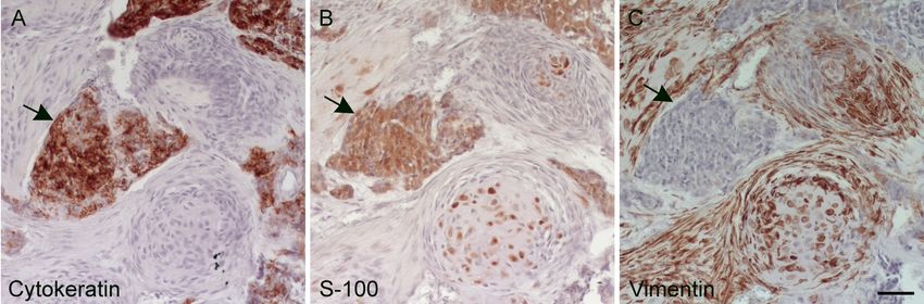
Stigen et al. Acta Veterinaria Scandinavica 2011, 53:55                                                                       Page 2 of 7
http://www.actavetscand.com/content/53/1/55

floor was not observed. Signs of discomfort or sponta-                General anaesthesia was induced by an iv injection of
neous pain were not registered.                                     4 mg/kg propofol (Propofol-® Lipuro B; Braun), main-
  On neurological examination, cranial nerve function               tained with 1·6 to 2 per cent isoflurane (Isoba vet;
was normal except for a bilateral absence of the menace             Schering Plough) in oxygen and air delivered by means
response that was explained by presence of keratitis and            of a circle patient breathing system. Computed tomogra-
cataract in both eyes. The proprioceptive positioning               phy (CT) of the cervical spine was performed with the
response was absent in all four limbs. The hopping reac-            dog in ventral recumbency. The CT machine was a GE
tion and the tactile placing response were delayed in the           highspeed Fxi single slice helical scanner (GE Health-
right versus the left thoracic limb. On flexion of the              care, Oslo Norway), the voltage and milliampere-sec-
neck, the dog showed signs of cervical pain.                        onds used were 120 kV and 150 mAs respectively and
  The results of clinical biochemistry and a complete               the slice thickness was 3 mm and 1 mm in both soft tis-
blood count were within the normal reference ranges                 sue (WW 400; WL 40) and bone (WW 1900; WL 580)
for the laboratory.                                                 algorithms.
                                                                      CT revealed an extradural heterogenous and amor-
Diagnostic imaging                                                  phous mass to the right of the vertebral canal just above
The dog was premedicated with an im administration of               the C3-C4 disc extending from mid C3 to mid C4 (Fig-
0·1 mg/kg acepromazine (Plegicil vet; Pharmaxim Swe-                ure 2A and 3A). The mass caused lateral compression
den AB) and 0·2 mg/kg methadone hydrochloride                       and displacement of two cervical cord segments to the
(Metadon; NAF) and lateral and ventrodorsal radio-                  left. Areas of high attenuation resembling calcification
graphs of the cervical and thoracic vertebral column                were scattered in the mass. In addition, there was calci-
were taken. In the ventrodorsal view, a smoothly margi-             fied material and increased soft tissue density in the
nated expansive bone lesion was seen on the right side              right intervertebral foramen around the C4 spinal nerve.
of the vertebral arch of C3 (Figure 1A). A moderate                 A lytic lesion, surrounded by sclerosis, was observed on
degree of calcification was identified in the intervertebral        the right dorsolateral side of the cranial part of the C4
disc spaces C7-T1 and T8-9.                                         body. A corresponding lytic area was observed in the
                                                                    C3-C4 disc. Mild atrophy of the epaxial muscles (m.
                                                                    splenius, m. semispinalis capitis and m. multifidus cervi-
                                                                    cis) on the right side of the neck was also observed.

                                                                    Surgical treatment
                                                                    Six days after the CT scan, surgery was performed. The
                                                                    dog was premedicated and anaesthesia inducted and
                                                                    maintained with the same procedure as presented in the
                                                                    Diagnostic Imaging section (above). For antimicrobial
                                                                    prophylaxis 40 mg/kg cephalothin (Cefalotin; ACS Dob-
                                                                    far Generics) was administered iv. Analgesic therapy

                                                                     Figure 2 CT scans in bone window through the C3-C4
 Figure 1 Ventrodorsal radiographs of the cervical spine at the      intervertebral disc, before surgery (A) and 25 months post-
 first presentation (A) and when the symptoms recurred 25            surgery (B). (A) The image demonstrates calcified material centrally
 months later (B). There are mottled mineralized opacities at C3     and to the right of the vertebral canal compressing the spinal cord
 and C4 and a mainly productive bone lesion (arrows) on the right    to the left. (B) The image demonstrates local recurrence about two
 side of the C3 vertebral arch.                                      years after subtotal resection of the cervical chordoma.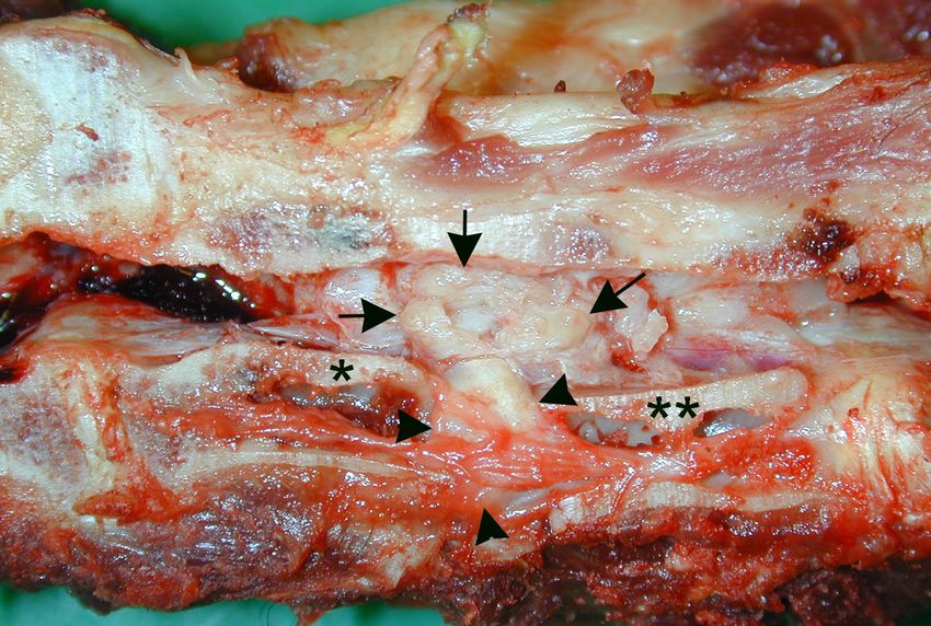
Stigen et al. Acta Veterinaria Scandinavica 2011, 53:55                                                                 Page 3 of 7
http://www.actavetscand.com/content/53/1/55

                                                                           The dog was discharged after six days. At that time,
                                                                         neurological function was similar to that observed preo-
                                                                         peratively. Exercise restriction was advised, and the use
                                                                         of harness instead of a collar was recommended.

                                                                         Histopathological examination
                                                                         The formalin fixed biopsy measuring 0·5 × 0·5 × 2 cm
                                                                         was decalcified and sectioned for histopathological
                                                                         examination. The morphological features together with
                                                                         immunohistochemical examinations indicated a chon-
                                                                         droid chordoma.

 Figure 3 CT scans in bone window through the cranial part of
                                                                         Postoperative development
 C4 at the first presentation (A) and 25 months later (B). (A)
 Note the radiolucent area with surrounding sclerosis (arrow) on the     The owner was questioned by telephone about the dog’s
 right dorsolateral side of the C4 body. (B) The C4 vertebral body has   condition twelve months after surgery. She reported that
 increased opacity and the radiolucent area is still visible (arrow).    the dog was doing well without observable gait distur-
                                                                         bances. The claws of the third and fourth digits of the
                                                                         right thoracic limb were normal. However, the owner
included transdermal administration of 4·5 μg/kg/h fen-                  commented that the dog tended to lie down when
tanyl (Durogesic; Janssen Cilag).                                        patted on his back and she presumed this behaviour was
  The dog was placed in ventral recumbency with the                      to avoid discomfort.
head and neck gently flexed in a neutral position. A dor-                  Twenty-five months following surgery, the owner tele-
sal midline skin incision extending from the occipital                   phoned and reported recurrence of clinical signs with
protuberance to the dorsal spinous process of C6 was                     gait disturbances and cervical pain. The dog had become
made. The laminae of vertebrae C2- C5 was exposed                        progressively worse over the last three months, and
through an approach with lateral retraction of epaxial                   despite treatment with carprofen, the owner felt the dog
musculature to the level of the articular processes [20].                now was suffering. On readmission to the Norwegian
Periosteal bone formation was identified on the right                    School of Veterinary Science, the dog was depressed
parts of the C3 and C4 laminae. A dorsal laminectomy                     and reluctant to walk. Worn claws were seen on both
extending from the cranial third of C3 to the caudal                     thoracic limbs. When walked on a lead, the dog showed
third of C4 was performed using a high-speed spinal                      weakness (tetraparesis) and a mild lameness in the right
bur. The laminae were irregularly thickened and a                        thoracic limb. Hands-on examination of the head and
mineralised mass was found extradurally to the right of                  neck revealed signs of pain, including attempts to bite
a compressed, but otherwise normal looking dura mater                    the examiner. A systolic murmur was heard on both
and cervical cord. Epidural fat was almost absent. A                     sides of the thorax.
sample of the mineralised mass and the adjacent lami-                      The dog was sedated and a second radiographic study
nae was collected for histopathology. Remaining abnor-                   of the cervical vertebral column was performed. A
mal tissue was removed to achieve satisfactory                           mixed (destructive and productive) bone lesion was
decompression, though some pathological tissue                           identified at C3 and C4, and included the body and arch
remained. A 2-3 mm thick layer of subcutaneous fat                       of both vertebrae (Figure 1B). Areas of increased radio-
was placed at the laminectomy site before routine                        density were observed on the right side of the vertebrae,
wound closure. A closed wound bandage was applied.                       and the borders to soft tissue were without a marked
  After surgery, the dog was monitored in the intensive                  periosteal response.
care unit and given an iv injection of 4 mg/kg carprofen                   CT of the cervical spine was repeated with the dog in
(Rimadyl; Pfizer, New York, USA). An im administration                   ventral recumbency. The slice thickness was 2 mm and
of 0·2 mg/kg methadone hydrochloride (Metadon; NAF)                      1 mm in both soft tissue and bone algorithms. The stu-
was repeated q4h in the immediate 12 hour-postopera-                     dies were performed before and after iv injection of 300
tive period. Anti-inflammatory treatment was continued                   mg I/ml iohexol (Omnipaque; GE Healthcare, Oslo,
by giving 2 mg/kg of carprofen po twice daily for two                    Norway) at a dose of 1·8 ml/kg bodyweight.
weeks. Two days post-surgery the dog was ambulatory                        The laminectomy of C3 and C4 was visible. The CT
with moderate ataxia and weakness in all four limbs.                     examination revealed a large mass to the right of the
Spontaneous pain was not registered and urination was                    vertebral canal involving the C3 and C4 vertebral arches
normal. The fentanyl patch was removed three days                        and the C4 vertebral body (Figure 2B and 3B). The mass
post-surgery.                                                            was heterogeneous and it was not possible to distinguish
Stigen et al. Acta Veterinaria Scandinavica 2011, 53:55 Page 4 of 7
http://www.actavetscand.com/content/53/1/55
fragments of possibly destroyed bone from tumour cal- borders. The cells had moderate to abundant eosino-
cification. Two cervical cord segments (C4 and C5) philic cytoplasm with small to medium sized nuclei
were laterally compressed and displaced to the left. that were round-, oval- or spindle shaped with a light
Owing to the size, location and history of the cervical to dark basophilic colour, and were interpreted as
mass, the prognosis was considered to be poor and with chordoid-like tumour cells (Figure 5A). In many areas
the owner’s consent the now twelve-year-old dog was of the tumour the cells contained cytoplasm that was
euthanased and submitted for post-mortem markedly vacuolated, often with a centrally placed
examination. nucleus (physaliferous cells) (Figure 5B). A basophilic
material was observed in a few areas and was inter-
Pathological examination preted as mucin matrix. There was chondroid differen-
At post-mortem examination of the vertebral column, tiation multifocally (Figure 5A, c), which in some areas
gross pathological findings were observed between cervi- had a dark basophilic colour interpreted as mineraliza-
cal vertebrae C3 and C4 (Figure 4). The intervertebral tion. The trabecular bone tissue observed multifocally
disc was replaced by tumorous tissue which extended could consist of either pre-existing bone or newly
into, and diffusely infiltrated adjacent bone and soft tis- formed bone tissue, indicating a remodelling process.
sues on the right lateral side of the vertebrae. The The tumour showed irregular infiltrative growth into
tumour measured approximately 3 × 3 × 4 cm, was rela- adjacent tissues, including preexisting trabecular bone
tively firm, multinodulated and with a glistening surface. tissue and the spinal cord (Figure 5C). The tumorous
On the right medial side of both C3 and C4 the tumour tissue also had a low mitotic index.
prominated into the vertebral canal and in an area of Immunohistochemical examination revealed strong
approximately 0·5 cm diameter it adhered to the spinal labelling for cytokeratin and S-100 in the chordoid
cord. The bodies of C3 and C4 were devoid of bone tumour cells (Figure 6A and 6B). Only a few of the cells
marrow, leaving a hollow cavity with only a string of showed weak labelling for vimentin (Figure 6C). The
soft tissue running through the length of the vertebral physaliferous cells contained PAS-positive material in
body. Metastases were not observed. In addition, the the cytoplasm.
necropsy showed bilateral valvular endocardiosis and an On the basis its location, the cellular components and
intracranial tumour mass measuring 1 × 2 × 2 cm. the immunohistochemical results, the tumour was defi-
Histopathological examination of the tissue from the nitively diagnosed as a chondroid chordoma.
area of C3 and C4 showed a poorly demarcated Histopathological and immunohistochemical examina-
tumour consisting of neoplastic cells in lacunae sepa- tion of the intracranial tumour mass showed a menin-
rated by fibrous septa. The neoplastic cells were poly- gioma with no resemblance to the tumour of the
gonal, moderately pleomorphic with poorly defined cell cervical spine.
Discussion
Following surgery, which included decompression of the
cervical spinal cord, ataxia and neck pain subsided and
the symptoms did not recur for almost two years. The
post-mortem examination revealed the chordoma to be
only 3 × 3 × 4 cm in size and no metastases were
observed. These findings are in accordance with pre-
vious reports that describe chordomas as slow-growing,
locally aggressive neoplasms with a high rate of recur-
rence, but with a limited potensial for metastasis [7,21].
In this case, the aim of the surgery was to obtain a
biopsy and to decompress the cervical spinal cord.
Because it was not possible to remove all the pathologic
(neoplastic) tissue, the decompression can be classified
as palliative. However, following treatment the dog lived
Figure 4 Gross pathology of the cervical vertebral column. The
vertebral column is sectioned in the median plane. Tumour tissue
for two more years, including 22 months with a very
(arrows) has replaced the intervertebral disc (arrowheads), joining good quality of life. This case shows that surgical treat-
the bodies of adjacent C3 (one asterisk) and C4 (two asterisks) ment of a chondroid chordoma significantly increased
vertebrae. The tumour extended laterally and dorsally on the right the life span of a dog despite incomplete resection of
side of the cervical column and protruded into the vertebral canal the tumour. This finding indicates that surgical treat-
where it adhered to the spinal cord (has been removed).
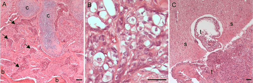
ment of chondroid chordomas in the dog is warranted.Stigen et al. Acta Veterinaria Scandinavica 2011, 53:55 Page 5 of 7 http://www.actavetscand.com/content/53/1/55 Figure 5 Histology of the tumour in the cervical vertebral column. (A) Section of tumour showing chordoid tumour cells in lacunaes (arrows) surrounded by connective tissue. The tumour infiltrated and replaced pre-existing, normally differentiated trabecular bone tissue (b). Multifocally there was chondroid differentiation in the tumour (c) (H&E, Bar 200 μm). (B) Physaliferous tumour cells, which are characteristic of chordomas, were observed multifocally (H&E, Bar 50 μm). (C) Section showing the tumour (t) to be locally invasive, here invading the spinal cord tissue (s) (H&E, Bar 200 μm). Surgical excision is considered to be the most impor- chondroid component in the chordoma presented tant mode of treatment for chordomas in humans. herein is unknown. Radiation therapy and chemotherapy are used as addi- In higher vertebrates, the nucleus pulposus is believed tional treatment modalities [22,23], but the delivery of to be the only derivative of notochordal tissue [25]. sufficiently high doses of radiation to the tumour tis- However, remnants of notochord may persist outside sue is a challenge [24]. In future, molecular targeted the intervertebral discs anywhere along the vertebral chemotherapies may improve the treatment of column [26]. The chordoma described in the present chondromas. case report included the C3-C4 disc space. The tumour In 55 cases of spheno-occipital chordomas in humans, could, therefore, have arisen from notochordal cells Heffelfinger and others [21] found that the prognosis within the nucleus pulposus of the C3-4 disc. Previous was better in 19 patients with chondroid chordomas ver- reports concerning chordomas in dogs have not sus in 36 patients with conventional chordomas. In dogs, described pathology of intervertebral discs, indicating distinct subtypes of chordomas have not been described that notochordal tissue elements outside the discs as the and, to the authors’ knowledge, no reports have origin of these tumours. included a follow-up after surgical treatment of canine In chondrodystrophoid dogs, intervertebral disc chordomas. Therefore, the prognostic importance of the degeneration is characterized by an early initiated and Figure 6 Immunohistochemical staining of tumour tissue. To differentiate this chondroid chordoma from chondrosarcoma, the tumour tissue was stained with vimentin, cytokeratin, and S-100. The chordoid tumour cells (arrows) showed a strong positive reaction for the cytokeratin- and S-100-antibody (A and B, respectively). The fibrous tissue and chondroid components were vimentin positive (C) (Bar 50 μm).
Stigen et al. Acta Veterinaria Scandinavica 2011, 53:55                                                                                          Page 6 of 7
http://www.actavetscand.com/content/53/1/55

very accelerated metamorphosis of the nucleus pulposus                         Author details
                                                                               1
                                                                                Department of Companion Animal Clinical Sciences, Norwegian School of
from a mucoid to a chondroid type [27]. From the age
                                                                               Veterinary Science, PO Box 8146, NO-0033 Oslo, Norway. 2Department of
of four years, the nuclei pulposi with very few excep-                         Animal Health, Section for Pathology, National Veterinary Institute, PO Box
tions are converted into the chondroid type, which                             750, NO-0106 Oslo, Norway. 3Department of Basic Sciences, Norwegian
                                                                               School of Veterinary Science, PO Box 8146, NO-0033 Oslo, Norway.
implies the replacement of the notochordal cells. Han-
sen [27] studied the pathology of intervertebral discs in                      Authors’ contributions
206 chondrodystrophoid dogs, aged from under two                               ØS carried out the clinical examination, performed the surgery and is the
                                                                               main author of the paper. NO carried out the diagnostic imaging. HG and
months to over seven years. In the 75 dogs over four
                                                                               CPÅ carried out the gross pathological, histopathological and
years of age, one seven year old dog was found to have                         immunohistochemical examinations. All authors read and approved the final
mucoid nuclei pulposi in the cervical region. The pre-                         manuscript.
sence of mucoid nuclei pulposi and thereby notochordal
                                                                               Competing interests
cells in some middle-aged chondrodystrophoid dogs                              The authors declare that they have no competing interests.
combined with the slow-growing nature of chordomas
                                                                               Received: 28 April 2011 Accepted: 21 October 2011
can explain why this neoplasm can be diagnosed in a
                                                                               Published: 21 October 2011
ten-year-old dachshund.
  Besides physaliferous cells and the nest formation of                        References
chordoid tumour cells, in chordomas and not in cartila-                        1. Zaki FA: Spontaneous central nervous system tumors in the dog. Vet Clin
                                                                                   North Am Small Anim Pract 1977, 7(1):153-163.
ginous tumours, immunohistochemical labelling is an
                                                                               2. Koestner A, Higgins RJ: Tumors of the nervous system. In Tumors in
important tool for differentiating these two tumour                                Domestic Animals.. 4 edition. Edited by: Meuten DJ. Ames: Iowa State Press;
types and to confirm the diagnosis [28]. As was                                    2002:697-738.
                                                                               3. Carpenter JL, Stein BS, King NWJ, Dayal YD, Moore FM: Chordoma in a cat.
observed in the presented case, chordoma tumour cells
                                                                                   J Am Vet Med Assoc 1990, 197:240-242.
label with the epithelial marker cytokeratin and S-100, a                      4. Carminato A, Marchioro W, Melchiotti E, Vascellari M, Mutinelli F: A case of
protein derived from the neural crest and chondrocytes                             coccygeal chondroid chordoma in a cat: morphological and
                                                                                   immunohistochemical features. J Vet Diagn Invest 2008, 20:679-681.
among others. Chondrosarcomas stain positively for S-
                                                                               5. Stefanski SA, Elwell MR, Mitsumori K, Yoshitomi K, Dittrich K, Giles HD:
100 and vimentin a marker for mesenchymally derived                                Chordomas in Fischer 344 rats. Vet Pathol 1988, 25:42-47.
tissues, but do not label with cytokeratin. Of the eight                       6. Hadlow WJ: Vertebral chordoma in two ranch mink. Vet Pathol 1984,
                                                                                   21:533-536.
chordomas or probable chordomas previously reported
                                                                               7. Dunn DG, Harris RK, Meis JM, Sweet DE: A histomorphologic and
in dogs, only three have been verified by immunohisto-                             immunohistochemical study of chordoma in twenty ferrets (Mustela
chemical analysis [15-17].                                                         putorius furo). Vet Pathol 1991, 28:467-473.
                                                                               8. Williams BH, Eighmy JJ, Berbert MH, Dunn DG: Brief communications and
                                                                                   case reports: cervical chordoma in two ferrets (Mustela putorius furo). Vet
Conclusions                                                                        Pathol 1993, 30:204-206.
In dogs, chondroid chordomas may arise in the cervical                         9. Munday JS, Brown CA, Richey LJ: Suspected metastatic coccygeal
                                                                                   chordoma in a ferret (Mustela putorius furo). J Vet Diagn Invest 2004,
spine and cause clinical signs from extradural masses
                                                                                   16(5):454-458.
that compress the spinal cord and spinal nerves. Both                          10. Meis JM, Raymond KA, Evans NL, Charles RE, Giraldo AA: “Dedifferentiated”
cervical vertebrae and discs may be subjected to neo-                              chordoma: a clinicopathologic and immunohistochemical study of three
                                                                                   cases. Am J Surg Pathol 1987, 11:516-525.
plastic tissue. Sclerotic and lytic lesions could be identi-
                                                                               11. Hruban RH, Traganos F, Reuter VE, Huvos AG: Chordomas with malignant
fied by diagnostic imaging. Chondroid chordomas                                    spindle cell components: a DNA flow cytometric and
localized in the vertebral column may not be possible to                           immunohistochemical study with histogenetic implications. Am J Pathol
                                                                                   1990, 137:435-447.
remove by complete surgical excision. However, an
                                                                               12. Unni KK, Inwards CY: Tumors of the osteoarticular system. In Diagnostic
incomplete resection including decompression of the                                Histopathology of Tumors.. 3 edition. Edited by: Fletcher CDM. Philadelphia:
spinal cord, may subsequently give a dog an excellent                              Elsevier Limited; 2007:1632-1652.
                                                                               13. Saliba AM, Neto LZ, Grecchi R, Mariano M, Martin BW, Migliano MF:
quality of life for almost two years.
                                                                                   Cordomas em cães (Chordomas in dogs). Arq Inst Biol (Sao Paulo) 1967,
                                                                                   34:45-50.
Consent                                                                        14. Jabara AG, Jubb KVF: A case of a probable chordoma in a dog. Aus Vet J
                                                                                   1971, 47:394-397.
Written informed consent was obtained from the owner
                                                                               15. Gruber A, Kneissl S, Vidoni B, Url A: Cervical spinal chordoma with
for publication of this case report and accompanying                               chondromatous component in a dog. Vet Pathol 2008, 45:650-653.
images. A copy of the written consent is available for                         16. Woo GH, Bak EJ, Lee YW, Nakayama H, Sasaki N, Doi K: Cervical chondroid
                                                                                   chordoma in a shetland sheep dog. J Comp Pathol 2008, 138:218-223.
review by the Editor-in-Chief of this journal.
                                                                               17. Munday JS, Brown CA, Weiss R: Coccygeal chordoma in a dog. J Vet Diagn
                                                                                   Invest 2003, 15(3):285-288.
                                                                               18. Pease AP, Berry CR, Mott JP, Peck JN, Mays MBC, Hinton D: Radiographic,
Acknowledgements                                                                   computed tomographic and histopathologic appearance of a presumed
The authors are most grateful to Dr. K. Hölling, Sørumsand Animal Clinic for       spinal chordoma in a dog. Vet Radiol Ultrasound 2002, 43:338-342.
referring this case and to Dr. Hannah Joan Jørgensen for language              19. Ball v, Auger L: Les chordomes ou tumeurs de la chorde dorsale chez
consultation.                                                                      l’homme et les animaux (Chordomas or tumors of the notochord in
                                                                                   man and animals). Rev Vét J Méd Vét Zootec 1933, 85:185-195.
Stigen et al. Acta Veterinaria Scandinavica 2011, 53:55 Page 7 of 7
http://www.actavetscand.com/content/53/1/55
20. Piermattei DL, Johnson KA: The vertebral column. An Atlas of Surgical
Approaches to the Bones and Joints of the Dog and Cat. 4 edition.
Philadelphia: Saunders; 2004, 47-105.
21. Hellefinger MJ, Dahlin DC, Maccarty CS, Beabout JW: Chordomas and
cartilaginous tumors at the skull base. Cancer 1973, 32(2):410-420.
22. Park L, Delaney TF, Liebsch NJ, Hornicek FJ, Goldberg S, Mankin H,
Rosenberg AE, Rosenthal DI, Suit HD: Sacral chordomas: impact of high-
dose proton/photon-beam radiation therapy combined with or without
surgery for primary versus recurrent tumor. Int J Radiat Oncol Biol Phys
2006, 65(5):1514-1521.
23. Hof H, Welzel T, Debus J: Effectiveness of cetuximab/gefitinib in the
therapy of a sacral chordoma. Onkologie 2006, 29:572-574.
24. Casali PG, Stacchiotti S, Sangalli C, Olmi P, Gronchi A: Chordoma. Curr Opin
Oncol 2007, 19:367-370.
25. Stemple DL: Structure and function of the notochord: an essential organ
for chordate development. Development 2005, 132:2503-2512.
26. Spoden JE, Bumsted RM, Warner ED: Chondroid chordoma: case report
and literature review. Ann Otol Rhinol Laryngol 1980, 89:279-285.
27. Hansen HJ: A pathologic-anatomical study on disc degeneration in dog.
Acta Orthop Scand Suppl 1952, 1-117.
28. Ishida T, Dorfman HD: Chondroid chordoma versus low-grade
chondrosarcoma of the base of the skull: can immunohistochemistry
resolve the controversy? J Neurooncol 1994, 18:199-206.
doi:10.1186/1751-0147-53-55
Cite this article as: Stigen et al.: Cervical chondroid chordoma in a
standard dachshund: a case report. Acta Veterinaria Scandinavica 2011
53:55.
Submit your next manuscript to BioMed Central
and take full advantage of:
• Convenient online submission
• Thorough peer review
• No space constraints or color figure charges
• Immediate publication on acceptance
• Inclusion in PubMed, CAS, Scopus and Google Scholar
• Research which is freely available for redistribution
Submit your manuscript at
www.biomedcentral.com/submitYou can also read