Co-transformation of a tropical maize endophytic isolate of Fusarium verticillioides(synonym F.moniliforme) with gusA and niagenes
←
→
Page content transcription
If your browser does not render page correctly, please read the page content below
Genetics and Molecular Biology, 27, 2, 253-258 (2004)
             Copyright by the Brazilian Society of Genetics. Printed in Brazil
             www.sbg.org.br

Research Article

Co-transformation of a tropical maize endophytic isolate of Fusarium
verticillioides (synonym F. moniliforme) with gusA and nia genes

João A. Pamphile1, Carmen Lúcia M.S.C. da Rocha1 and João L. Azevedo2
1
  Universidade Estadual de Maringá, Departamento de Genética e Biologia Celular,
Maringá, Paraná, Brazil.
2
  Universidade de São Paulo, Escola Superior de Agricultura ‘Luiz de Queiroz’, Piracicaba, SP, Brazil.

Abstract
A tropical endophytic isolate of the fungus Fusarium verticillioides (synonym Fusarium moniliforme) obtained from
Zea mays was co-transformed with plasmid pNH24 containing the Fusarium oxysporum nitrate reductase nia gene
and plasmid pNOM 102 carrying the Escherichia coli β-glucuronidase gusA gene. Transformation frequency for the
nia marker was 75 transformants µg-1 vector DNA and introduction of the gusA gene by co-transformation was 57.2%
                                          +
as indicated by the presence of the GUS phenotype on plates. Southern analyses confirmed the integration of both
                                       +
plasmids into the genome of ten GUS transformants. All co-transformants showed mitotic stability in respect of the
GUS+ phenotype. To assess the potential of transformed endophytic fungi as vectors for introducing desirable
characteristics into host tropical plants of biotechnological and agricultural importance we successfully infected
maize roots and detected GUS+ phenotype”.
Key words: co-transformation, gus, nia, Fusarium verticillioides, endophyte.
Received: March 11, 2003; Accepted: October 24, 2003.

Introduction                                                        and plants in the tropics. Furthermore, genetically manip-
       The fungus Fusarium verticillioides (synonym F.              ulated fungal endophytes have the potential to introduce
moniliforme) is often found in maize, and constitutes an            important biotechnological characteristics (e.g. insect-
important source of inoculum in soil. Fusarium spp., asso-          resistance) into the host plants.
ciated with assymptomatic plants living within their host,                The β-D-glucuronidase (GUS) gene (gus) is widely
may be considered an endophyte. According to Azevedo                used as a histochemical marker in genetic transformation
et al. (2000), much of the research on endophytes has been          experiments, the Escherichia coli GUS gene fusion system
conducted using hosts from temperate climates in the                (Jefferson, 1989) being a powerful tool for both the assess-
Northern Hemisphere and from New Zealand, with data                 ment of gene activity in transgenic plants and the develop-
on endophytes from tropical climates being scarce be-               ment of molecular genetic analysis systems. In fungi, GUS
cause only a few studies have been carried out on fun-              transformation has been performed on Fusarium
gus-host interactions in tropical regions. This may be              moniliforme (Yates et al. 1999) and Fusarium culmorum
important because endophyte-plant interactions become               (Doohan et al. 1998), albeit with very low transformation
more complex in regions having a wide variety of organ-             frequencies (≈1 transformant µg-1 DNA), while Couteau-
isms, including many different plant-attacking insects.             dier et al. (1993) obtained very high co-transformation fre-
Fungal endophytes isolated from tropical plants could be            quencies of up to 75% in Fusarium oxysporum using the
used in genetic manipulation experiments (especially                plasmid vectors pNOM102 (containing the E. coli gusA
those involving genetic transformations with morphologi-            gene) and pAN301 (carrying the Aspergillus nidulans niaD
cal markers) which would result in a better understanding           gene).
of the interactions occurring between endophytic fungi                    The aim of our research was to use the co-trans-
                                                                    formation procedure employed by Couteaudier et al.
                                                                    (1993) to obtain an increased transformation frequency in a
Send correspondence to João A Pamphile. Universidade Estadual
de Maringá, Departamento de Genética e Biologia Celular, Avenida    tropical endophytic strain of F. verticillioides using the F.
Colombo 5790, 87020-900 Maringá, PR, Brazil. E-mail:                oxysporum nitrate reductase nia gene instead of the A.
japamphile@uem.br.                                                  nidulans niaD gene as the main transformation marker.
254 Pamphile et al.
Material and Methods 4 x 10⁷ per 100 µL of TMSC and the suspension kept on ice
for 20 min before adding plasmids pNOM102 and pNH24
Fungal strains and culture media
(5 µg each in 60 µL of a solution containing 10 mM Tris,
In our experiments we used Fusarium verticillioides 1 mM EDTA and 40 mM CaCl2, pH7,5) and allowing the
strain S68 (Pamphile et al., 1996; Hua-Van et al., 2001), a mixture to stand on ice for a further 20 min and then adding
nia- mutant derived from the endophytic F. verticillioides 160 µL of MS solution containing 60% PEG 4000 (Koch-
strain L7 which had been isolated from surface disinfected Light) and incubating at room temperature for 15 min after
seeds collected from asymptomatic maize growing in Bra- which 1 mL of TMSC was added and the protoplasts
zil (Pamphile and Azevedo, 2002). Strain S68 was grown pelleted by centrifugation at 4,500 g for 5 min, re-sus-
on the complete medium (CM) and minimal medium (MM) pended in 200 µL of TMSC and mixed with 3 mL of MM
described by Pontecorvo et al. (1953) or potato-dextrose supplemented with 20% sucrose, 0.4 % (w/v) Oxoid agar
agar (PDA) (Smith and Onions, 1983). As a positive con-
and 100 µL of 100 mM glutamine solution. To test the effi-
trol for β-D-glucuronidase (GUS) activity we used F. ciency of transformation a positive control was made under
oxysporum strain MT5 which was grown on the same me- the same conditions except that only plasmid pNH24 was
dia as strain S68. For protoplast preparation and DNA ana- used. The pUC plasmid was used as a negative control to
lysis the strains were grown in PD broth (PDA without
test for strain stability. Transformants exhibiting the NIA+
agar) supplemented with 1% (w/v) each of yeast extract
phenotype conferred by pNH24 were selected on MM with
and casein (PD-YC).
nitrate as the sole nitrogen source and purified by isolation
Vectors of uninucleated conidia according to standard techniques.
The vectors used were plasmid pNOM 102 (Roberts
et al., 1989) containing the E. coli β-glucuronidase gene DNA isolation and manipulation
(gusA) flanked by a glyceraldehyde 3-phosphate promoter
(gpd) from Aspergillus nidulans upstream and the A. For both transformed and untransformed isolates,
nidulans trpC transcription termination signal downstream Roux flasks containing 100 mL of PD-YC liquid medium
and plasmid pNH24 (Diolez et al., 1993) carrying the F. were inoculated with approximately 107 spores and incu-
oxysporum nitrate reductase gene (nia) subcloned into bated for 72-96h, after which the mycelium was collected
plasmid pUC19. The pUC plasmid (without the F. by filtration using a sterile sintered glass filter, washed with
oxysporum nia gene) was used as a negative control to test sterile distilled water and lyophilized. To isolate the DNA,
for strain stability. Millipore pincers were used to grind the lyophilized myce-
lia to a powder, 100 mg of which was then suspended in
Protoplast preparation buffer (50 mM EDTA, 0.2% (w/v) SDS, pH 8.5) containing
Protoplasts were isolated from germinated conidia. 10 µL mL-1 of a 10 mg mL-1 proteinase K solution and the
Conidial suspensions from 1 week-old cultures were incu- mixture incubated for 30 min at 37 °C and 15 min at 70 °C
bated (1 x 10⁸ conidia/ 10 mL PDA + 1% yeast extract and before adding a 1/10 volume of 5 M potassium acetate solu-
1% caseine) and shaken (150 revs min-1) at 26 °C for 17 h. tion and holding on ice for 30 min. The mixture was centri-
After incubation, the mycelia were collected by centrifu- fuged at 10,000 g for 15 min and the supernatant separated
gation, washed and re-suspended in 10 mL of 1.2 M MgSO4 and washed three times with an equal volume of phe-
containing 10 mM Na2HPO4 (pH 5.8) and 50 mg mL-1 of nol-chloroform-isoamyl alcohol (48-48-4) saturated with
Glucanex (Novo Nordisk Ferment, Dittingen, Switzerland) 1 M Tris (pH 8.0) and then mixed with phenol-isoamyl al-
and incubated at 26 °C for 2 h until protoplasts formed. cohol (48-2) and centrifuged at 10,000 g for 5 min after
Following incubation, 10 mL of MS solution (10 mM which 1/2 volume of 7.5 M ammonium acetate was added
MOPS plus 0.6 M Sorbitol, pH 6.3) was added to separate and the mixture left to stand on ice for 15 min and then cen-
the cell debris from the protoplasts (which collected at the trifuged at 10,000 g for 15 min at 4 °C after which the
interface) and the protoplasts transferred to 30% sucrose supernatant was mixed with one volume of isopropanol and
solution and centrifuged at 3000 g, the protoplasts being centrifuged at 12,000 g for 15 min. After centrifugation the
collected and washed three times in TMS solution (1 M supernatant was discarded and the DNA pellet re-
Sorbitol plus 10 mM MOPS, pH 6.3) and then re-suspended suspended in TE buffer (10 mM Tris HCl, 1 mM EDTA, pH
in 200 µL TMSC solution (40 mM CaCl2 in TMS). 8) and 2 volumes of ethanol added and the mixture centri-
fuged at 10,000 g for 15 min, the supernatant being dried
Transformation procedure using a speed vac apparatus and re-suspended in 200 µL of
We transformed the F. verticillioides protoplasts us- TE buffer. Restriction enzyme digestions of fungal geno-
ing calcium chloride/polyethylene glycol (PEG) according mic DNA and plasmids were performed according to the
to a modified Malardier et al. (1989) procedure. For trans- manufacturer procedures and standard protocols
formation, the number of protoplasts was adjusted to (Sambrook et al., 1989).Gus/nia co-transformation of Fusarium 255
Southern blot analysis Results
To determine the fate of plasmid DNA in transfor-
mants, Southern blotting and hybridization was carried out Primary selection in the co-transformation experi-
according to the method of Daboussi et al. (1992). Briefly, ment with the pNOM 102 and pNH24 plasmids was per-
Genomic DNA was extracted from the recipient F. formed using MM plates which allowed only F.
verticillioides strain (S68) and ten GUS+ co-transformants verticillioides colonies harboring the pNH24 plasmid
and digested to completion with the EcoRI restriction en- (nia+) to grow, this being shown by the fact that when the
zyme which cuts plasmid pNOM102 once and probed with recipient strain (F. verticillioides nia-) was transformed
the 4.5-kb HindIII-EcoRI plasmid pNOM102 fragment with empty pUC vector (the negative control) no trans-
containing the entire GUS construct and the pnH24 plasmid formants were recovered on MM plates, thus confirming
containg the nia gene labeled with 32P. the stability of the recipient strain. We recorded 75 NIA+
transformants per µg of vector DNA, the MUG microtiter
Histochemical localization of β-glucuronidase activity plate β-glucuronidase (GUS) assay showing that 57.20% of
on maize roots infected with Fusarium verticillioides these transformants had the GUS+ phenotype as the result
GUS+ transformant of co-transformation with the gusA gene. Ten co-transfor-
mants exhibiting the GUS+ phenotype on MUG microtiter
Maize (Zea mays) seeds were washed in running tap
plates were tested for mitotic stability and analyzed by
water with a soft brush and then vigorously washed twice for
Southern blotting. The fluorometric assay results for the six
15 min in 0.1% Tween 80 solution and then surface disin-
F. verticillioides co-transformants (TG1, TG3, TG4, TG5,
fected by immersion in 70% ethanol for 1 min, then 3% so-
TG7, TG8), F. oxysporum strain MT5 (the positive control)
dium hypochloride solution for 25 min and then 70% ethanol
and the F. oxysporum S68 parent strain are given in Table 1.
for 30 s. The disinfected seeds were washed in distilled ster-
ile water, transferred to sterilized petri plates containing The mitotic stability of individual transformants
moist filter paper and germinated for 32 h at 26 °C under tested in the MUG microtiter assay indicated that all the
aseptic conditions. For inoculation with fungi the germinat- transformants analyzed retained the GUS activity of the
ing seeds were aseptically immersed into a suspension of F. original transformant after five serial transfers on non-
verticillioides GUS+ conidia (1.6 x 10⁷ conidia mL-1) and im- selective medium. Although the F. verticillioides recipient
mediately transferred to flasks containing Murashige & strain (S68) showed basal GUS activity, both histochemical
Skoog (MS) medium (Murashige & Skoog, 1962) and incu- staining and the MUG microtiter assay were negative. All
bated at 26 °C using a 13 h-light and 11 h-dark photoperiod. GUS+ transformants were positive in histochemical stain-
After 10 days the roots were collected, washed and stained as ing and, as shown in Figure 1, it was possible to localize
described by Couteaudier et al. (1993), GUS activity being β-glucuronidase activity on maize roots infected with a
indicated by a blue precipitate in the mycelium of F. verticil- GUS+ F. verticillioides transformant.
lioides GUS+ transformants colonizing the maize roots.
Southern hybridization, performed to determine the
Assay of β-glucuronidase activity and assessment of fate of plasmid DNA in the transformants, detected se-
genetic stability quences homologous with those of the pNOM102 plasmid
in all of the F. verticillioides GUS+ transformants (Figure
A qualitative β-glucuronidase activity assay was con- 2A). The GUS+ transformant TG1 showed a DNA fragment
ducted on the NIA+ transformants using microtiter plates, corresponding to a linearized copy of plasmid pNOM102, a
each well containing 200 µL of MM and 1 mM 4-methyl- pattern which may be explained by the integration of re-
umbelliferyl β-d-Glucuronide (MUG, Sigma) as substrate, peated and unmodified copies of the plasmids in tandem.
fluorescence of GUS+ co-transformants being observed Most of the GUS+ transformants (TG2, TG3, TG4, TG5,
with a UV transluminator. A fluorometric β-glucuronidase TG6 and TG9) showed multiple plasmid integration, with
activity assay was also performed on six of the transfor- transformant TG5 presenting multiple in tandem integra-
mants (TG1, TG3, TG4, TG5, TG7, TG8) according to the tion. Transformant TG7 showed two plasmid DNA bands,
method of Couteaudier et al. (1993) using MUG as sub- neither of which was the same size as the plasmid, indicat-
strate, the results being expressed as the concentration (in ing a simple copy integration into the chromosome by ei-
nanomoles) of 4-methylumbelliferyl (MU) produced min-1 ther a rearrangement of the GUS sequence construct or by
mg-1 of protein. integration of two single copies. The hybridization pattern
The mitotic stability of the GUS+ transformants was observed in transformant TG8 may indicate simple integra-
tested by making five serial transfers of each transformant tion. Transformant TG10 possessed a DNA fragment the
on non-selective PDA medium and testing each using the same size as the original plasmid plus two other plasmid
MUG microtiter assay and a sample with no less than 25 DNA fragments suggesting the integration of multiple cop-
colonies, an isolate being considered stable if all of the co- ies of the plasmid in tandem plus integration at other sites or
lonies showed the GUS+ phenotype. rearrangements.256 Pamphile et al.
Table 1 - Morphological, biochemical and molecular characteristics of transformant and recipient Fusarium sp. strains.
Straina GUS phenotypeb Mitotic stabilityc MU Productiond Copy number of the
pNOM102 plasmide
TG1 GUS+ transformant Stable 1631.90 >1
TG2 GUS+ transformant Stable nd >1
TG3 GUS+ transformant Stable 1167.82 >1
TG4 GUS+ transformant Stable 754.03 >1
+
TG5 GUS transformant Stable 998.35 >1
TG6 GUS+ transformant Stable nd >1
TG7 GUS+ transformant Stable 279.33 1 or 2
TG8 GUS+ transformant Stable 1427.41 1
TG9 GUS+ transformant Stable nd >1
TG10 GUS+ transformant Stable nd >1
MT5 GUS+ positive control Stable 935.28
S68 GUS- recipient 115.55 0
a +
F. verticillioides GUS co-transformants are designated as TG1 to TG10; MT5 is the F. oxysporum GUS transformant obtained by Couteaudier et al.
(1993) which was used as the positive control; Strain S68 is Fusarium verticillioides NIA- recipient strain.
b
GUS = β-D-glucuronidase, this phenotype being determined on 4-methylumbelliferyl β-d-Glucuronide (MUG) medium microtiter plates using UV illu-
mination. GUS+ = fluorescent colonies GUS- = non-fluorescent colonies.
c
Mitotic stability was determined using a sample with no less than 25 colonies in a microtiter assay in which MUG was the substrate. Stable, i.e. all of the
colonies had the GUS+ phenotype.
d
MU = 4-methylumbelliferyl concentration expressed as nanomoles of MU produced min-1 mg-1 of protein. nd = not-determined.
e
Determined by Southern blotting.
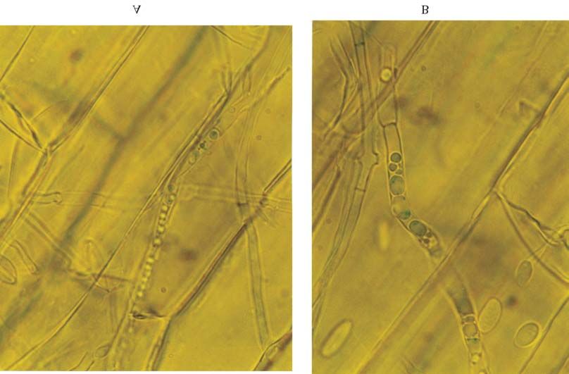
Figure 1 - Histochemical analysis of β-D-glucuronidase (GUS) activity in mycelia of Fusarium verticillioides co-transformant TG3 in maize roots.
Histochemical analysis at (A) 400x and (B) 1000x magnification. Intracellular GUS activity is visible as discrete blue rounded bodies.
The hybridization pattern using the nia probe (Figure sites. A DNA fragment of about 5 kb, which did not corre-
2B) showed integration of the pnH24 plasmid at several spond to the pUC vector (data not shown), was observed inGus/nia co-transformation of Fusarium 257
Figure 2 - Southern blot analysis of Fusarium verticillioides transformants. DNA was extracted from recipient and co-transformed strains, digested to
completion with EcoRI and probed with (A) the 4.5-Kb Hind III-EcoRI pNOM102 fragment containing the entire GUS construct and (B) the pNH24
plasmid linearized with EcoRI and labelled with 32P. Lane 1, F. verticillioides recipient strain; lanes 2 to 11, F. verticillioides co-transformant TG1, TG2,
TG3, TG4, TG5, TG6, TG7, TG8, TG9 and TG10.
both the S68 recipient strain an all transformants and may the gene in the plant and demonstrating the effectiveness of
correspond to a resident copy of the nitrate reductase gene endophytic fungi as ‘vectors’ for the introduction of char-
of F. verticillioides. acteristics of biotechnological or agricultural interest into
A blue precipitate was clearly observable in the my- tropical host plants. The result showing co-transformant
celium of F. verticillioides GUS+ transformant in the maize expressing the gene in the plant is important because, as
roots (Figure 1). shown by Pamphile and Azevedo (2001), endophytic F.
verticillioides strains may be specific to particular maize
Discussion populations or lines. Direct histochemical examination of
the endophyte in situ can aid studies on fungal competition,
Our co-transformation procedure resulted in a trans-
endophyte colonization of plant tissues, endophyte-plant
formation efficiency some 75 times higher than that ob-
relationships and investigations involving the relationship
tained by Yates et al. (1999) who performed GUS
of endophytes with other components of the plant-
transformation of F. moniliforme using a pHPG plasmid
associated microbial community.
containing the gusA reporter gene and the selectable
marker gene hph encoding for hygromycin-resistance
(hygr), while our procedure was about 750 times of that de- Acknowledgments
scribed by Doohan et al. (1998) who transformed Fusarium We thank Catherine Gerlinger for technical support
culmorum using a plasmid harboring the E. coli gusA gene and the Brazilian agency CNPq for financial support. We
and the hph gene. Taking into account only the GUS trans- would like also to thank Dr. M.J. Daboussi allowing Dr.
formants, co-transformant efficiency was high (57.20%) in J.A. Pamphile to do the transformation experiments at the
our assays, with transformation efficiency attaining a 43 to Université Paris-Sud, Orsay, France.
430 fold increase as compared to previous reports. Bao et
al. (2000) obtained a co-transformation efficiency of References
46.30% in F. oxysporum using the plasmid vector pCF20
containing the E. coli gusA gene and pAN7-2 encompass- Azevedo JL, Maccheroni Jr W, Pereira JO and Araújo WL (2000)
ing E. coli hph gene. Endophytic microorganisms: a review on insect control and
Some of our F. verticillioides transformants gave recent advances on tropical plants. EJB: Electronic Journal
of Biotechnology [online]. 15 April 2000, v. 3, n. 3. Avail-
higher fluorometric GUS activities than the positive control
able at http://www.ejb.org/content/vol3/issue1/full/4/index.
strain M-T5 (Table 1) which had originally been obtained html. ISSN 0717-3458.
by Couteaudier et al. (1993) and had given the best GUS Bao JR, Velema J, Dobinson KF and Lazarovits G (2000) Using
activity in their assay, the GUS activity of strain M-T5 in GUS expression in a nonpathogenic Fusarium oxysporum
our study (Table 1) being similar to that reported by strain to measure fungal biomass. Can J Plant Pathol 22:70-
Couteaudier et al. (1993). 78.
A blue precipitate was clearly observable in the my- Couteaudier Y, Daboussi MJ, Eparvier A, Langin T, and Orcival J
celium of F. verticillioides GUS+ transformants colonizing (1993) The GUS gene fusion system (Escherichia coli β-D-
maize roots, indicating that the co-transformant expressed Glucuronidade gene), a useful tool in studies of root coloni-258 Pamphile et al.
zation by Fusarium oxysporum. Appl Environ Microbiol Pamphile JA and Azevedo JL (2002) Molecular characterization
59:1767-1773. of endophytic strains of Fusarium verticillioides (=
Daboussi MJ, Langin T and Brygoo Y (1992) FOT1, a new family Fusarium moniliforme) from maize (Zea mays. L). World J
of fungal transposable elements. Mol Gen Genet 232:12-16. Microbiol Biotechnol 18:391-396.
Diolez A, Langin T, Gerlinder C, Brygoo Y and Daboussi MJ Pamphile JA, Azevedo JL, Langin T and Daboussi MJ (1996)
(1993) The nia gene of Fusarium oxysporum: isolation, se- Transformação de Fusarium moniliforme usando-se o gene
quence and development of a homologous transformation nia de F. oxysporum. Braz J Genet (suppl):19. XLII
system. Gene 131:61-67. Congresso Nacional de Genética, Caxambu, Brazil.
Doohan FM, Smith P, Parry DW and Nicholson P (1998) Trans- Pontecorvo G, Roper JA, Hemmons LM, McDonald KD and
formation of Fusarium culmorum with the β-D-glucuro- Bufton AWJ (1953) The genetics of Aspergillus nidulans.
nidase (GUS) reporter gene: A system for studying Adv Genet 5:141-148.
host-pathogen relationships and disease control. Physiol Roberts IN, Oliver RP, Punt PJ and Vandenhondel CAMJJ (1989)
Mol Plant Pathol 53:253-268. Expression of the Escherichia coli β-glucuronidase gene in
Hua-Van A, Pamphile JA, Langin T and Daboussi M-J (2001) industrial and phytopathogenic filamentous fungi. Curr
Transposition of autonomous and engineered impala trans- Genet 15:177-180.
posons in Fusarium oxysporum and related species. Mol Sambrook J, Fritsch EF and Maniatis, T (1989) Molecular clon-
Gen Genet 264:724-731. ing: a laboratory manual. v.1. 2nd ed. Cold Spring Harbour
Jefferson RA (1988) Plant reporter genes: The GUS gene fusion Laboratory, New York, NY, 621 pp.
system. In: Setlow, JK (eds) Genetic Engineering. Principles Smith D and Onions AHS (1983) The preservation and mainte-
and Methods. v. 10. New York Plenum Press, p. 247-263. nance of living fungi. Page Bros, Norwick, 51 pp.
Malardier L, Daboussi MJ, Julien J, Roussel F, Scazzocchio C and Yates IE, Hiett KL, Kapczynski DR, Smart W, Glenn AE, Hinton
Brygoo Y (1989) Cloning of the nitrate reductase gene DM, Bacon CW, Meinersmann R, Liu S and Jaworski AJ
(niaD) of Aspergillus nidulans and its use for transformation (1999) GUS transformation of the maize fungal endophyte
of Fusarium oxysporum. Gene 78:147-156. Fusarium moniliforme. Mycol Res 103:129-136.
Murashige T and Skoog F (1962) A revised medium for rapid
grown and bioassays with tobacco tissue culture. Physiol
Plant 15:473-497. Editor: Sérgio Olavo Pinto da CostaYou can also read