COMPARATIVE STUDY: FREE RADIAL FOREARM FLAP PHALLOPLASTY VERSUS ANTERIOR LATERAL THIGH FLAP PHALLOPLASTY
←
→
Page content transcription
If your browser does not render page correctly, please read the page content below

Academic Year 2015 - 2017

COMPARATIVE STUDY: FREE RADIAL FOREARM
  FLAP PHALLOPLASTY VERSUS ANTERIOR
   LATERAL THIGH FLAP PHALLOPLASTY

                          Rani CAPELLE
                         Elise MISSAULT

                   Promotor: Prof. Dr. S. Monstrey
                      Co-promotor: Dr. S. D’Arpa

   Dissertation presented in the 2nd Master year in the programme of
                 MASTER OF MEDICINE IN MEDICINE

Academic Year 2015 - 2017

COMPARATIVE STUDY: FREE RADIAL FOREARM
  FLAP PHALLOPLASTY VERSUS ANTERIOR
   LATERAL THIGH FLAP PHALLOPLASTY

                          Rani CAPELLE
                           Elise MISSAULT

                   Promotor: Prof. Dr. S. Monstrey
                      Co-promotor: Dr. S. D’Arpa

   Dissertation presented in the 2nd Master year in the programme of
                 MASTER OF MEDICINE IN MEDICINE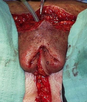

Deze pagina is niet beschikbaar omdat ze persoonsgegevens bevat.
Universiteitsbibliotheek Gent, 2021.

This page is not available because it contains personal information.
Ghent University, Library, 2021.

ACKNOWLEDGEMENTS
Firstly, we would like to express our gratitude towards our supervisor and promotor Prof. Dr.
Monstrey for all his help and remarks during our master thesis. He allowed this paper to be our
own, but guided us during the learning process. Furthermore, we would like to thank our co-
promotor Dr. D’Arpa for his time, remarks and helping hand during the making of the master
thesis. We would also like to thank Jan Smeyers for all his help. We could not have done this
without them.
Also, we would like to thank all participants for their time especially the ones who filled in our
survey. Their responses were of great value.

Special thanks, goes out to the experts who helped with the drafting of the survey; Els Elaut,
Prof. Dr. Hoebeke and Prof. Dr. Tsjioen.
We would also like to thank Mrs. Sabine Van Overloop for her help translating the survey.
Furthermore, we would like to thank the secretaries in the plastic surgery office, for their
logistic help. Finally, we would like to thank our family and friends for their help, comments
and support during this process.

ABREVIATIONS

RFF      Radial Forearm Flap
ALT      Anterior Lateral Thigh Flap
FTM      Femal To Male
EEC      Exstrophy- Epispadias Complex (EEC).
BXO      Balanitis Xerotica Obliterans
DSM-5    Diagnostic and Statistical Manual of Mental Disorders edition 5
WHO      World Health Organisation

ICD-11   International Classification of Disease
SRS      Sex Reassignment Surgery
CBE      Classic Bladder Exstrophy
LCFA     Lateral Femoral Circumflex Artery
SCIAP    Superficial Circumflex Iliac Artery Perforator
MDCT     Multidetector CT
PICO     Patient, Intervention, Control and Outcome
EPF      Electronic Patient File
STG      Split Thickness skin Graft
POSAS    Patient and Observer Scar Assessment Scale
IPSS     International Prostate Symptom Score
BPH      Benign Prostatic Hyperplasia
FGSIS    Female Genital Self Image Scale
CI       Confidence Interval
SD       Standard Deviation

CONTENTS
ACKNOWLEDGEMENT........................................................................................................
ABBREVIATIONS .................................................................................................................

ABSTRACT .......................................................................................................................... 1

INTRODUCTION ................................................................................................................. 3

   1.1.       Phalloplasty indications .......................................................................................... 3

      1.1.1. FTM population .................................................................................................... 4

      1.1.2. Exstrophy .............................................................................................................. 5

   1.2.       Phalloplasty surgery ............................................................................................... 6

      1.2.1. Flap specifics ........................................................................................................ 7

      1.2.2. Indications............................................................................................................. 9

      1.2.3. Additional procedures ......................................................................................... 10

      1.2.4. Advantages ......................................................................................................... 10

      1.2.5. Disadvantages ..................................................................................................... 11

      1.2.6. Complications ..................................................................................................... 12

RESEARCH QUESTION .................................................................................................... 13

METHODS .......................................................................................................................... 14

   3.1        Literature study resources ..................................................................................... 14

   3.2        Study population................................................................................................... 15

   3.3. Surgical techniques .................................................................................................... 16

      3.3.1      Radial forearm flap ........................................................................................... 16

      3.3.2      Anterolateral thigh flap ..................................................................................... 18

   3.4. Study design .............................................................................................................. 20

      3.4.1.         Short- term results ......................................................................................... 20

      3.4.2.         Long- term results ......................................................................................... 21

   3.5. Statistical Analyses .................................................................................................... 24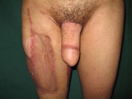
RESULTS ............................................................................................................................ 25

   4.1. Short- term results/ Postoperative period .................................................................... 25

   4.2. Long- term results ...................................................................................................... 27

      4.2.1. Long- term results: EPF. ...................................................................................... 27

      4.2.2 Long- term results: survey .................................................................................... 29

DISCUSSION ...................................................................................................................... 38

   5.1. Statement of principal findings .................................................................................. 38

      5.1.1.         Short- and long- term follow-up evaluated by the EPF .................................. 38

      5.1.2           Long- term follow-up evaluated by a questionnaire ...................................... 41

   5.2. Study strengths .......................................................................................................... 46

   5.3. Study limitations and remarks .................................................................................... 46

   5.4. Future research .......................................................................................................... 48

CONCLUSION.................................................................................................................... 49

REFERENCES .................................................................................................................... 50

ADDENDUM I: Dutch summary ........................................................................................ i
ADDENDUM II: Clearance EC Capelle Rani ..................................................................... iii
ADDENDUM II: Clearance EC Missault Elise ................................................................... v
ADDENDUM IV: Request to participate in study ............................................................... vii
ADDENDUM V: Request to complete the survey ............................................................... ix
ADDENDUM VI: Informed Consent .................................................................................. x
ADDENDUM VII: Survey .................................................................................................. xii
ADDENDUM IX: Data acquired out of the EPF ................................................................. xxii
ADDENDUM X: Data acquired out of the survey ............................................................... xxv
ABSTRACT
Introduction: The free radial forearm flap (RFF) is the most frequently used operative
technique in phalloplasty surgery. It is considered to be the golden standard and has let to great
results. Yet, a mayor inconvenience of the RFF procedure is the presence of an unavoidable
and distinct scarring on the forearm. This can be experienced as a stigma, as the procedure is
growing more recognizable within the general population. For 12 years, the anterior lateral
thigh flap (ALT) has been introduced for phalloplasty surgery and is used in one out of 3
patients at the University Hospital of Ghent (UZ Ghent). The largest series of RFF and ALT
phalloplasty surgeries are performed in this hospital. It follows that this hospital is in a good
position to initiate a comparative study between both techniques.
Aim: Can the ALT phalloplasty be considered as an equivalent alternative of the RFF
phalloplasty?
Methods: Health records of patients, who underwent a RFF or ALT phalloplasty surgery since
2005, were reviewed. Only female-to-male and extrophia- epispadias complex patients were
included in this study. In these records data regarding short- term and long- term complications
such as strictures, flap failure, urinary fistulas, delayed wound healing etc. were analyzed. Each
participant in this study received a survey as well. This survey contains a series of questions
and validated scoring systems about urological and sexual functioning as well as questions
regarding the satisfaction of aesthetic appearance of the donor site and neophallus.
Results: An EPF-analysis of 74 patient files was performed; 49 patients underwent a RFF
phalloplasty and 22 an ALT phalloplasty. Note that 3 patients were classified in both groups,
since both procedures were performed. The data found in the patient files led to only one
statistically significant result. The ALT phalloplasty is associated with a higher occurrence rate
of flap failure when the cases with postoperative complications were isolated. When the cases
of flap failure were further investigated and subdivided into marginal, minor and major flap
failure; ALT phalloplasty was also associated with a higher occurrence rate of major flap failure
compared to the distribution in the RFF group. No statistically significant differences between
ALT and RFF phalloplasty regarding the presence of urinary fistulas, strictures, wound healing
and discharge with a urinary diversion were found.
1The survey was completed by 49 patients; of which 32 RFF and 17 ALT. Based on the survey, the difference in erogenous sensitivity seemed to be statistically significant with ALT reporting inferior sensitivity compared to RFF. Also, ALT patients reported significantly more frequent to suffer from the sensation of an incompletely voided bladder. Conclusion: Few significant differences were discovered in this study. The erogenous sensitivity of the neophallus seems to be inferior after an ALT phalloplasty. In addition, it was found that ALT phalloplasty is associated with a higher risk of flap failure when the cases with postoperative complications were isolated. Since the sample size is small, it is difficult to elicit conclusions. The only assumption that can be made, based on this research, is that the ALT technique can be seen as a good alternative for the RFF technique. Yet we cannot conclude if they are equivalent or not, further research is necessary to provide an answer. 2
INTRODUCTION
The Free Radial Forearm Flap (RFF) is the most commonly used operative technique in
phalloplasty surgery and therefore considered as the gold standard. However, the noticeable
scarring at the forearm is considered as a major disadvantage, since it is identified with this
operation. For 12 years, the anterior-lateral thigh (ALT) flap has been introduced in phalloplasty
surgery and is used in one out of 3 patients at the University Hospital of Ghent.

The goal of this study is to compare both techniques and evaluate if the ALT phalloplasty can
be considered as an equivalent alternative for RFF. For a better understanding of this subject,
we will clarify some crucial information in the following introduction.

1.1.       Phalloplasty indications
Phalloplasty surgeries are performed for numerous indications of penile reconstruction or penile
absence/ insufficiency. The main indications for this procedure can be found in table 1. Not all
indications are included in this study. The study population is limited to female-to-male (FTM)
transsexuals and patients who suffer from the Exstrophy- Epispadias Complex (EEC).

 Congenital absence, hypoplasia or malformation      Self- inflicted penile amputations
 - Aphallia                                          - Mentally incapacitated patients
 - Penile hypoplasia or micropenis                   - Drug addicts
 - Epispadias/ Exstophy vesicae complex
                                                     Ambiguous genitalia
 - Hypospadia
                                                     Gender identity disorder
 Mutilating Trauma                                   - Female to male transsexuals
 - Avulsing injuries                                 Infection
   Heavy moving machinery accidents                  - Balanitis
   Road traffic accidents                            - Necrotizing fasciitis
 - Blast injuries                                    - BXO (Balanitis Xerotica Obliterans)
   Mineblast injuries                                Iatrogenic
   Improvised explosive device injuries              - Circumcision-related injuries
 - Criminal injuries                                 - Penile loss following tumour extirpation
   Sexual partner- inflicted injuries
 - Burns

Table 1: Indications for phalloplasty surgery (1).

                                                                                                  3
1.1.1. FTM population 1.1.1.1. Modern definition of transsexualism: gender dysphoria and gender incongruence The Diagnostic and Statistical Manual of Mental Disorders edition 5 (DSM-5) uses the term gender dysphoria defined by “marked incongruence between one’s experienced/ expressed gender and assigned gender for at least 6 months” and “clinically significant distress or impairment in social, school, or other important areas of functioning”. The presence of distress and dysfunction are the central rationale for classifying these conditions as mental disorders (2). Yet, over the years, different terms and diagnostic criteria were used by different classification systems. The World Health Organisation (WHO) is in the process of developing the eleventh version of the International Classification of Disease (ICD-11). The ICD-11 proposes the term gender incongruence. The associated diagnostic requirements include the continuous presence for at least several months of at least two of the following features: “a) a strong dislike or discomfort with primary or secondary sex characteristics due to their incongruity with the experienced gender; b) a strong desire to be rid of some or all of one’s primary or secondary sex characteristics (or, in adolescence, anticipated secondary sex characteristics); c) a strong desire to have the primary or secondary characteristics of the experienced gender; and d) a strong desire to be treated (to live and be accepted as) a person of the experienced gender”. The ICD-11 will no longer classify categories related to gender identity as a part of its classification of mental disorders. This is the main difference between the proposals for ICD-11 and the DSM-5 (2). 1.1.1.2. Prevalence of transsexualism: FTM Arcelus et al. (2015) performed a meta-analysis of prevalence studies in transsexualism worldwide. The findings within this study revealed that there has been a clear increase in the prevalence of individuals diagnosed with transsexualism over time. The study showed that the prevalence of trans men is 2.6/100.000 (3). De Cuypere et al. performed a study on the prevalence and demography of transsexualism in Belgium. In 2003 the prevalence of female-to-male transsexuals was 2.96/ 100.000. It should be noted that the target population of this study consisted of all Belgian transgender individuals who had undergone Sex Reassignment Surgery (SRS) since 1985 (4). Therefore, this number is an underestimation of the transgender population since not every transgender undergoes SRS. 4
1.1.1.3. Indications for phalloplasty
As stated above the number of patients suffering from gender dysphoria has increased the last
few years, this resulting in more people requesting for gender reassignment surgery (3, 5).
Despite SRS is an efficient treatment for FTM transsexuals, it must be noted that not all patients
with gender dysphoria require an surgical treatment (5).
The positive consequences of this treatment were established by multiple studies. research
showed that patients who underwent gender reassignment surgery had increased psychosocial
outcomes in comparison with the still-waiting control-group (7) G. De Cuypere et al. (2006)
performed a study on phychosocial outcome of Belgian transsexuals after SRS. Their research
showed that none of the patients showed any regrets about the SRS (6).

The following surgical interventions can be executed in FTM gender reassignment surgery:

   1. Voice surgery;
   2. Several cosmetic procedures (lipofilling, liposuction, facial masculinisation, etc.);
   3. Subcutaneous mastectomy;
   4. Genital surgery:
       - hysterectomy/ ovariectomy (performed before phalloplasty)
       - phalloplasty or metoidioplasty
       - vaginectomy
       - scrotoplasty
       - implantation of an erection and/ or testicular prostheses (5, 7).

However, this research solely focuses on phalloplasty and the associated scrotoplasty.

1.1.2. Exstrophy
1.1.2.1. Modern view of the Exstrophy- Epispadias complex
The Exstrophy- Epispadias Complex (EEC) is a rare spectrum of congenital abnormalities
which are probably the most challenging multisystem birth defects in urology and paediatric
surgery. It presents as a series of defects affecting the genitourinary and gastrointestinal tract,
the musculoskeletal system, the pelvic floor musculature and the bony pelvis (8-10).

The three most common manifestations of this EEC are epispadias, Classic Bladder Exstrophy
(CBE) and cloacal exstrophy (9). These three forms differ in severity as well as prevalence,
anatomic anomalies and approach.

                                                                                                 5
1.1.2.2. Prevalence and etiopathogenesis of ECC
Although all three forms are extremely rare, there is still a big range in prevalence between
these three forms. The most common presentation of EEC is the CBE which occurs in
approximately one every 10,000 to 50,000 births (9, 11). The second most common form is
complete epispadias, it occurs in one in every 117,000 male births and one in every 477,000
female births (9, 11). The third form of ECC is cloacal exstrophy, this is the most severe and
rare form, occurring in approximately one out of 200,000 to 400,000 births, with higher rates
in females (9, 11, 12).
1.1.2.3. Indications for phalloplasty
Male patients born with bladder and cloacal exstrophy are born with demure or even absent
genitalia, such as micropenis and epispadias, (13, 14). The penis is at least 50% shorter than the
average sized penis. The phallus is wedge-shaped as a result of the fact that the corpora
cavernosa only fuse at the distal part of the shaft and because the crura are inserted on the caudal
aspect of the ischiopubic rami (15).
Since patients are often left with a shortened phallus, many males with EEC desire a genital
reconstruction. Many patients undergo phalloplasty surgery, which is usually performed in their
late adolescence or early adulthood (13). Usually, the RFF is the method of choice, yet in some
institutions the ALT is used, such as in the University Hospital of Ghent (13, 15).
1.2. Phalloplasty surgery
In case of FTM transsexuals, the construction of a penis is particularly challenging because of
the additional need for vaginectomy, scrotoplasty, and reconstruction of the horizontal part
(pars fixa) of the urethra (16). In the patient group of boys without a penis (bladder exstrophy),
no scrotoplasty is necessary, since the scrotum is normal.
Phallic reconstruction remains a major surgical challenge. There are different cosmetic and
functional requirements that ideally shall be met. The requirements are stated below.
1. One- stage procedure;
2. aesthetic phallus;
3. normal scrotum;
4. voiding while standing;
5. protective and erogenous sensation;
6. minimal mortality and morbidity;
7. sexual intercourse (16, 17).
6The presence of protective and erogenous sensation is of great importance for numerous
reasons. One, protective sensation is necessary to prevent genital injuries and is imperative to
support the insertion of an erection prosthesis after a phalloplasty. The other, erogenous
sensitivity, is essential in order to reach an orgasm during sexual activities such as masturbation
and intercourse. Erogenous sensation is obtained by microsurgical coaptation of nerves from
the donor flap to the recipient nerves in the perineum (dorsal clitoral nerve), as well as the
preservation of the clitoris (18). The surgical techniques evaluated in this study are the free
radial forearm flap (RFF) and the anterolateral thigh flap (ALT). Flap specifics, indications,
advantages, disadvantages and complications of each technique are stated below.
1.2.1. Flap specifics
1.2.1.1. RFF
The RFF phalloplasty requires a free tissue transfer from the radial forearm to create the
neophallus. The RFF flap is used to create a neophallus and neo-urethra by using the tube-
within- a- tube technique. This technique is based on 2 steps; first the neo-urethra is formed by
creating a tube using a small portion of the flap. The tube is formed by rolling up this flap, skin
faced inwards. Afterwards the outer tube is formed with a bigger portion of the flap, wrapped
around the earlier formed neo-urethra. In the outer tube the skin is faced outwards. Picture and
figure 1 show both parts of the RFF flap and how the flaps are tubed respectively.
Once the phallus is formed, the flap is freed from the forearm and placed at the groin. The blood
supply of the neophallus depends on microvascular anastomoses between the vessels of the flap
and the acceptor site. Nerve coaptation between cutaneous nerves of the donor flap and the ilio-
inguinal and dorsal clitoral nerve are responsible for tactile and erogenous sensitivity
respectively (18).
The flap is generally harvested from the volar and dorso-radial side of the non- dominant
forearm. In presence of tattoos or extensive scarring on the non- dominant forearm, another
donor site is required, such as the dominant forearm or the thigh. The flap is harvested in the
suprafascial plane of the forearm. Proximally, the incision is carried ou to the antecubital fossa
which enables the proximal dissection of the vessels and nerves (19).
7A sufficient surface of the flap is required in order to reconstruct a functional and normal sized neophallus. The width of the flap is approximately 16 cm, this encircles and often involves the entire forearm (19, 20). The length of the flap is often 14 cm, with the addition of 2 cm on the proximal ulnar aspect to allow for the pars pendulans of the urethra. The flap is harvested based on a drawing made on the skin based on a template, a smaller template can be used in case the patient has a narrow forearm (19). Picture 1: Radial forearm flap. This picture shows both Figure 1: Tube-within-a-tube. The urethral flap is parts of the RFF flap: the flap used to tube the neo- tubed first (skin faced inwards). Then the outer tube is urethra (the upper flap in the picture) and the flap used wrapped around the earlier formed urethra (skin faced to form the outer part of the neophallus (the lower flap outwards). in the picture). 1.2.1.2. ALT The anterolateral thigh flap is a pedicled perforator flap supplied by the descending branch of lateral femoral circumflex artery (LCFA) (17). Hence the blood supply is not interrupted, there is no need for vascular anastomoses. It is important that there is no traction on the pedicle, therefore the pedicle has to be long enough. The ALT flap meets almost all of the following qualities: safe, sensate, with a long pedicle and large amount of soft tissues. Also, it can be harvested in a single procedure and with a low donor site morbidity (21). In most cases, the flap is used as a wrap-around flap to cover a urethral reconstruction which can be provided by a tube or a variety of flaps. The following flaps are used for this urethra reconstruction: a skin graft prelamination, a superficial circumflex iliac artery perforator (SCIAP) flap/ groin flap, a narrow radial artery forearm flap or material from a previous penile reconstruction (22). In the literature, the “tube-within-a-tube” technique is described in which the penis and urethra are constructed with a single flap (22, 23). Though this technique is only feasible with very thin patients. 8
Knowledge of the precise location of the perforators (branch of the LCFA) is essential for
adequate shaping of the flap and the need of transferring the shaped flap on its pedicle to the
pubic area. Therefore, before surgery, preoperative perforator mapping is performed. The
current instrument of choice is the multidetector CT (MDCT). It allows locating the perforators
in combination with an evaluation of their supra- and subfascial quality and pedicle course. The
latter is important to estimate the pedicle length and to determine if it will allow the flap to
reach the pubic area. It is essential to avoid any traction on the pedicle because this can
compromise the blood supply and lead to ischemia and flap failure (22). In most cases the defect
of the donor site is closed by using split thickness skin grafts, but sometimes preoperative skin
expansion is used to achieve primary closure of the donor site (24). Before placement of the
tissue expanders it is crucial to have knowledge of where the perforators are located. The pivot
point of the pedicled ALT flap is the origin of the LCFA from the femoral artery. By using the
MDCT the position of the perforators can be measured from a reference point which is the
anterior superior iliac spine (ASIS), the branching pattern of the perforator can be visualized
and it is possible to measure the thickness of the subcutaneous fat tissue layer of the ALT flap.
The latter is one of the most important criteria for choosing the reconstructive option
(RFF/ALT) as well as the accompanying urethra reconstruction. After measurement of the
thickness of the subcutaneous tissue of the ALT flap, the exact dimensions of the flap can be
calculated. This to make sure it can be tubed around the reconstructed urethra without
compression of the urethral flap and closure without tension (22). Generally, the choice of the
donor limb rests with the patient (21). However, it should be noted that the quality of the
perforator is superior to the preference of the patient.

1.2.2. Indications
1.2.2.1. RFF
This procedure is widely accepted as the best donor site for penile reconstruction. RFF
phalloplasty has shown to be a very reliable technique for the creation of a normal looking penis
(16, 17, 25). It is therefore currently considered as the gold standard for phalloplasty surgeries
on patients with penile insufficiency and FTM transsexuals (16, 17).

The RFF technique is used in up to 90% of all published phalloplasty surgeries and for
numerous indications (16). Yet some contra indications must be mentioned. One is the presence
of intensive scarring or tattoos on both forearms. Another reason is the absence of adequate
perfusion of the forearm. Before performing a RFF phalloplasty, the mixed radial- ulnar

                                                                                                9
vascular supply must be tested in order to ensure adequate perfusion of the donor site after phalloplasty. This is done by the Allen test. Absence of mixed vascular supply can be an indication for using the thigh as a donor site (13, 16, 20). 1.2.2.2. ALT The ALT flap is used for FTM transsexuals and patients with severe penile insufficiency who refuse the RFF flap due to the major residual scar and FTM transsexuals in whom the RFF flap is contraindicated (abnormal Allen test). Also, the ALT flap can be used. In cases of severe penile insufficiency (‘boys without a penis’), like in bladder exstrophy patients, the reconstruction of the urethra is seldom required because these patients underwent earlier surgery where in most of these a Mitrofanoff channel was created (22, 26). 1.2.3. Additional procedures The following statements are valid for both techniques. The average hospitalization period after phalloplasty surgery is about 2,5 weeks. After 3 to 6 months, before the return of the sensation of the penis, tattooing of the glans can be implemented. For the implantation of a penile prosthesis, return of sensation to the top of the neophallus is obligated. This usually takes about one year. Sufficient protective sensation is needed in preventing breakdown and erosion of the stiffener. A second requirement before implanting a stiffener is that the urethra must be free of strictures or fistulas (17). After a 12-month period, implantation of testicular prostheses can be performed in accordance to the patients’ wishes (16). 1.2.4. Advantages 1.2.4.1. RFF The following advantages of RFF phalloplasty are stated in the literature. First, the flap is thin and pliable allowing the construction of a normal-sized tube-within-tube penis (27). Second, the flap is easy to dissect and is predictably well vascularised making it safe to perform a coronaplasty at the distal end of the flap in the same procedure. Third, research shows that of all flaps used for phalloplasty surgeries, the RFF has the greatest sensitivity. Another advantage is that his procedure gives a satisfying result regarding the erogenous and tactile sensitivity of the phallus. This can be explained by the coaptation of the cutaneous nerves and the ilio- inguinal and clitoral nerves and the preservation of the clitoris (18). Van Caenegem et al. (2013) and Selvaggi et al. (2006) showed that patients operated with the RFF procedure observed no functional limitations on daily life activities (19, 20). And finally, a research performed by G. 10
Selvaggi et al. (2007) reported that all of the included patients who underwent RFF phalloplasty
were able to achieve an orgasm afterwards (18).

1.2.4.2. ALT
The current literature mentions some advantages of the use of the ALT flap compared to the
use of the RFF flap. First, there is no need for a microsurgical technique as the flap is pedicled
(21, 28). Second, the color of the anterolateral thigh matches more that of the perineum (28,
29). Third, in shorter individuals who have shorter forearms, the reconstructed penis can be
longer with an ALT compared to a RFF (21). Fourth, there is no sacrifice of major blood vessels
(23). Another advantage of this technique is that there are no scars at noticeable sites (23, 28).
This is a major advantage as the visibility of the donor site scar is the leading drawback of the
radial forearm flap (22). The volume of the neophallus created by this flap is rather large and
the volume remains the same over time, meaning atrophy of the neophallus does not occur. This
is a major advantage of the ALT flap since the phallus created by a RFF flap does atrophy. It
follows that in some ALT phalloplasties, there is no need for the implantation of a penile
prosthesis in order to have sexual intercourse.

1.2.5. Disadvantages
1.2.5.1. RFF
One of the disadvantages of this technique is the residual and unavoidable scarring on the
forearm. This is considered as a stigma for many patients (22). Potential drawbacks of this
procedure are the possibility of volume loss over time (due to atrophy of the tissue) and the
need for a rigidity prosthesis to ensure penetrative sexual intercourse (27). Another
disadvantage of the RFF technique is the risk of additional complications. The RFF is based on
a free flap, which requires microvascular transfer for penile reconstruction. Additional
complications can occur due to problems with this microvascular transfer (27). The occurrence
of complications is further elaborated (section 1.2.6).

1.2.5.2. ALT
Some disadvantages of using the ALT flap have been described. First, there is a high anatomical
variability in ALT flap perforators (23). The MDCT examination allows to overcome this
problem by preoperative evaluation of anatomic variations in order to determine if the patient
is suitable for ALT flap phalloplasty (22). Second, thick flaps are obtained from women and
obese patients due to the presence of more fat tissue (23). This often results in a bigger volume
penis and is thus cosmetically less pleasing. It is a principal facet to select patients of whom the

                                                                                                 11
subcutaneous fat layer of the thigh allows the surgeons to establish a penis that is acceptable in
volume. Patients with a thick subcutaneous fat layer are not candidates for an ALT phalloplasty
unless the flap can be thinned (22). Few research was done on the sensitivity of both flaps. One
study mentioned the sensitivity is a major concern in the ALT flap (17).
vs RFF flap
Picture 2: Thin neophallus and Picture 3: Thick neophallus Picture 4&5: Phallus after ALT
distinct scarring on forearm and scarring on thigh after ALT surgery (left). Phallus after RFF
after RFF phalloplasty. phalloplasty. surgery (right). These pictures
clearly show the difference in
volume.
1.2.6. Complications
A number of complications can occur after a phalloplasty. These complications include the
general ones that can occur during every surgical intervention, combined with several
complications that are specific for these procedures. A few are mentioned below in table 2.
Flap-related Various
Anastomotic revision Pulmonary embolism
Complete flap loss Regrafting of defect on arm/leg
Partial necrosis Nerve compression (early cases)
Delayed wound healing
Urologic
Fistula treated conservatively Erectile prosthesis
Stricture treated conservatively Revision surgery (mechanical failure, infections,
Fistula/Stricture requiring urethroplasty exposure and malposition)
Incapacity to perform sexual intercourse
Table 2: Complications (16).
Multiple research has been performed exploring the possible complications following the RFF
procedure. For example, it is known that the occurrence of urinary fistulas is frequent. Other
long-term urological complications are also associated with the RFF technique (27). In table 3,
a selection of important complications is stated and their frequency in the RFF group. These
data are based on research performed at the University hospital of Ghent on 562 RFF patients.
Yet, for ALT phalloplasty a lack of specific data is seen since no research has been performed
12on a large scale. Although no specific data is found, the amount of several complications is
expected to be larger following ALT phalloplasty (30).
COMPLICATIONS RFF PHALLOPLASTY N %
Flap failure
- Complete flap failure 6 1.07%
- Partial flap failure 43 7.65%
Compression syndrome 4 0.71%
Delayed wound healing 58 10.32%
Urinary fistula 197 35.05%
Urinary strictures 78 13.88%
Transient Ischemia 15 2.67%
Table 3: Complications observed in RFF phalloplasty surgery (30).
Picture 2: Partial flap Picture 3: Fistula with its Picture 4: Flap failure of the
failure at the tip of the opening at the basis of the urethral flap in ALT
neophallus. phallus. phalloplasty.
RESEARCH QUESTION
The main research question is if the ALT phalloplasty can be considered as an equivalent
alternative to the RFF phalloplasty. In order to give a correct answer to the main research
question, it has to be divided into different sub-questions.
1. Is there a difference regarding short- term follow- up/ complications between the two
techniques? If so, which technique presents the best results?
2. Is there a difference regarding long- term follow- up/ complications found between the
two techniques? If so, which technique presents the best results?
3. Is there a difference regarding subjective satisfaction between the two techniques?
If so, which technique presents the best results?
13METHODS
3.1. Literature study resources
First, articles of Prof. Dr. Monstrey and/ or other members of the University of Ghent
Transgenderteam were reviewed. These articles seemed most relevant to this study because the
performed surgical technique is the same as in our study population, which makes comparison
more relevant and accurate.
Second, relevant articles were searched in PubMed, WoS and Google Scholar with the PICO-
strategy (Patient, Intervention, Control and Outcome).
P: FTM or Exstrophy patients
I: RFF or ALT phalloplasty surgery
C: /
O: Evaluation of complications, functionality.
The inclusion and exclusion criteria used are found in table 4.
Selection Criteria Inclusion Criteria Exclusion Criteria
Population FTM Other pathology, non-human
Exstrophy (bladder and cloacal)
Intervention Phalloplasty surgery RFF & ALT Other phalloplasty surgeries
Outcome Complications, functionality /
Design Studies, review Comment
Language English, Dutch Other languages
Other Abstract, Full text available No abstract or full text available
in Ugent Library
Table 4: Inclusion and exclusion criteria.
Note; all pictures included in this masterthesis were placed at our disposal by Prof. Dr. Monstrey and
Dr. D’Arpa.
143.2. Study population
All FTM and exstrophia vesicae patients who had undergone RFF or ALT phalloplasty surgery
since 2005, were eligible to take part in this study. Every patient received a personal letter from
Prof. Dr. Monstrey. The letter contained information about the study and a formal request for
permission to extract data out of their Electronic Patient File (EPF). Non- responders received
a reminder. Every patient included in this study gave permission by sending an email or a letter
to the Plastic Surgery office. Data of non-responders and patients who refused to take part in
this study remained unknown to the researchers. A total of 74 patients gave permission to
analyse their data for this study, of which 52 patients are classified in the RFF group and 25 in
the ALT group. Note that 3 patients first underwent a RFF phalloplasty, but due to
complications a revision was obliged and an ALT phalloplasty was performed in a later stage.
Therefore, these 3 patients were included in both categories.
An additional survey and informed consent was sent to the patients who had agreed to
participate in the study. A total of 49 patients completed the survey of whom 32 RFF patients
and 17 ALT patients. The number of responders and non-responders is shown in flow chart 1.
FTM and ECC patients who underwent a phalloplasty
surgery identified:
RFF = 146 & ALT = 70
Non- responders identified:
RFF = 94 & ALT = 45
Responders that gave permission to review their EPF:
RFF = 49& ALT = 22
both RFF and ALT = 3
Non- responders identified:
RFF = 17 & ALT = 8
Responders that filled in the additional survey.
RFF = 32 & ALT = 17
Flow chart 1: Response rate.
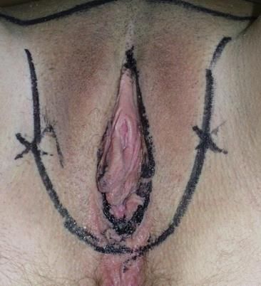
153.3. Surgical techniques All patients included in the study underwent a phalloplasty at the University Hospital of Ghent. The specific procedure used to perform this surgery is described below. This information was obtained out of surgical reports drafted after every phalloplasty surgery. 3.3.1 Radial forearm flap 3.3.1.1. Preoperative preparation RFF Preoperatively in every patient, an Allen test was performed and a drawing was made on the skin, based on a standard template. 3.3.1.2. Surgical procedure (as performed at the UZ Gent-hospital) RFF For the genitoperineal transformation two surgical teams are required. A team of urologists and a team of plastic surgeons are working simultaneously. The urologists perform a vaginectomy, scrotoplasty, reconstruction of the fixed part of the urethra, they prepare the clitoris for burial and harvest the dorsal clitoral nerve for coaptation. This is performed while the plastic surgeons dissect the radial forearm flap and construct a-tube-within-a-tube phallus. Urological team The patient is brought into a gynecological position. When the patient is positioned correctly a local injection with a solution of xylocain and adrenalin (1/2 diluted) is administered in the vaginal introitus. Then the surgeon starts with an incision around the dorsal part of the labia majora, another incision is made around the vaginal introitus. The vaginal mucosa is removed to the level of the previously performed hysterectomy. The inner parts of the labia minora are used to reconstruct the urethra and sutured around a silicon drain. The right dorsal nerve of the clitoris is identified and dissected in preparation for coaptation with a nerve of the flap. The mucosa of the glans of the clitoris is completely resected to allow the clitoris to be buried (after being transposed ventrally). The clitoris and the terminal part of the pars fixa of the urethra are relocated to a more ventral position, the definitive place for future anastomosis with the pars pendulans of the urethra (neo-urethra). The scrotoplasty is performed with two labia majora flaps. The flaps are rotated medially and bent on themselves. The tips of the two triangular labia majora flaps are sutured to each other along the midline. They are both elevated, while the skin of the dorsum of the clitoris is pulled down. This is the so called V-Y plasty (Hoebeke’s technique). 16
Picture 5a(left): Before scrotoplasty. Picture 5b(right): V-Y scrotoplasty. Two
labia majora flaps are rotated medially and bent on themselves. The tips of the
two triangular labia majora flaps are sutured to each other along the midline. They
are both elevated, while the skin of the dorsum of the clitoris is pulled down.

Plastic surgeons
While the urologists prepare the recipient area, the plastic surgeons start with dissecting the
radial forearm flap. The flap is harvested in the suprafascial plane and according to a drawing
made on the skin based on a template. The radial artery is dissected proximally to its take-off
from the brachial artery and the connection between the superficial and the deep venous
systems. The medial and lateral antebrachial cutaneous nerves are identified and included in
the flap, as well as the cephalic vein.

The next step is to reconstruct the urethra and form a phallus with the flap. The flaps stays
pedicled to the forearm while the a-tube-within-a-tube phallus is formed as described above
(section 1.2.1.1.). A coronaplasty is also performed at this stage with a small flap and a skin
graft. When the urological team has finished, the patient is placed into a supine position to make
harvest of the femoral vessels and ilio-inguinal nerve possible. Once the recipient site is ready,
split thickness skin grafts are harvested to reconstruct the forearm donor site and the whole flap
is transported to the perineal region after pedicle division.

The innervation and vascularisation of the flap is assured by anastomosis between arteries,
nerves and veins of the donor site and the recipient site. Two vascular anastomosis are required,
one between the great saphenous vein and the superficial cephalic vein. The second anastomosis
is mostly performed by an end-to-side anastomosis between the radial artery and the common,
or superficial femoral artery or end-to-end to one of its branches. The flap nerves are connected
with one inguinal nerve and one dorsal clitoral nerve. Finally, the groin is closed with an
absorbable suture.

                                                                                               17
3.3.1.3. Postoperative care
All patients are provided with a suprapubic urinary diversion intraoperatively. The patients need
to remain in bed for 10 days after which the transurethral catheter is removed. The day after,
the suprapubic catheter is clamped and voiding is started. When the patient is able to efficiently
empty the bladder, the suprapubic catheter is removed as well.
3.3.2 Anterolateral thigh flap
3.3.2.1. Preoperative perforator mapping
The flap is drawn on the thigh based on the MDCT scan that allows to detect the perforator.
The second flap to reconstruct the urethra is drawn preoperatively as well.
Picture 6a: The preoperative design of the ALT flap. “X” Picture 6b: The preoperative design of the ALT flap
marks the perforator selected with MDCT. The (wrap-around) on the left thigh and the SCIAP- flap
rectangular shape is the template of the ALT flap. (urethra) on the right groin. “X” marks the perforator
selected with MDCT.
3.3.2.2. Surgical procedure
Urological team
The surgical procedure performed by the urological team within the ALT phalloplasty surgery
is identical to the one performed within the RFF phalloplasty surgery. However, due to the
proximity of both donor and recipient site, both surgical teams cannot work simultaneously.
The plastic surgeons only start when the urologists have finished.
Plastic surgeons
The dissection of the ALT flap is started proximally to identify two sensory nerves, branches
of the lateral femoral cutaneous nerve. Then the lateral and distal incisions are performed and
lateral-medial dissection continues until the perforator is identified. The fascial opening that
gives passage to the perforator is extended distally and proximally and dissection of the
perforator (intraseptal or intramuscular) is performed to the origin of the descending branch of
18the LCFA from the LCFA itself. All side branches are divided after ligation and all motor nerves
are preserved. Then the flap is completely incised and tunneled underneath the rectus femoris
muscle and the sartorius muscle and then subcutaneously to reach the pubic area. Any pull on
the pedicle should be avoided. The flap is then ready to be wrapped around the neo- urethra that
has already been reconstructed at this point with a RFF flap or a pedicled SCIAP flap
(exceptionally a free SCIAP flap). One nerve branch of the ALT flap is coaptated to the
ilioinguinal nerve for tactile sensation, the other is coaptated to one of the dorsal clitoral/penile
nerves for erogenic sensation. The defect on the donor area is covered with split thickness skin
grafts which are harvested from the medial and anterior thigh. If the area has been pre-expanded,
the donor site can be closed directly.
Picture 7: ALT flap. The ALT flap (with subcutaneous fat) is Picture 8: ALT-flap and neo-urethra. The neo-
completely harvested from the thigh. Only the pedicles stays urethra is already formed. The ALT-flap is
intact. completely harvested from the thigh. Only the
pedicle stays intact. The ALT-flap is now ready
to be tunneled underneath the rectus femoris
muscle and sartorius muscle and then
subcutaneously to reach the pubic area.
3.3.2.3. Postoperative care
The postoperative care is the same as after RFF phalloplasty (section 3.3.1.3). There is one
substantial difference compared to the RFF phalloplasty. In RFF, the coronaplasty is performed
within the same procedure because the flap has the advantage of being vascularized by multiple
branches of the radial artery. In the ALT flap coronaplasty is performed after a 7- day delay,
this to ensure sufficient blood supply before further manipulation. Manipulation of the flap in
the days shortly after phalloplasty might increase the risk of flap failure since the blood supply
relies on the one perforator.
193.4. Study design The study is a comparative study that consists out of two parts: a retrospective evaluation of the patient files and evaluation of the responses given to the survey. This research was granted clearance by the Ethics Committee of the University of Ghent. 3.4.1. Short- term results Based on preliminary research, a variety of complications is known to occur. The EPF was consulted for evaluation of the complications mentioned below. Postoperative complications Numerous complications can occur during a phalloplasty surgery. Only complications specific for this procedure that required revision and operative correction shortly after the operation were investigated. The following postoperative complications were examined during this step: revision of the anastomosis, partial and complete flap failure, presence of infection and hematoma (that required drainage). Fistulas were not included into this comparison because operative correction is only performed after a six- month period. These postoperative complications were compared in numerous ways. First, the presence of complications requiring surgical intervention were compared. Subsequently the postoperative complications were divided into the above-mentioned categories. Partial and Complete flap failure Since flap failure is a severe complication, further exploration deemed necessary. The presence of flap failure was therefore compared between both study populations. Since the ALT neophallus is constructed out of 2 different flaps, necrosis can occur in either one of the flaps. The location of flap failure is of crucial importance and will therefore be further analyzed. In order to receive additional information, the data were divided into three groups: a group concerning the necrosis where no additional surgery was necessary, a group where only a minor surgery was required (debridement and split thickness skin graft (STG)) and a group that needed a new flap. Fistulas The presence of a fistula is a frequent complication which is described following RFF and ALT phalloplasty. Fistulas as result of the phalloplasty surgery may heal spontaneously or require surgical intervention. First, the presence of a fistula was evaluated in both groups. Subsequently, the fistulas that healed spontaneously in the first six months and the fistulas that required an operation were compared separately. 20
Wound healing
Data about delayed wound healing were not explicitly reported in the EPF, an assumption of
the wound healing was made based on the duration of the hospitalization after the phalloplasty
surgery. The cutting point of uncomplicated wound healing was set to 18 days. A stay longer
than 18 days was classified as a prolonged stay and therefore categorized as prolonged/
insufficient wound healing.

Discharge with a urinary diversion
In an optimal situation discharge from the hospital occurs not only within the course of an 18
day stay, but along with the ability to urinate independently (which means without a urinary
diversion). This is not the case for every patient and the difference between both subgroups was
assessed.

3.4.2. Long- term results
Long- term results were evaluated based on information obtained from the EPF. This was
however only possible if the patient was further observed in the University hospital of Ghent.

Strictures
An analysis of the presence of strictures in both study populations was performed, since
strictures are a frequently reported complication in the literature.

Complications associated with the presence of an erection prosthesis
To ensure sexual intercourse an erection prosthesis can be implanted. First, it was investigated
whether a penile prosthesis is present. Within this group it was further explored whether
complications occurred or not and if so, it was evaluated which complication. This way
complications were divided into four groups: mechanical failure, infection, exposure, and
malposition.

Additional procedures
Following phalloplasty other procedures can be necessary to provide a satisfying aesthetic
neophallus and donor site. Possible additional procedures are corrections of the position of the
phallus as well as adjustments of the phallus (narrowing or thickening). The donor site may
require additional surgery as well such as scar correction or lipofilling of the donor site. Note
that these additional procedures are mostly performed to improve aesthetics.

                                                                                              21
Besides the EPF- data, a survey was used to further evaluate long- term results. The survey
contains questions that can be divided into four subgroups: the general questions, questions
evaluating the aesthetic result and those evaluating urological and sexual functioning.
General
In the first part of the survey general questions were asked. This part contains questions about
smoking habits and their motivation for the chosen operative technique. To determine which
reasons influenced the choice of operative technique, the patient was asked to tick off the
reasons that applied to them. The following answer possibilities were drafted:
1. The possibility of being recognized as a transgender.
2. The assumed difference in aesthetic appearance of the neophallus.
3. The assumed difference in recovery process.
4. The presence of tattoos or signs of auto- mutilation.
5. The chosen operation technique was recommended by the surgeon.
6. Other reasons (not further explored).
Evaluation of aesthetic result
To evaluate the aesthetic appearance of the donor site as well as the neophallus, the Patient and
Observer Scar Assessment Scale (POSAS) was used. POSAS is a scale that measures scar
quality. Hence this study solely contains a survey that was sent to the patient, only the patient
scale was included in the survey. This patient scale contains seven questions; all questions give
an idea of the subjective perception of the donor site scarring. A score from 1 to 10 was given
to each question, where a score of 1 indicates that the skin of the scar is no different from normal
skin whereas a score of 10 corresponds to the worst scar. The last question of the P(O)SAS
‘Overall Opinion’ is not part of the total score of the POSAS.
In order to make evaluation of the skin/ scar of the neophallus possible, the scale was slightly
modified.
Urological functioning
The first question in the survey explores whether voiding while standing is possible. The second
question inquires the patient about loss of drops after voiding. Third, the patient is asked about
suffering of incontinence (apart from the drop loss). Another important question is if the patient
suffers from pain while voiding. The fifth question asks about urinary tract infections
experienced after phalloplasty surgery as well as the frequency. Following questions are also
part of the urologic section of the questionnaire. Does the patient suffer from malodour of the
22urine? Did the patient suffer from strictures after surgery and has the patient had any surgery at
the level of the urethra after phalloplasty surgery?
The International Prostate Symptom Score is also included in the questionnaire. It is a validated
scoring system. It is the most commonly used scoring system for the quantification of the benign
prostatic hyperplasia symptoms (BPH) (31). The use of this score was advised by Prof. Dr.
Hoebeke. The IPSS consists out of the following questions:
1. How often have you had the sensation of not completely emptied your bladder?
2. How often have you had to urinate less than every two hours?
3. How often have you found you stopped and started again several times while urinating?
4. How often have you found it difficult to postpone urination?
5. How often have you had a weak urinary stream?
6. How often have you had to strain to start urination?
7. How many times did you typically get up at night to urinate?
8. If you were to spend the rest of your life with your urinary condition just the way it is now, how
would you feel about that?
The possible answers to these questions are: never, less than one in five times, less than half the
time, about half the time, more than half the time and almost always. Each answer possibility
is related to a score respectively from zero to five. The sum of the scores results in the general
IPSS-score. Based on the individual result, the patients are put in one of the following
categories: mild, moderate or severe suffering from the urological symptoms.
Sexual functioning
Different questions relating to the sexuality and sexual functionality of the neophallus were
included in the questionnaire. The questions range from sexual preference to fear of damaging
of the neophallus. The reason these questions were included in our survey are stated below.
Sexual preference and current sexual partner
The study subject could answer the question by pointing out where they find themselves in the
Kinsey Scale of Sexuality. This scale was used because research findings had showed that not
all people fit into the categories solely heterosexual or solely homosexual (32).
Female genital self-image scale
The Female Genital Self Image Scale (FGSIS) is also included in the survey. This scale can be
used as a way to objectively compare the subjective feeling/ satisfaction of the patients about
their genitals. The scale consists out of 7 questions, which are stated below:
23You can also read