Construction of esterase-responsive hyperbranched polyprodrug micelles and their antitumor activity in vitro
←
→
Page content transcription
If your browser does not render page correctly, please read the page content below
e-Polymers 2022; 22: 513–521
Research Article
Jianxia Qiao#, Shufen Li#, Haoyu Yuan#, Yujie Wang, Jianhong Li, Peilong Wang, and
Xiao Duan*
Construction of esterase-responsive
hyperbranched polyprodrug micelles and
their antitumor activity in vitro
https://doi.org/10.1515/epoly-2022-0047 could distribute to cell nuclei during 8 h and induce cell
received February 21, 2022; accepted May 06, 2022 apoptosis during 48 h. Overall, these hyperbranched
Abstract: This research constructs an esterase-responsive polyprodrug micelles prepared by one-pot method could
hyperbranched polyprodrug nano pharmaceutical and be esterase-responsive disrupted and release the anti-
investigates their antitumor activity. Polyprodrug micelle tumor drugs in a high esterase environment for cancer
was prepared by one-pot method based on glutathione therapy in vitro. These results confirm that DOX-GSH-PEG
(GSH), doxorubicin (DOX), and polyethylene glycol (PEG) is an effective nanomedicine in vitro and the endo-
under the catalyst of N,N-dicyclohexylcarbodiimide (DCC), genous-based strategy with one-pot synthesis to construct
4-dimethylaminopyridine (DMAP), and 1-hydroxybenzo- esterase-responsive polyprodrug would probably be a pre-
triazole (HOBt). The polyprodrug was characterized by ferred choice in the future.
nuclear magnetic resonance (NMR), Fourier transform infrared Keywords: doxorubicin, polyprodrug micelles, esterase-
spectrometer (FT-IR), ultraviolet-visible spectrophotometer responsiveness, drug controlled-release, antitumor
(UV-Vis), dynamic light scattering (DLS), and transmission
electron microscope (TEM), respectively. The antitumor
activity of polyprodrug micelle was evaluated by Hela
cell and the distributions of micelles in cells were observed 1 Introduction
by fluorescent microscope. The NMR and FT-IR confirmed
that the DOX-GSH-PEG polyprodrug was successfully Chemotherapeutic is the most important and common
synthesized. The drug loading rate is 10.21% and par- method for cancer therapy in clinical application. However,
ticle size is 106.4 ± 1 nm with a narrowed polydispersity the severe side effects on normal tissues are the major reason
(PDI = 0.145). The DLS showed that the micelles were stable for the failure of cancer therapy (1–3). Many different smart-
during 7 days at 25°C. The drug release results showed that responsive drug delivery systems (DDS) were reviewed by
the micelles could be esterase-responsive disrupted, and researchers (4) and these smart-responsive DDS, including
the drug release rate could reach 43% during 72 h. Cell pH (5), temperature (6), enzymes (7), redox-reduction (8),
uptake and cell viability demonstrated that the micelles etc., can significantly decrease the side effects of chemother-
apeutics to normal tissues (9–11). Meanwhile, there are still
many challenges of nanomedicines for clinical translation
(12,13). One of the most important factors is the massive
# These authors contributed equally to this work.
number of un-approved biocompatible materials in DDS
(14–17) for developing the novel nanomedicines for clinical
use (18). Although many biocompatible materials, like poly-
* Corresponding author: Xiao Duan, Department of Pharmacy,
Changzhi Medical College, Changzhi, 046000, China, lactic acid (19), poly(lactic-co-glycolic acid) (20), and polyca-
e-mail: duanxiao0211@czmc.edu.cn prolactone (6), showed good biocompatibility in vitro
Jianxia Qiao, Yujie Wang, Jianhong Li, Peilong Wang: Department of and in vivo, the endogenous-based biomaterials (21–26)
Endoscopy, Heji Hospital Affiliated to Changzhi Medical College, are the perfect carriers to construct novel nanomedicine
Changzhi, 046000, China
for the possible clinical application. Some endogenous
Shufen Li: Department of Physiology, Changzhi Medical College,
Changzhi, 046000, China
organic or inorganic materials (27,28) were successfully
Haoyu Yuan: Department of Pharmacy, Changzhi Medical College, used in constructing novel nanomedicines with higher
Changzhi, 046000, China biocompatibility.
Open Access. © 2022 Jianxia Qiao et al., published by De Gruyter. This work is licensed under the Creative Commons Attribution 4.0
International License.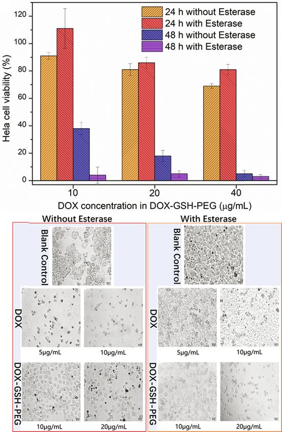
514 Jianxia Qiao et al.
Glutathione (GSH) is an endogenous molecule in the 2.2 Instruments
human body. The GSH molecule could be detected in
tumor cells and normal cells, especially the tumor cells. Vacuum freeze drier (FD-2), Ultraviolet-visible spectro-
The concentration range of GSH in different tumor cells is photometer (UV-Vis; TU-1901), fluoro spectrophotometer
2–20 mM (29,30). GSH-based prodrug nanomedicine is (RF-6000), Fourier transform infrared spectrometer (FT-IR)
probably the better choice for addressing the problem (Tensor 27, Bruker), dynamic light scattering (DLS) (Malvern,
of carriers’ safety in vivo. Fortunately, the carboxyl groups Zetasizer Nano ZSE), clean bench (SW-CJ-2FD), CO2 incubator
in GSH could be reacted with hydroxyl in DOX by east- (CQ-80L), fluorescent inverted microscope (IX53, Olympus),
ernization reaction for constructing esterase-responsive multifunctional enzyme marker (Infinite 200Pro, Tecan), nuclear
polyprodrug. The formation and disruption of the ester magnetic resonance (NMR) (Bruker Avance 400 MHz), transmis-
bond could be triggered under the catalyst of enzymes sion electron microscope (TEM) (FEI Tecnai F20).
(7,31), supporting the application of esterase-responsive
drug-releasing in drug delivery field.
Given the main consideration of carriers’ safety in 2.3 Preparation of hyperbranched
clinical application, the hyperbranched polyprodrug micelle polyprodrug micelle (DOX-GSH-PEG)
was fabricated with Food and Drug Administration (FDA)-
approved material of polyethylene glycol, an endogenous The GSH (127.5 mg) and DCC (186.7 mg) were added to a
molecule of GSH and the anticancer drug of doxorubicin round flask with 10 mL dried DMF at temperature. One
(DOX) by one-pot method. The hydroxyl and amino groups hour later, the DOX·HCl (100 mg) and DMAP (25.3 mg)
in DOX could be reacted with carboxyl groups in GSH to were added to the same flask for reacting for 12 h in the
form enzyme-responsive ester bonds and amido bonds. darkroom. After that, the mPEG-NH2 (172.8 mg) and HOBt
The ultimate product of hyperbranched polyprodrug (9.3 mg) were added to the flask for continuously reacting
(DOX-GSH-PEG) could be assembled into nano micelle for 5 days. The reaction solution was put into centrifuge
in water or PBS and disrupted with a high concentra- tubes and centrifuged at high speed. Then, the superna-
tion of esterase. This endogenous-based strategy with tant was transferred to a dialysis bag (MWCO 3500) and
one-pot synthesis to construct esterase-responsive immersed in a beaker with 200 mL of fresh DMF (change
polyprodrug would probably be a preferred choice fresh DMF 3 times during 3 days). After that, the dialysis
in the future. bag was immersed in water (change fresh water multiple
times). The reaction solution in the dialysis bag was
centrifuged at high speed and the supernatant was fil-
tered by a 0.45 mm membrane. The filtered solution was
2 Materials and methods freeze-dried, and the red powder was obtained. The struc-
ture of DOX-GSH-PEG was confirmed by FT-IR and NMR
2.1 Materials (yield: 40%).
Doxorubicin hydrochloride (DOX·HCl, purity 98%), dicy-
clohexylcarbodiimide (DCC, purity 98%), 4-dimethylami- 2.4 Measurement of polyprodrug micelle’
nopyridine (DMAP, purity 99%), 1-hydroxybenzotrizole particle and stability
(HOBt, purity 97%), and GSH (purity 98%) were pur-
chased from Macklin biocompany (Shanghai, China). One milligram red powder of DOX-GSH-PEG was dissolved
Amino-polyethylene-glycol monomethyl ether (mPEG-NH2, in 3 mL of pure water under sonic conditions. Then, the
purity 95%) was purchased from Yarebio company (Shanghai, DOX-GSH-PEG solution was filtered using a 0.45 mm mem-
China). Esterase (lyophilized powder, ≥15 units‧mg−1 solid) brane and prepared for particle measurement. The 1 mg red
was obtained from Xianding biotech company (Shanghai, powder of DOX-GSH-PEG was dissolved in 12 mL of pure
China). Cell counting Kit-8 (CCK-8), Hoechst 33258 Staining water. Then, 1 mL of micelle solution was put into a vial
Kit, and trypsin cell digestion solution (0.25%) were purchased and 1 mg esterase was put into the same vial to measure
from Beyotime Biotechnology Company (China). Fetal bovine the micelle particles’ change. Another vial with micelle was
serum was purchased from Tianhang biotech company prepared without adding esterase as the control group. The
(Zhejiang, China). All other organic solvents were obtained micelle particles’ stability was evaluated using DLS at dif-
from Fuchen chemical company (Tianjin, China). ferent time points.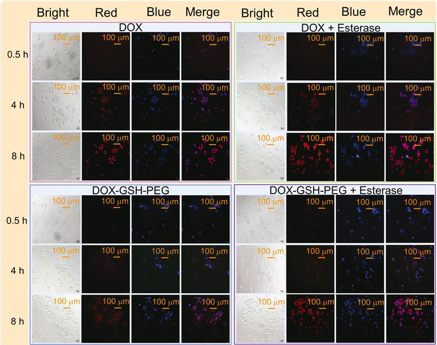
Esterase-responsive hyperbranched polydoxorubicin prodrug 515
2.5 Measurement of drug loading and drug media is discarded. Then, the DOX and DOX-GSH-PEG
release were dissolved into media with 2% fetal bovine serum
and added to 96-well plate. The concentration of DOX
The standard curve of DOX was measured by UV-Vis at in DOX-GSH-PEG is set up as 10, 20, and 40 μg·mL−1,
481 nm. The standard curve equation is A = 0.0186 C. The respectively. The DOX-GSH-PEG group with 7.5 U‧mL−1
UV-Vis curves at 481 nm did not significantly change esterase and without esterase and its control groups
between free DOX and DOX-GSH-PEG. Based on the curves were set up. After the Hela cells were treated with DOX
of free DOX and DOX-GSH-PEG at 481 nm, the approxi- and DOX-GSH-PEG for 24 and 48 h, the media were dis-
mate drug loading could be determined by the standard carded and the fresh PBS (100 μL) with 5 μL CCK-8 was
curve equation. added to the 96-well plate for continuously cultivating for
Three groups of DOX-GSH-PEG (6 mg) were dissolved 1 h. Finally, the 96-well plate was measured by Microplate
into PBS, PBS with 2.5 U‧mL−1 esterase and PBS with Reader at 450 nm.
15 U‧mL−1 esterase, respectively, and put into corre-
sponding dialysis bags (MWCO 3500). Then, the dialysis
bags were put into the flasks and immersed in corre-
sponding media. After that, the fixed volumes (3 mL) 3 Results and discussion
were taken out from flasks and added corresponding
fresh media (PBS with 2.5 U‧mL−1 esterase and PBS with 3.1 Synthesis and characterization of
15 U‧mL−1 esterase) to the flasks at fixed time points for
hyperbranched polyprodrug
72 h. The absorbance of fixed volumes was measured by
UV-Vis and the drug release rate could be calculated and
The synthesis route of GSH-based hyperbranched poly-
determined depending on the standard curves of DOX. The
doxorubicin prodrug is shown in Figure 1a. In Figure 1b,
drug release experiments were conducted twice.
the characteristic peaks of ether bond (1,200 cm−1) in
mPEG-NH2 and DOX-GSH-PEG are observed. The C]O
peaks in DOX, GSH, and DOX-GSH-PEG are shown in
2.6 Cell uptake experiment Figure 1b. The carboxyl groups in GSH could be reacted
with hydroxyl and amino groups in DOX to form a
Hela cells were seeded in a 24-well plate with a density of polymer of DOX-GSH with massive ester bonds and amido
1 × 10⁵ cells per well. Twelve hours later, the media is bonds. The excess carboxyl groups could be on the sur-
discarded. Then, the prepared media of free DOX, DOX face of DOX-GSH for further conjugating with mPEG-NH2
with 7.5 U‧mL−1 esterase, DOX-GSH-PEG, and DOX-GSH-PEG to form the amphiphilic hyperbranched polyprodrug
with 7.5 U‧mL−1 esterase were added to 24-well plate for (DOX-GSH-PEG). The NMR and FT-IR confirmed the struc-
continuously cultivating 30 min, 4 h, and 8 h, respec- ture of DOX-GSH-PEG. We can observe the characteristic
tively. The concentration of DOX in DOX-GSH-PEG and peak of the benzene ring (7.5–8.0 ppm) in DOX-GSH-PEG
free DOX was set up for 20 μg·mL−1. The media with the (Figure 1c) and free DOX (Figure 1d), meaning that the
drug were discarded and the dye solution of Hoechst DOX was conjugated with GSH in DOX-GSH-PEG. The
33258 was added to 24-well for 30 min. After that, the characteristic peak of –CH2CH2O– (3.6 ppm) in PEG can be
cells were rinsed with fresh PBS three times. Finally, the observed in Figure 1c compared with free DOX in Figure 1d.
distributions in cells of DOX and DOX-GSH-PEG were These NMR and FT-IR results fully demonstrated that the
observed under inverted fluorescence microscope. DOX-GSH-PEG was successfully synthesized.
3.2 The particle size and stability of
2.7 Cell viability experiment hyperbranched polyprodrug
The Hela cell viabilities were evaluated by CCK-8 kit The polyprodrug of DOX-GSH-PEG could be assembled
treated by DOX and DOX-GSH-PEG micelle. The detailed into nano micelle in PBS or water. Assembled nano micelle
process: Hela cells were seeded in a 96-well plate with a of DOX-GSH-PEG displayed a distinguished Tyndall phe-
density of 8 × 10³ cells per well. Twelve hours later, the nomenon in pure water (Figure 2a). The particle size was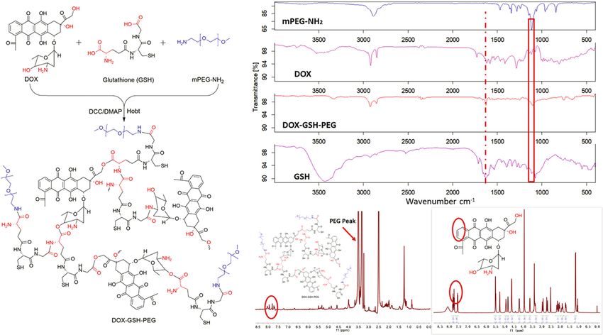
516 Jianxia Qiao et al.
(a) (b)
(c) (d)
Figure 1: (a) Synthesis route of hyperbranched polyprodrug; (b) FT-IR spectra of DOX, GSH, PEG, and DOX-GSH-PEG; NMR spectra of
(c) DOX-GSH-PEG and (d) free DOX in DMSO-d6.
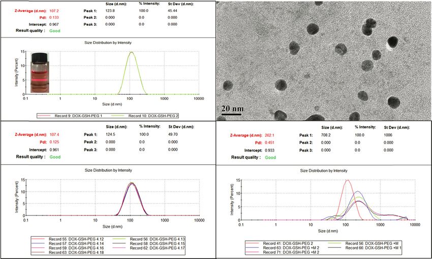
107.2 nm measured by DLS (Figure 2a) and 15 nm mea- 3.3 The drug loading and release properties
sured by TEM (Figure 2b). The significant differences in of hyperbranched polyprodrug
particle results between DLS and TEM are attributed to
the status of hydrophilic PEG. The long chain of PEG could To measure the drug loading of DOX in hyperbranched
be swollen in water, resulting in the bigger particles polyprodrug, we measured the UV-Vis curves of esterase,
measured by DLS. Otherwise, the PEG would be shown DOX, and DOX-GSH-PEG in PBS, discovering that the
in shrinkable status on dried copper-mesh measured by absorbance peaks at 481 nm were observed and not signif-
TEM. These results were consistent with the previous icantly changed in the curves of DOX and DOX-GSH-PEG
paper (32). (Figure 3a). Based on these results, the drug loading of
The stability of DOX-GSH-PEG micelle was evaluated DOX in DOX-GSH-PEG could be determined according to
by DLS. Polyprodrug micelles were measured 7 times by the standard curve of DOX (C = A/0.0186, R2 = 0.9999).
DLS during 1 week in PBS without esterase, showing that The drug loading of DOX in DOX-GSH-PEG is 10.21%. To
the stability of DOX-GSH-PEG micelle was pretty good in further precisely determine the drug release rate in vitro,
non-esterase circumstances (Figure 2c). The statistical we set up three groups of no esterase, low concentration of
particles’ data of DOX-GSH-PEG is 106.4 ± 1 nm (size) esterase (2.5 U‧mL−1), and high concentration of esterase
and 0.145 ± 0.020 (PDI). Conversely, the DOX-GSH-PEG (15 U‧mL−1) to evaluate the esterase-responsive drug release
micelles were responsive-disrupted in PBS with esterase property. Esterase is an overexpressed enzyme with high
circumstances and the average particles’ size signifi- concentration and activity in tumor cells compared with
cantly increased from 108.8 to 262.1 nm with more exten- normal cells (33,34). The DOX-GSH-PEG micelle with mas-
sive polydisperse index during 24 h (Table 1). The increased sive ester bonds could be disrupted in the circumstance of
particle size and more extensive polydisperse index indi- a high concentration of esterase according to previous
cated that the partial ester bonds in hyperbranched poly- enzyme-responsive drug delivery systems (DDS) (7,35,36).
prodrug were disrupted in esterase circumstances. These In Figure 3b, we discovered that the DOXs were slightly
results were consistent with the drug release experiment released from DOX-GSH-PEG micelle under the environ-
in Figure 3b. ment of pH 6.0 during 72 h without esterase and the drug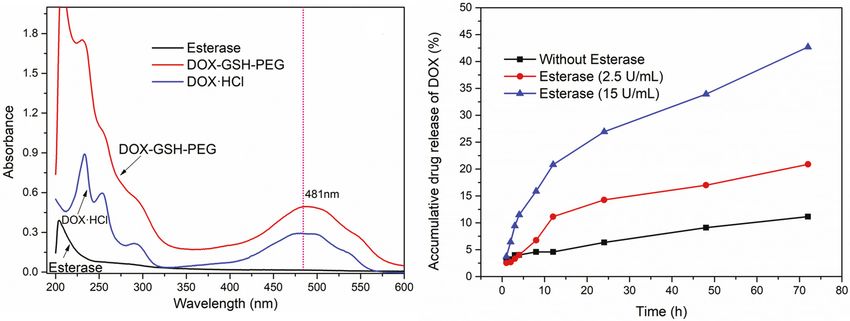
Esterase-responsive hyperbranched polydoxorubicin prodrug 517
(a) (b)
(c) (d)
Figure 2: The particle size of DOX-GSH-PEG micelle measured by (a) DLS and (b) TEM; (c) the stability of DOX-GSH-PEG micelle in PBS during
1 week and (d) the esterase-responsive un-stability of DOX-GSH-PEG micelle in esterase environment.
Table 1: The particle size of DOX-GSH-PEG micelle in esterase 3.4 The distribution of DOX-GSH-PEG in Hela
circumstance cells
Time (h) Z-average size (d.nm) PDI Temperature (°C)
The DOX-GSH-PEG micelle (red fluoresce) could be swal-
0 108.8 ± 44.9 0.141 25.0 lowed by cells via endocytosis and located by inverted
1 187.2 ± 162.7 0.393 25.0 fluorescence microscope. The nuclei were dyed by Hoechst
3 187.6 ± 205.7 0.379 25.0
33258 (blue fluoresce). The free DOX and DOX-GSH-PEG
12 224.7 ± 230.9 0.478 25.0
24 262.1 ± 1006.0 0.451 25.0
micelle were co-cultured with Hela cells for 0.5, 4, and 8 h
in culture media with esterase or without esterase. We
discovered that the distribution of free DOX has no distin-
guishable differences between the with esterase group and
release rate only reached 11%. The experiment group the without esterase group during 8 h (Figure 4a and b).
showed that the drug release rate reached 43% under Meanwhile, the distribution of DOX-GSH-PEG micelle
the high concentration of esterase environment (15 U‧mL−1) groups with esterase or without esterase showed sig-
with pH 6.0 for 72 h and the low concentration of esterase nificant differences. In the merged channel images of
environment (2.5 U‧mL−1) group showed that the 20% DOX DOX-GSH-PEG at the time point of 4 h, we can clearly
was released from DOX-GSH-PEG. These results further indi- notice that the blue fluoresce (cell nuclei) was surrounded
cated that the DOX-GSH-PEG micelle indeed could be dis- by red fluoresce (DOX-GSH-PEG) in the group of without
rupted in the high concentration of an esterase environment. esterase, meaning that most of the DOX-GSH-PEG did not
Combining with the cell viability results of the DOX-GSH-PEG distribute to cell nuclei during 4 h (Figure 4c). In the cor-
group (without esterase) at 24 and 48 h (Figure 5a), we can responding group (4 h) of DOX-GSH-PEG with esterase,
speculate that DOXs were esterase-responsive triggered and the partial red fluoresce overlapped with blue fluoresce,
released from DOX-GSH-PEG in tumor cells with overex- indicating that the partial DOXs were released from
pressed esterase, resulting in the Hela cells apoptosis. DOX-GSH-PEG micelle in esterase circumstance in culture518 Jianxia Qiao et al.
(a) (b)
Figure 3: (a) The UV-Vis curves of esterase, DOX·HCl, and DOX-GSH-PEG in PBS and (b) the accumulative drug release rate of DOX from
DOX-GSH-PEG in PBS (pH = 6.0) with esterase environment.
(a) (b)
(c) (d)
Figure 4: The distributions of (a) free DOX, (b) free DOX with esterase, (c) DOX-GSH-PEG and (d) DOX-GSH-PEG with esterase in culture
media at 0.5, 4, and 8 h (blue fluoresce means Hoechst 33258, red fluoresce means DOX).
media and more quickly distributed into cells’ nuclei. In esterase (Figure 4d), demonstrating that the massive ester
the time point of 8 h groups, the partial blue fluoresce cannot bonds in DOX-GSH-PEG could be disrupted in esterase
be overlapped with red fluoresce in the DOX-GSH-PEG group circumstance and the released free DOX from disrupted
without esterase and the almost blue fluoresce was over- DOX-GSH-PEG distributed more quickly into cell nuclei.
lapped with red fluoresce in DOX-GSH-PEG group with These results further confirmed that the DOX-GSH-PEGEsterase-responsive hyperbranched polydoxorubicin prodrug 519
(a) differences of DOX-GSH-PEG between esterase group and
without esterase group are shown in Figure 5 at the DOX
concentration of 10 mg‧mL−1. This result strongly indicated
that the DOXs were released in an esterase environment
(extra addition of esterase) from DOX-GSH-PEG at 48 h
in vitro, resulting in lower cell viability compared with
without esterase group. The higher concentration of DOX
in DOX-GSH-PEG (20 and 40 mg‧mL−1) between esterase
and without esterase group showed no significant difference
at 48 h. This result should probably be attributed to itself,
i.e., toxicity of the DOX-GSH-PEG in higher concentrations.
The cell viability of DOX-GSH-PEG (including 10 mg‧mL−1
DOX) without esterase group at 48 h was 38%. The lower
cell viability of DOX-GSH-PEG (including 10 mg‧mL−1 DOX)
(b)
without esterase group at 48 h (38%) compared with
DOX-GSH-PEG (including 40 mg‧mL−1 DOX) without
esterase group at 24 h (69%) is probably attributed to
that DOXs were released from DOX-GHS-PEG in Hela cells
with overexpressed esterase for inducing the cell apoptosis
(Figure 5a). To observe the cells’ status, the images were
obtained by microscopy. We can observe that the cells
were almost killed by free DOX at the concentration of
5 and 10 mg‧mL−1 at 48 h. Meanwhile, the massive cells
showed a pretty good status at the DOX concentration of
10 mg‧mL−1 in the DOX-GSH-PEG group without esterase.
Conversely, the shapes of the cells changed when treated
by DOX-GSH-PEG with esterase, indicating that the esterase
in culture media could speed up the disruption of
DOX-GSH-PEG and the released DOXs can result in
Figure 5: (a) The Hela cell viabilities treated with DOX-GSH-PEG
during 24 and 48 h and (b) the cells’ status imaged by different the death of massive Hela cells compared with the
concentrations of DOX and DOX-GSH-PEG with or without esterase group of DOX-GSH-PEG without esterase in culture media
at 48 h (scale: 20×). (Figure 5b).
could be disrupted in a higher esterase environment and
strongly supported the esterase-responsive drug release
properties in vitro (Figure 3b). 4 Conclusion
In this article, an endogenous-based hyperbranched poly-
prodrug was developed using the anticancer drug of DOX,
3.5 Cell viability experiment endogenous molecule of GSH, and FDA-approved material
of PEG by one-pot method. This polyprodrug micelle could
To evaluate the drug efficacy in vitro of esterase-respon- be assembled into micelles with 106.4 ± 1 nm in PBS,
sive DOX-GSH-PEG, we added the esterase to media which can be stable for 1 week. The massive ester bonds
for co-culturing with Hela cells. The cell viabilities of in polyprodrug could be responsively disrupted in a high
DOX-GSH-PEG at 24 h have no significant differences concentration of esterase circumstances. The drug release
between esterase group and without esterase group. experiment showed that the DOX can be released from
The probable reasons are the drug release rate of DOX polyprodrug micelle and reach 43% during 72 h. Cell via-
from DOX-GSH-PEG is 30% and the released DOXs were bility results demonstrated that the group of DOX-GSH-PEG
distributed into cells’ nuclei after 8 h, turning out that with esterase could significantly inhibit the growth of the
cell apoptosis cannot be instantly triggered by released cells compared with no esterase group in 48 h and the
and nuclei-distributed DOX. While the vast cell viability cell uptake experiment showed that the DOX-GSH-PEG520 Jianxia Qiao et al.
could be swallowed by cells and the released DOX from late congestive heart failure in breast cancer survivors aged
DOX-GSH-PEG in esterase environment can be distributed between 50 and 59 at diagnosis: A nationwide study. Breast.
to cell nuclei in 8 h. These in vitro results indicated that 2020;53:125–9. doi: 10.1016/j.breast.2020.07.006.
(4) Chen J, Jiang Z, Zhang YS, Ding J, Chen X. Smart transformable
this hyperbranched polyprodrug can be esterase-respon-
nanoparticles for enhanced tumor theranostics. Appl Phys
sive disrupted in higher esterase environment. This endo- Rev. 2021;8:041321. doi: 10.1063/5.0061530.
genous-based strategy is a better choice for developing (5) Zheng P, Liu Y, Chen J, Xu W, Li G, Ding J. Targeted pH-
novel nanomedicines. responsive polyion complex micelle for controlled intracellular
drug delivery. Chin Chem Lett. 2020;31:1178–82. doi: 10.1016/
j.cclet.2019.12.001.
Acknowledgment: We thank the Central Experiment
(6) Carrillo-Castillo TD, Luna-Velasco A, Zaragoza-Contreras EA,
Platform and Laboratory of Molecular Drug Design and Castro-Carmona JS. Thermosensitive hydrogel for in situ-con-
New Pharmaceutical Preparation in Changzhi Medical trolled methotrexate delivery. e-Polymers. 2021;21:910–20.
College and a teacher called Wenjuan Li in Department doi: 10.1515/epoly-2021-0085.
of Pharmacy. (7) Surnar B, Jayakannan M. Structural engineering of biode-
gradable PCL block copolymer nanoassemblies for enzyme-
controlled drug delivery in cancer cells. ACS Biomater Sci Eng.
Funding information: This work was supported by “1331”
2016;2(11):1926–41. doi: 10.1021/acsbiomaterials.6b00310.
Project in Shanxi Province, Natural Science Foundation (8) Feng X, Xu W, Xu X, Li G, Ding J, Chen X. Cystine proportion
for Young Scientists of Shanxi Province, China (no. regulates fate of polypeptide nanogel as nanocarrier for che-
201901D211473), Scientific and Technological Innovation motherapeutics. Sci China Chem. 2021;64:293–301.
Programs of Higher Education Institutions in Shanxi Province, doi: 10.1007/s11426-020-9884-6.
(9) Zhou N, Zhi Z, Liu D, Wang D, Shao Y, Yan K, et al. Acid-
China (no. 2021L349), Academic leader project of Changzhi
responsive and biologically degradable polyphosphazene
Medical College (no. XSQ202102), and National College nanodrugs for efficient drug delivery. ACS Biomater Sci Eng.
Student’ Innovation and Entrepreneurship Training Plan 2020;6(7):4285–93. doi: 10.1021/acsbiomaterials.0c00378.
Program, China (no. 20210540 and 20210527). (10) Li X, Sun W, Zhang Z, Kang Y, Fan J, Peng X. Red light-triggered
polyethylene glycol deshielding from photolabile cyanine-
Author contributions: Jianxia Qiao and Shufen Li: writing modified mesoporous silica nanoparticles for on-demand drug
release. ACS Appl Bio Mater. 2020;3(11):8084–93.
– original draft; Haoyu Yuan: conducted the experiment;
doi: 10.1021/acsabm.0c01160.
Yujie Wang, Jianhong Li, and Peilong Wang: formal ana- (11) Chen J, Cao H, Li S, Luo L, Yu P, Li M. Research progress of
lysis; Xiao Duan: writing – review and editing, supervi- novel nano-drug delivery systems for cancer treatment. Chin
sion, project administration, and resources. Pharma J. 2020;55:1749–56. doi: 10. 11669/cpj. 2020. 21. 001
(12) Zheng C, Li M, Ding J. Challenges and opportunities of nano-
medicines in clinical translation. Bio Integr. 2021;2:57–60.
Conflict of interest: Authors state no conflict of interest.
doi: 10.15212/bioi-2021-0016.
(13) Wei L, Chen J, Ding J. Sequentially stimuli-responsive antic-
Data availability statement: All data generated or ana- ancer nanomedicines. Nanomedicine. 2021;16(4):261–4.
lysed during this study are included in this published doi: 10.2217/nnm-2021-0019.
article. (14) Liu Y, Liu YQ, Zang J, Isam Abdullah AA, Li Y, Dong H. Design
strategies and applications of ROS-responsive phenylborate
ester-based nanomedicine. ACS Biomater Sci Eng.
2020;6(12):6510–27. doi: 10.1021/acsbiomaterials.0c01190
(15) Zhang Y, Ma S, Liu X, Xu Y, Zhao J, Si X, et al. Supramolecular
References assembled programmable nanomedicine as in situ cancer
vaccine for cancer immunotherapy. Adv Mater.
(1) Furlanetto J, Marmé F, Seiler S, Thode C, Untch M, 2021;33(7):2007293. doi: 10.1002/adma.202007293.
Schmatloch S, et al. Chemotherapy-induced ovarian failure in (16) Wang WQ, Jin YL, Liu X, Chen F, Zheng X, Liu T, et al.
young women with early breast cancer: Prospective analysis of Endogenous stimuli-activatable nanomedicine for immune
four randomised neoadjuvant/adjuvant breast cancer trials. theranostics for cancer. Adv Funct Mater.
Eur J Cancer. 2021;152:193–203. doi: 10.1016/ 2021;31(26):2100386. doi: 10.1002/adfm.202100386.
j.ejca.2021.04.038. (17) Operti MC, Bernhardt A, Grimm S, Engel A, Figdor CG, Tagit O.
(2) Banke A, Fosbøl EL, Ewertz M, Videbæk L, Dahl JS, Poulsen MK, PLGA-based nanomedicines manufacturing: Technologies
et al. Long-term risk of heart failure in breast cancer patients overview and challenges in industrial scale-up. Int J Pharm.
after adjuvant chemotherapy with or without trastuzumab. 2021;605:120807. doi: 10.1016/j.ijpharm.2021.120807.
J Am Coll Cardiol HF. 2019;7:217–24. doi: 10.1016/ (18) Su C, Liu Y, Li R, Wu W, Fawcett JP, Gu J. Absorption, distri-
j.jchf.2018.09.001. bution, metabolism and excretion of the biomaterials used in
(3) Chung IY, Lee JW, Moon HG, Shin KH, Han W, Son BH, et al. nanocarrier drug delivery systems. Adv Drug Deliv Rev.
Effect of standard low-dose anthracycline chemotherapy on 2019;143:97–114. doi: 10.1016/j.addr.2019.06.008.Esterase-responsive hyperbranched polydoxorubicin prodrug 521
(19) Chen S, Jia F, Zhao L, Qiu F, Jiang S, Ji J, et al. Electrospun fiber (28) Zheng P, Ding J. Calcium ion nanomodulators for mitochondria
membrane with asymmetric NO release for the differential targeted multimodal cancer therapy. Asian J Pharm Sci.
regulation of cell growth. Bio-Des Manuf. 2021;4:469–78. 2022;17:1–3. doi: 10.1016/j.ajps.2021.10.004.
doi: 10.1007/s42242-021-00131-w. (29) Roshchupkina GI, Bobko AA, Bratasz A, Reznikov VA,
(20) He Z, Bao K, Zhang J, Ju D, Luo M, Liu L, et al. Multifunctional Kuppusamy P, Khramtsov VV. In vivo EPR measurement of
nanoparticles for targeted delivery of apoptin plasmid in glutathione in tumor-bearing mice using improved disulfide
cancer treatment. e-Polymers. 2022;22:342–56. doi: 10.1515/ biradical probe. Free Radic Biol Med. 2008;45:312–20.
epoly-2022-0020. doi: 10.1016/j.freeradbiomed.2008.04.019.
(21) Schwartz-Duval AS, Wen R, Srivastava I, Moitra P, Pan D. (30) Cheng R, Feng F, Meng F, Deng C, Feijen J, Zhong Z.
A simplistic single-step method for preparing biomimetic Glutathione-responsive nano-vehicles as a promising platform
nanoparticles from endogenous biomaterials. ACS Appl Mater for targeted intracellular drug and gene delivery. J Control Rel.
Interfaces. 2021;13(39):46464–77. doi: 10.1021/ 2011;152:2–12. doi: 10.1016/j.jconrel.2011.01.030.
acsami.1c17302. (31) Xiao D, Jin X, Song Y, Zhang Y, Li X, Wang F. Enzymatic acyla-
(22) Hatakeyama H. Recent advances in endogenous and exo- tion of proanthocyanidin dimers from acacia mearnsii bark:
genous stimuli-responsive nanocarriers for drug delivery and effect on lipophilic and antioxidant properties. J Bioresour
therapeutics. Chem Pharm Bull. 2017;65:612–7. doi: 10.1248/ Bioprod. 2021;6:359–66. doi: 10.1016/j.jobab.2021.03.001.
cpb.c17-00068. (32) Duan X, Bai T, Du J, Kong J. One-pot synthesis of glutathione-
(23) Liao W, Du Y, Zhang C, Pan F, Yao Y, Zhang T, et al. Exosomes: responsive amphiphilic drug self-delivery micelles of doxoru-
the next generation of endogenous nanomaterials for bicin-disulfide-methoxy polyethylene glycol for tumor therapy.
advanced drug delivery and therapy. Acta Biomater. J Mater Chem B. 2018;6:39–43. doi: 10.1039/C7TB02817B.
2019;86:1–14. doi: 10.1016/j.actbio.2018.12.045. (33) Wells A, Grandis JR. Phospholipase C-Gammal in tumor pro-
(24) Raza A, Rasheed T, Nabeel F, Hayat U, Bilal M, Iabal HMN. gression. Clin Exp Metastas. 2003;20(4):285–90.
Endogenous and exogenous stimuli-responsive drug delivery doi: 10.1023/A:1024088922957.
systems for programmed site-specific release. Molecules. (34) Niu R, Jing H, Chen Z, Xu J, Dai J, Yan Z. Differentiating malig-
2019;24:1117. doi: 10.3390/molecules24061117. nant colorectal tumor patients from benign colorectal tumor
(25) Zhang LY, Bi Q, Zhao C, Cheng JY, Cai MH, Chen XY. Recent patients by assaying morning urinary arylsulfatase activity.
advances in biomaterials for the treatment of bone defects. Asia-pac J Clin Onco. 2012;8(4):362–367. doi: 10.1111/j.1743-
Organogenesis. 2020;16(4):113–25. doi: 10.1080/ 7563.2012.01545.x.
15476278.2020.1808428. (35) Aluri R, Jayakannan M. Development of l-tyrosine-based enzyme-
(26) Fenton OS, Olafson KN, Pillai PS, Mitchell MJ, Langer R. responsive amphiphilic poly(ester-urethane) nanocarriers for
Advances in biomaterials for drug delivery. Adv Mater. multiple drug delivery to cancer cells. Biomacromolecules.
2018;30:1705328. doi: 10.1002/adma.201705328. 2017;18(1):189–200. doi: 10.1021/acs.biomac.6b01476.
(27) Zheng P, Ding B, Shi R, Jiang Z, Xu W, Li G, et al. A multichannel (36) Lin FC, Yu Q, Zink JI. Self-contained nanocapsules carrying
Ca2 + nanomodulator for multilevel mitochondrial destruction- anticancer peptides for magnetically activated and enzyme-
mediated cancer therapy. Adv Mater. 2021;33:2007426. cleaved drug delivery. ACS Appl Nano Mater.
doi: 10.1002/adma.202007426. 2021;4(10):10771–83. doi: 10.1021/acsanm.1c02209.You can also read