Correlation between optic disc deformation and retinal vasculature in non pathological high myopia
←
→
Page content transcription
If your browser does not render page correctly, please read the page content below
EXPERIMENTAL AND THERAPEUTIC MEDICINE 21: 380, 2021

           Correlation between optic disc deformation and retinal
               vasculature in non‑pathological high myopia
                                       JIAO SUN, JIALIN WANG and YANLING WANG

                                   Department of Ophthalmology, Beijing Friendship Hospital
                                Affiliated to Capital Medical University, Beijing 100050, P.R. China

                                      Received January 23, 2020; Accepted August 13, 2020

                                                   DOI: 10.3892/etm.2021.9811

Abstract. The aim of the current study was to investigate           particularly in the radial peripapillary capillaries and the
the correlation between optic disc deformation and retinal          deep retinal plexus. Therefore, optic disc deformation may
vasculature in high myopia. A total of 130 eyes with                be used to predict the retinal vasculature in high myopia.
non‑pathological high myopia were included in the current
cross‑sectional study. β‑zone parapapillary atrophy (β‑PPA),        Introduction
optic disc tilt ratio, and horizontal and vertical disc diameters
were analyzed using fundus color photography. A 3x3 mm              Myopia is one of the most commonly reported ocular disor‑
grid and a 4.5x4.5 mm grid were used to scan parafoveal and         ders worldwide, and there has recently been a significant
peripapillary regions, respectively, using optical coherence        increase in the myopic population in China (1). The costs
tomography angiography. Vessel flow density (VFD) and               of examinations and surgical corrections of myopia are
fractal dimension of the retina, as well as the foveal avas‑        significant, and this disorder has been associated with other
cular zone (FAZ), were analyzed and quantified using en face        pathological eye conditions. High myopia is associated with
projection images. Optic disc parameters that were associ‑          reduced retinal perfusion. In fundus photography, retinal
ated with vascular changes were determined using multiple           vessel density and blood flow are markedly reduced in
linear regression analysis. The results from the multivariate       highly myopic eyes (2). Using a dynamic vessel analyzer,
analysis revealed that β‑PPA was negatively correlated with         it was previously demonstrated that there was a narrowing
the VFD of the superficial retinal plexus (R= ‑2.805; P= 0.006),    of retinal large vessels in high myopia (3). Since the retinal
deep retinal plexus (R=‑2.801; P= 0.006), radial parapapillary      microvasculature directly supplies O 2 and nutrients to
capillaries (R=‑3.936; P
2 SUN et al: CORRELATION OF OPTIC DISC DEFORMATION WITH RETINAL VASCULATURE IN MYOPIA
Materials and methods using Bennett's formula (10). Two independent examiners
(JS and JW) reviewed each image. Poor‑quality images were
Participants. The current study was approved by the Beijing excluded based on the following criteria: i) Evidence of poor
Friendship Hospital Affiliated to Capital Medical University fixation, including a double vessel pattern and motion artifacts;
(Beijing, China), and was conducted in accordance with the ii) presence of motion artifacts that could not be corrected by
ethical standards stated in the Declaration of Helsinki and motion correction technology; iii) media opacity, as exhibited
the Health Insurance Portability and Accountability Act. by shadowing or obscuration of the vessel signal in the field of
Written informed consent was obtained from all the exam‑ view or a signal strength index of 21 mmHg or evidence of retinal disease (other than Statistical analysis. Statistical analysis was performed using
myopic degeneration) that affected the retinal or choroidal a commercially available statistical software program (SPSS
vasculature as evidenced by history or examination were for Microsoft; version 24.0; IBM Corp.). Firstly, the mean
excluded. Additionally, eyes that exhibited diffuse retinal and standard deviation of the main outcome parameters
pigment epitheliopathy (RPE) atrophy due to high myopia or were calculated. Following this, a regression analysis using
any structural changes, including myopic choroidal neovascu‑ the angiographic parameters as the dependent variables was
larization, were excluded from the analyses. performed. The parameters that were significantly associated
with the angiographic parameters following the univariate
Image acquisition and analysis. OCTA imaging was analysis were used as independent variables. For all analyses,
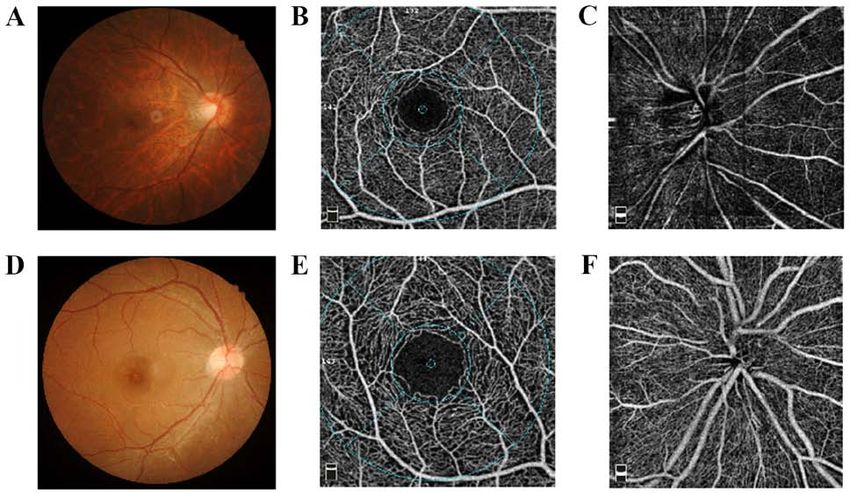
performed using an RTVue XR Avanti system with AngioVue PEXPERIMENTAL AND THERAPEUTIC MEDICINE 21: 380, 2021 3 Table I. Demographic and ocular characteristics of the participants. Characteristic Mean ± SD (n=77) Median (n=77) Range (n=77) Age (years) 35.24±8.45 34.50 20.00‑55.00 Sex Male 37 Female 40 SE (D) 10.03±3.57 8.94 6.15‑20.13 AL (mm) 27.43±1.68 27.28 26.10‑34.46 SP (mmHg) 119.95±11.90 122.00 91.00‑149.00 DP (mmHg) 74.67±8.75 75.00 56.00‑103.00 HR (bpm) 76.11±8.33 76.00 59.00‑93.00 IOP (mmHg) 15.62±3.25 15.15 9.90‑20.80 Optic tilt ratio 1.29±0.18 1.26 0.91‑1.76 Horizontal optic disc diameter (mm) 1.33±0.17 1.33 1.01‑2.04 Vertical optic disc diameter (mm) 1.05±0.19 1.05 0.67‑1.57 β‑PPA (mm2) 1.09±0.62 0.88 0.19‑2.85 RNFL (µm) 95.69±9.49 95.00 72.00‑117.00 C/D 0.28±0.18 0.28 0.03‑0.65 Disc area (mm2) 2.01±0.59 1.93 0.94‑4.38 SE, spherical equivalent; D, diopters; AL, axial length; SP, systolic pressure; DP, diastolic pressure; HR, heart rate; IOP, intraocular pressure; β‑PPA, β‑peripapillary atrophy; RNFL, retinal nerve fiber layer; C/D, cup‑to‑disc area ratio. Figure 1. Image of healthy volunteer and high myopia with tessellated fundus and optic disc deformation. (A) Color fundus photograph, (B) foveal avascular zone and vessel image and (C) image of the radial peripapillary capillaries in the healthy volunteer. (D) Color fundus photograph, (E) foveal avascular zone and vessel image and (F) image of the radial peripapillary capillaries in a patient with high myopia. P= 0.006), nasal (R=‑2.743; P= 0.008) and temporal sectors (R=‑3.667; P
4                       SUN et al: CORRELATION OF OPTIC DISC DEFORMATION WITH RETINAL VASCULATURE IN MYOPIA

Table II. Correlation between age, optic disc deformation, superficial retinal plexus perfusion parameters and FAZ.

                                        Mean                    Temporal                    Superior                     Nasal                    Inferior                      FAZ
	-------------------------------	-------------------------------	-------------------------------	-------------------------------	-------------------------------	------------------------------
Parameter                           R        P‑value           R        P‑value           R        P‑value           R        P‑value           R        P‑value           R        P‑value

Age (years)                     ‑1.271        0.207        ‑1.064         0.291 ‑0.702              0.485       ‑2.116          0.038 ‑1.672               0.099 1.020               0.311
Optic tilt ratio                ‑0.365        0.723        ‑0.377         0.707 ‑0.431              0.668        0.336          0.738 ‑0.750               0.456 ‑0.592              0.556
Horizontal optic                 0.134        0.894         0.162         0.871 0.559               0.578        0.724          0.472 0.245                0.807 0.879               0.382
disc diameter (mm)
AL (mm)                         ‑0.307        0.001        ‑0.346
EXPERIMENTAL AND THERAPEUTIC MEDICINE 21: 380, 2021 5
recruited participants whose SE are ‑10.03±3.57 D and may occur during eye growth within the retina. This may
performed regression analysis separately in each direction result in retinoschisis and subsequently reduce perfusion in
(superior, inferior, nasal and temporal). the macula.
The results demonstrated that the optic disc tilt ratio was There is interplay between genetic factors and envi‑
correlated with the mean and inferior vessel density of the ronmental stressors in the development of myopia. Myopia
DRP. Furthermore, since disc tilt and torsion were signifi‑ is typically exhibited with apparent familial aggregation;
cantly more frequent in the inferior direction, it is possible however, genetic factors alone cannot explain the rapid
that changes in optic disc morphology may be associated increase in the prevalence of myopia over the past one or
with changes in inferior scleral thinning (12). Since it is two generations (23). Bredrup et al (24) reported that the
difficult to precisely measure the true horizontal diameter clinical characteristics of a family with a distinctly excavated
of the optic disc, previous study has evaluated the amount optic disc anomaly exhibited optic nerve dysplasia, high‑grade
of tilt by calculating the ratio between the minimum and myopia and increased ALs. Additionally, genetic analysis
maximum diameters of the nerve, a value termed the index revealed a co‑segregation of this optic disc anomaly with a
of tilt (13). mutation in the MYC‑binding protein 2 gene. Overall, the
In the current study, the superior vessel density of the underlying mechanisms of myopia remain to be elucidated.
DRP became lower as the horizontal disc diameter increased. The current study had certain limitations. Firstly, the
However, Dai et al (14) reported that β‑PPA and γ‑PPA were present study was limited by its cross‑sectional design.
associated with vertical disc diameter, and that the associa‑ Therefore, additional studies that include frequent follow‑ups
tions between β‑PPA or γ‑PPA and horizontal disc diameter of these patients are warranted. Secondly, participants did not
were unclear and not significant. By contrast, Guo et al (15) present with pathological myopia. Thus, further studies are
demonstrated that the horizontal and vertical disc diameters needed.
were positively associated with the enlargement of γ‑PPA. In conclusion, a correlation between optic disc deforma‑
The results of the present study revealed that vessel density tion and retinal vasculature in non‑pathological highly myopic
in the RPC was negatively correlated with β‑PPA. However, eyes was observed using OCTA. This association may explain
Fan et al (5) demonstrated that there were no differences the reduced peripapillary and macular vessel density in high
in vascular density in the optic disc region among the three myopia. According to the results of the present study, disc
groups (control, moderate and high myopia), and vascular deformation (particularly optic disc tilt and β ‑PPA) may
density in the optic disc region was not associated with AL, occur earlier than changes in the macular region in myopia
spherical equivalent or RNFL thickness. retinopathy. Therefore, changes in the optic disc may be early
Furthermore, the current study demonstrated that signs of retinal changes in myopic eyes.
FAZ was not correlated with optic disc deformations.
Wang et al (6) did not identify any differences in the area Acknowledgements
and diameter of the FAZ in healthy Chinese volunteers. This
finding may indicate that the FAZ is not a suitable outcome to Not applicable.
study changes in the microvessel network density of myopic
eyes. Notably, the FAZ area did not significantly change in Funding
response to hyperoxia. However, most of the O2 supplied to
the retina from the FAZ area is derived from the choroidal The current study was supported by the Research Foundation
vessel, rather than from the retinal circulation, which may of Beijing Friendship Hospital Affiliated to Capital Medical
explain the lack of changes in the FAZ area in response to University, Beijing, China (grant no. yyqdkt2019‑29).
hyperoxia (16).
Garg et al (17) reported that choroidal thinning was Availability of data and materials
associated with β‑PPA. The current study demonstrated that
subfoveal choroidal thickness was thinner in eyes with higher The datasets used and/or analyzed during the current study
β ‑PPA than that in eyes with lower β ‑PPA. Furthermore, are available from the corresponding author on reasonable
Wang et al (6) revealed that the density of the macular vascular request.
networks in superior and deep layers and the choriocapillaris
decreased with age. Authors' contributions
β ‑PPA is associated with myopic eyeball axial elongation
and temporal pulling of the optic nerve. The adjacent retinal YW contributed to the acquisition and analysis of data. JW
tissue extends externally, and this mechanical stretching designed the current study and revised the manuscript. JS
results in morphological changes in vessel and tissue thick‑ designed the study, analyzed data and drafted the manuscript.
ness (18). During β ‑PPA, the shape of vessels becomes All authors read and approved the final manuscript.
straighter and thinner, which may affect the vessel flow in the
macular region (19). Furthermore, changes in vessel thick‑ Ethics approval and consent to participate
ness may damage endothelial cells and subsequently reduce
the concentration of VEGF (20,21). Chui et al (22) revealed The current study was approved by the Ethics Committee
that retinal stretching may not mirror scleral growth, and of the Beijing Friendship Hospital (Beijing, China). Written
that there is a difference between the photoreceptor margin informed consent was obtained from all the examined patients
and RPE margin in certain eyes, indicating that slippage and volunteering participants.6 SUN et al: CORRELATION OF OPTIC DISC DEFORMATION WITH RETINAL VASCULATURE IN MYOPIA
Patient consent for publication 11. He J, Chen Q, Yin Y, Zhou H, Fan Y, Zhu JF, Zou HD and
Xu X: Association between retinal microvasculature and optic
disc alterations in high myopia. Eye (Lond) 33: 1494‑1503,
Not applicable. 2019.
12. Ohno‑Matsui K, Lai TY, Lai CC and Cheung CM: Updates of
pathologic myopia. Prog Retin Eye Res 52: 156‑187, 2016.
Competing interests 13. Tay E, Seah SK, Chan SP, Lim AT, Chew SJ, Foster PJ and
Aung T: Optic disk ovality as an index of tilt and its relation‑
The authors declare that they have no competing interests. ship to myopia and perimetry. Am J Ophthalmol 139: 247‑252,
2005.
14. Dai Y, Jonas JB, Huang H, Wang M and Sun X: Microstructure
References of parapapillary atrophy: Beta zone and gamma zone. Invest
Ophthalmol Vis Sci 54: 2013‑2018, 2013.
1. Jonas JB, Xu L, Wei WB, Wang XY, Jiang WJ, Bi HS and Jonas SP: 15. Guo Y, Liu LJ, Tang P, Feng Y, Lv YY, Wu M, Xu L and Jonas JB:
Myopia in China: A population‑based cross‑sectional, histological, Parapapillary gamma zone and progression of myopia in school
and experimental study. Lancet 388 (Suppl 1): S20, 2016. children: The Beijing children eye study. Invest Ophthalmol Vis
2. Shimada N, Ohno‑Matsui K, Harino S, Yoshida T, Yasuzumi K, Sci 59: 1609‑1616, 2018.
Kojima A, Kobayashi K, Futagami S, Tokoro T and Mochizuki M: 16. Xu H, Deng G, Jiang C, Kong X, Yu J and Sun X: Microcirculatory
Reduction of retinal blood flow in high myopia. Graefes Arch responses to hyperoxia in macular and peripapillary regions.
Clin Exp Ophthalmol 242: 284‑288, 2004. Invest Ophthalmol Vis Sci 57: 4464‑4468, 2016.
3. La Spina C, Corvi F, Bandello F and Querques G: Static char‑ 17. Garg A, Blumberg DM, Al‑Aswad LA, Oll M, Yzer S,
acteristics and dynamic functionality of retinal vessels in longer Forbes M, Allikmets RL and Bearelly S: Associations between
eyes with or without pathologic myopia. Graefes Arch Clin Exp β ‑peripapillary atrophy and reticular pseudodrusen in early
Ophthalmol 254: 827‑834, 2016 age‑related macular degeneration. Invest Ophthalmol Vis Sci 58:
4. Li M, Yang Y, Jiang H, Giovanni G, Luiz R, Zheng F, Ke B, Qu DY 2810‑2815, 2017.
and Wang JH: Retinal microvascular network and microcirculation 18. Kwon JW, Choi JA, Kim JS and La TY: Ganglion cell‑inner
assessments in high myopia. Am J Ophthalmol 174: 56‑67, 2017. plexiform layer, peripapillary retinal nerve fiber layer, and
5. Fan H, Chen HY, Ma HJ, Chang Z, Yin HQ, Ng DS, Cheung CY, macular thickness in eyes with myopic β ‑zone parapapillary
Hu S, Xiang X, Tang SB and Li SN: Reduced macular vascular atrophy. J Ophthalmol 2016: 3746791, 2016.
density in myopic eyes. Chin Med J (Engl) 130: 445‑451, 2017. 19. Lee KM, Choung HK, Kim M, Oh S and Kim SH: Positional
6. Wang Q, Chan S, Yang JY, You B, Wang YX, Jonas JB and change of optic nerve head vasculature during axial elongation
Wei WB: Vascular density in retina and choriocapillaris as as evidence of lamina cribrosa shifting: Boramae myopia cohort
measured by optical coherence tomography angiography. Am J study report 2. Ophthalmology 125: 1224‑1233, 2018.
Ophthalmol 168: 95‑109, 2016. 20. Zhang M, Hwang TS, Campbell JP, Bailey ST, Wilson DJ,
7. Samarawickrama C, Mitchell P, Tong L, Gazzard G, Lim L, Huang D and Jia Y: Projection‑resolved optical coherence
Wong TY and Saw SM: Myopia‑related optic disc and tomographic angiography. Biomed Opt Express 7: 816‑828,
retinal changes in adolescent children from Singapore. 2016.
Ophthalmology 118: 2050‑2057, 2011. 21. Landa G and Rosen RB: New patterns of retinal collateral
8. Ohno‑Matsui K: Proposed classification of posterior staphy‑ circulation are exposed by a retinal functional imager (RFI). Br J
lomas based on analyses of eye shape by three‑dimensional Ophthalmol 94: 54‑58, 2010.
magnetic resonance imaging and wide‑field fundus imaging. 22. Chui TY, Zhong Z and Burns SA: The relationship between peri‑
Ophthalmology 121: 1798‑1809, 2014. papillary crescent and axial length: Implications for differential
9. Wang X, Kong X, Jiang C, Li M, Yu J and Sun X: Is the peri‑ eye growth. Vision Res 51: 2132‑2138, 2011.
papillary retinal perfusion related to myopia in healthy eyes? A 23. Cai XB, Shen SR, Chen DF, Zhang Q and Jin ZB: An overview of
prospective comparative study. BMJ Open 6: e010791, 2016. myopia genetics. Exp Eye Res 188: 107778, 2019.
10. Sampson DM, Gong P, An D, Menghini M, Hansen A, 24. Bredrup C, Johansson S, Bindoff LA, Sztromwasser P, Kråkenes J,
Mackey DA, Sampson DD and Chen FK: Axial length variation Mellgren AE, Brurås KR, Lind O, Boman H, Knappskog PM and
impacts on superficial retinal vessel density and foveal avascular Rødahl E: High myopia‑excavated optic disc anomaly associated
zone area measurements using optical coherence tomography with a frameshift mutation in the MYC‑binding protein 2 gene
angiography. Invest Ophthalmol Vis Sci 58: 3065‑3072, 2017. (MYCBP2). Am J Ophthalmol 159: 973‑979.e2, 2015.You can also read