CVA to BPA: Using Balloon Pulmonary Angioplasty to Treat Chronic Thromboembolic Pulmonary Hypertension Post Stroke
←
→
Page content transcription
If your browser does not render page correctly, please read the page content below
ISSN: 2378-2951
                                                                                              Kanda and Prins. Int J Clin Cardiol 2021, 8:229
                                                                                                        DOI: 10.23937/2378-2951/1410229
                                                                                                                           Volume 8 | Issue 3
                   International Journal of                                                                                     Open Access

                   Clinical Cardiology
Case Report

CVA to BPA: Using Balloon Pulmonary Angioplasty to Treat
Chronic Thromboembolic Pulmonary Hypertension Post
Stroke
Adinan Kanda, MD1* and Kurt Prins, MD, PhD2                                                                                     Check for
                                                                                                                                updates
Internal Medicine Department, University of Minnesota, USA
1

Assistant Professor of Medicine, Cardiovascular Division, Lillehei Heart Institute, University of Minnesota, USA
2

*Corresponding author: Adinan Kanda, MD, Internal Medicine Department, University of Minnesota, 401 East River
Parkway, Minneapolis, MN 55455, USA, Tel: 765-409-6575

    Abstract                                                            Learning Objectives
    In patients with CTEPH who are not eligible for PTE, BPA            • To realize the importance of early diagnosis in CTE-
    may be a treatment option. It is important to diagnose and            PH given the association with better outcomes.
    treat patients early since delays are associated with worse
    clinical outcomes. We present a case of CTEPH where ear-            • To highlight that early diagnosis of CTEPH can be
    ly diagnosis and treatment resulted in normalization of PA            challenging.
    pressures.
                                                                        • To recognize BPA as a treatment option for patients
                                                                          not eligible for PTE.
Introduction
    Chronic thromboembolic pulmonary hypertension                       Case Presentation
(CTEPH) is characterized by chronic thromboemboli-                          A 35-year-old male with a history of hereditary sphe-
sm in the proximal pulmonary arteries and small-ves-                    rocytosis with splenectomy fifteen years prior, presen-
sel disease in the pulmonary capillary system [1]. This                 ted to cardiology clinic for evaluation of patent foramen
results in progressive remodeling of the pulmonary                      ovale (PFO), right ventricular enlargement and elevated
vasculature, increased pulmonary vascular resistan-                     right-sided pressures observed on echocardiogram.
ce (PVR), pulmonary hypertension, right ventricular                     Two months prior, he experienced an acute ischemic
remodeling and ultimately right ventricular failure                     stroke, and during the work-up for his CVA an echo-
[2]. Early diagnosis of CTEPH is associated with im-                    cardiogram revealed severe pulmonary hypertension
proved outcomes, however, this can be challenging                       with a right ventricular systolic pressure (RVSP) of 80
since patients may be asymptomatic for many years                       mmHg. His right ventricle (RV) was dilated, had reduced
or present with non-specific symptoms [3,4]. Pulmo-                     systolic function, and there was intraventricular septal
nary thrombo endarterectomy (PTE) is the current                        flattening during systole and diastole consistent with
gold standard treatment for CTEPH butmay not be an                      pressure and volume overload. The study also revea-
option for up to one-third patients. For such patien-                   led a patent PFO. Transesophageal echocardiography
ts, balloon pulmonary angioplasty (BPA) has emerged                     later confirmed the presence of a moderate-sized PFO,
as an efficacious alternative to PEA [5-9]. Here, we                    which was successfully closed percutaneously with a
report the case of a patient with CTEPH who was dia-                    25 mm Gore Cardioform device. Right heart cathete-
gnosed early and treated successfully with BPA.                         rization (RHC) performed at the time of PFO closure
                                                                        showed mean pulmonary artery pressure (mPAP) of 39

                                       Citation: Kanda A, Prins K (2021) CVA to BPA: Using Balloon Pulmonary Angioplasty to Treat Chronic
                                       Thromboembolic Pulmonary Hypertension Post Stroke. Int J Clin Cardiol 8:229. doi.org/10.23937/2378-
                                       2951/1410229
                                       Accepted: May 29, 2021: Published: May 31, 2021
                                       Copyright: © 2021 Kanda A, et al. This is an open-access article distributed under the terms of the
                                       Creative Commons Attribution License, which permits unrestricted use, distribution, and reproduction
                                       in any medium, provided the original author and source are credited.

Kanda and Prins. Int J Clin Cardiol 2021, 8:229                                                                            • Page 1 of 4 •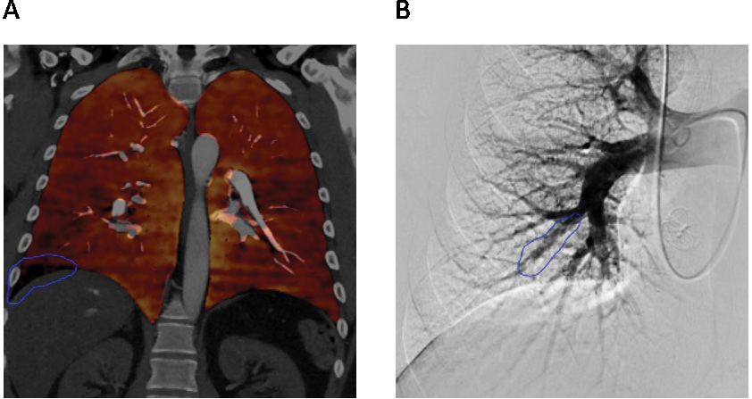
DOI: 10.23937/2378-2951/1410229 ISSN: 2378-2951
Table 1: Values from right heart catheterization (RHC) before and after BPA.
Pre-BPA Post-BPA
Mean Pulmonary Artery Pressure (mPAP) 31 mmHg 21 mmHg
Pulmonary Capillary Wedge pressure (PCWP) 17 mmHg 13 mmHg
Pulmonary Vascular Resistance (PVR) 2.53 Woods Units 1.16 Woods Units
Figure 1: A) CT pulmonary angiogram showing perfusion defect in the right lower lobe (blue outline); B) Invasive pulmonary
angiogram showing chronic obstruction of the A8 branch of the right pulmonary artery.
mmHg, pulmonary capillary wedge pressure (PCWP) of 17 mmHg and PVR of 2.53 Woods Units (Table 1). An
10 mmHg and pulmonary vascular resistance (PVR) of invasive pulmonary angiogram revealed chronic ob-
3 Woods Units. A computed tomography pulmonary struction of the A8 branch of the right pulmonary artery
angiogram (CTPA) revealed possible isolated subseg- with poor filling of distal branches (Figure 1B).
mental right lower lobe pulmonary embolus (PE) and
The patient was not a candidate for pulmonary
a ventilation-perfusion (VQ) scan showed small to mo-
thromboendarterectomy (PTE) since he had an isola-
derate-sized mismatched perfusion defects in the right
ted distal lesion. The decision was therefore made to
lower lobe superior segment and anterior segment right
perform a balloon pulmonary angioplasty (BPA) of the
upper lobe.
A8 branch of the right pulmonary artery. For the proce-
He underwent a repeat RHC about 8 months later, dure, the patient’s right femoral vein was accessed un-
which showed mean pulmonary (mPAP) of 36 mmHg, der ultrasound guidance without any difficulty. A long
PCWP of 17 mmHg and PVR of 3 Woods Units. VQ scan 8 French sheath was then placed the right pulmonary
at that time redemonstrated multiple perfusion defects artery. Using this sheath, a 6 French JR4 guiding cathe-
in the right lung, including the superior segment of the ter was used to engage the right A10 segment and a
right lower lobe. High-resolution computed tomography weblike lesion in the proximal A10 segment was noted.
(CT) showed no evidence of acute or interstitial lung di- A Sion blue wire was used to cross the A10 lesion and
sease. The patient’s cardiologist suspected a diagnosis the lesion was serially dilated using 3.0 × 15 mm, 3.5 ×
of chronic thromboembolic pulmonary hypertension 15 mm and 4.0 × 12 mm emerge balloons. Subsequent-
(CTEPH) and referred the patient to a pulmonary hyper- ly, a 6 French multipurpose guide was used to engage
tension (PH) referral center. At the time of referral, the the right A8 segment. A Sion blue wire was used to cross
patient had minimal limitations with his exercise capabi- the A8 lesion and the lesion was serially dilated using
lities, consistent with World Health Organization (WHO) 3.0 × 15 mm, 3.5 × 15 mm and 4.0 × 12 mm emerge
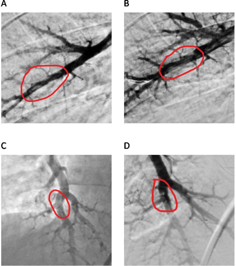
functional class II. balloons with good results (Figure 2A, Figure 2B, Figure
2C and Figure 2D). There were no periprocedural com-
His initial work-up was remarkable for a normal na-
plications.
triuretic peptide (NT-pro-BNP) concentration of 73 pg/
mL. Echocardiography showed mild PH with RVSP of 43 RHC performed one month after BPA showed mPAP
mmHg. LV function and morphology were preserved. of 21 mmHg (decreased from 31 mmHg), PCWP of 13
CTPA showed a perfusion defect in the right lower lobe mmHg (decreased from 17 mmHg) and PVR of 1.16 Wo-
(Figure 1A). RHC showed mPAP of 31 mmHg, PCWP of ods Units (decreased from 2.53 Woods Units) (Table 1).
Kanda and Prins. Int J Clin Cardiol 2021, 8:229 • Page 2 of 4 •
DOI: 10.23937/2378-2951/1410229                                                                           ISSN: 2378-2951

    Figure 2: A) A8 segmental pulmonary artery pre-BPA and B) Post-BPA; C) A10 segmental pulmonary artery pre-BPA
    and D) Post-BPA.

Echocardiography showed normal global RV and LV fun-         cific symptoms [3]. The presence of thromboemboli on
ction. Patient is now WHO functional class 1.                CTPA and mismatched perfusion defects on VQ scan in a
                                                             patient who has received at least 3 months of therapeu-
Discussion                                                   tic anticoagulation is diagnostic of CTEPH [5].
    CTEPH, classified within group 4 PH, is characterized
                                                                 PTE is the current gold standard treatment for CTE-
by chronic thromboembolism in proximal pulmonary
                                                             PH, however, about a third of patients may be deemed
arteries and small-vessel disease involving both the pul-
                                                             ineligible due to high-risk comorbidities, persistent pul-
monary venous and pulmonary arterial capillary systems
                                                             monary hypertension post-PTE or presence of chronic
[1]. These changes result in progressive remodeling of
                                                             thromboembolism in the distal pulmonary arteries [6-
the pulmonary vasculature, increased PVR, pulmonary
                                                             9]. For such patients, BPA has emerged as an efficacious
hypertension, RV remodeling and ultimately RV failure
                                                             alternative to PTE, and has been reported to improve
[2]. CTEPH should be considered in the evaluation of pa-
                                                             symptoms, hemodynamics, exercise capacity and right
tients with pulmonary hypertension since it is treatable
                                                             ventricular function when performed at an expert me-
and potentially curable [8]. It is especially important to
                                                             dical center [6,7]. In a meta-analysis by Kalra, et al. whi-
diagnosis and treat CTEPH earlier in the course of the di-
                                                             ch included a total of 1604 patients (755 patients with
sease, as delays in treatment have been associated with
                                                             inoperable CTEPH treated with BPA; 849 patients with
worse outcomes [4]. Making an early diagnosis of CTE-
                                                             inoperable CTEPH treated with pulmonary vasodilators)
PH, however, can be challenging since patients may be
                                                             across 34 studies, BPA was associated with greater im-
asymptomatic for many years or present with non-spe-

Kanda and Prins. Int J Clin Cardiol 2021, 8:229                                                           • Page 3 of 4 •
DOI: 10.23937/2378-2951/1410229 ISSN: 2378-2951
provement in 6-minute walk distance, PVR and mPAP 4. Held M, Hesse A, Gött F, Holl R, Hubner G, et al. (2014) A
compared to pulmonary vasodilator therapy. BPA was symptom-related monitoring program following pulmonary
embolism for the early detection of CTEPH: A prospective
notably associated with more complications due to its observational registry study. BMC Pulm Med 14: 141.
invasive nature [9].
5. Memon HA, Lin CH, Guha A (2016) Chronic thromboembo-
As discussed above, CTEPH can be difficult to diagno- lic pulmonary hypertension: Pearls and Pitfalls of Diagno-
se earlier in the course of the disease. The patient in sis. Methodist DeBakey Cardiovasc J 12: 199-204.
this case was diagnosed at an early stage as part of the 6. Ogo T (2015) Balloon pulmonary angioplasty for inoperable
work-up for his CVA, which portends better outcomes. chronic thromboembolic pulmonary hypertension. Curr
He had no prior history of PE, similar to 21.2% of patien- Opin Pulm Med 21: 425-431.
ts with CTEPH in a study by Martinez, et al. who were 7. Kim NH, Delcroix M, Jais X, Madani MM, Matsubara H, et
found to have no history of PE [10]. Interestingly, this al. (2019) Chronic thromboembolic pulmonary hyperten-
sion. Eur Respir J 53: 1801915.
patient had a history of splenectomy, which is a known
risk factor for CTEPH [1]. He was not a candidate for PTE 8. Kim NH, Delcroix M, Jenkins DP, Channick R, Dartevelle P,
et al. (2013) Chronic thromboembolic pulmonary hyperten-
since his initial evaluation showed an isolated distal le-
sion. J Am Coll Cardiol 62: D92-D99.
sion. He, instead, underwent BPA which normalized his
PA pressures. 9. Kalra R, Duval S, Thenappan T, Raveendran G, Pritzker M,
et al. (2020) Comparison of balloon pulmonary angioplasty
References and pulmonary vasodilators for inoperable chronic throm-
boembolic pulmonary hypertension: A systematic review
1. (2020) Group 4 Pulmonary Hypertension - Clinical Key. and meta-analysis. Sci Rep 10: 8870.
2. Delcroix M, Noordegraaf AV, Fadel E, Lang I, Simonneau 10. Martinez C, Wallenhorst C, Teal S, Cohen AT, Peacock AJ
G, et al. (2013) Vascular and right ventricular remodelling in (2018) Incidence and risk factors of chronic thromboembo-
chronic thromboembolic pulmonary hypertension. Eur Re- lic pulmonary hypertension following venous thromboem-
spir J 41: 224-232. bolism, a population-based cohort study in England. Pulm
3. Nishiyama KH, Saboo SS, Tanabe Y, Jasinowodolinski D, Circ 8: 2045894018791358.
Landay MJ, et al. (2018) Chronic pulmonary embolism: Dia-
gnosis. Cardiovasc Diagn Ther 8: 253-271.
Kanda and Prins. Int J Clin Cardiol 2021, 8:229 • Page 4 of 4 •You can also read