Design of photoacoustic microscope system based on labVIEW platform
←
→
Page content transcription
If your browser does not render page correctly, please read the page content below
MATEC Web of Conferences 309, 04016 (2020)                              https://doi.org/10.1051/matecconf/202030 904016
CSCNS2019

         Design of photoacoustic microscope system
         based on labVIEW platform
         Yang Bai, Chuncheng Zhang, Lvming Zeng and Guodong Liu*
         Key Lab of Optic-Electronic and Communication, Jiangxi Sciences and Technology Normal University,
         Nanchang 330038, China

                       Keywords: Virtual instrument, Digital oscilloscope, Photoacoustic
                       imaging.

                       Abstract. A photoacoustic microscope system based on virtual instrument
                       development environment is presented, including ultrasonic sensor, digital
                       oscilloscope, laser diode, personal calculation and other hardware platforms.
                       and developed supporting software and image reconstruction algorithms. In
                       the subcutaneous angiography experiment, the distribution characteristics of
                       the ear blood vessels in mouse were completely reproduced perfectly, and
                       the spatial resolution of the system can reach 14um. The system and method
                       can potentially to develop into a non-invasive biological tissue structure and
                       functional imaging technique.

         1 Introduction
             Pure optical imaging has the advantages of fast imaging, non-invasive, high resolution, etc.
         [1,2], but with the increase of light penetration depth, the strong spatial scattering of light
         causes a rapid decrease in imaging spatial resolution; ultrasound imaging has the advantage of
         high-penetration type of tissue. It is easy to cause misdiagnosis because of imaging contrast is
         low for early lesions. The positron emission tomography has an important position in the
         current medical imaging research [3], but due to its complicated equipment, slow imaging
         speed, and existing dyeing. The development of the type of agent is limited, and its promotion
         has received restrictions; MRI has the advantages of less loss lessness and high resolution, and
         can obtain information such as tissue anatomy and physiological functions at the same time
         [4], but its sensitivity is low (micromolar level), and the cost is expensive, so researching and
         developing a low-cost, high-contrast and high-resolution non-destructive medical imaging
         method is a problem to be solved in the field of clinical medicine.
             Photoacoustic imaging is a non-destructive bio-photonic medical imaging technology
         based on the difference in optical absorption between biological tissues and ultrasound [5-8].
         It combines the advantages of high contrast of pure optical imaging and high penetration
         depth of pure blood imaging [9-10]. As one of the important branches of photoacoustic
         imaging, photoacoustic microscopy also has the above advantages, which can realize the

         *   Corresponding author: Alexbaiy@163.com

   © The Authors, published by EDP Sciences. This is an open access article distributed under the terms of the Creative
   Commons Attribution License 4.0 (http://creativecommons.org/licenses/by/4.0/).
MATEC Web of Conferences 309, 04016 (2020)                          https://doi.org/10.1051/matecconf/202030 904016
CSCNS2019

        imaging image of millimeter-scale detection depth and micron-level imaging accuracy
        [11-13], but the existing photoacoustic microscopy system usually uses volume. A large
        solid-state laser or an OPO laser is used as an excitation source for the system and is
        constrained in practical applications [13-15]. In this paper, a "virtual instrument" (VI) is used
        to design a small-scale high-resolution photoacoustic microscopy imaging system. The
        excitation source uses laser diodes with small size, low price and high repetition rate [15-18].
        It was initially applied to the imaging of subcutaneous vascular imaging in mouse ears.

        2 Hardware design
        The whole experimental system is mainly composed of PC, high-definition digital
        oscilloscope and 3D mobile platform. As shown in Figure 1, the main hardware includes: 1.
        Ultrasonic sensor, with a center frequency of 500KHz and a relative bandwidth of 50% at
        -6dB, the relative echo sensitivity is -36.82dB, and the receiving area diameter is 15mm; 2.
        The signal amplifier includes a preamplifier and a main amplifier, where in the bandwidth of
        the main amplifier (Olympus, 5662) is 50K-5MHz, there are 34dB and 54dB two gears; 3,
        The high-definition digital oscilloscope (KEYSIGHT, DSOS054A) has a maximum sampling
        frequency of 20GS/s, a bandwidth of 500 MHz, four analog channels, and a built-in USB
        interface; 4, 3D Mobile platform (Daheng Optoelectronics, GCD-202050M), which uses a
        stepper motor controller (Daheng Optoelectronics, GCD-040101M). The light source is a
        custom diode laser with a repetition rate of 1 kHz. The output wavelength is 650 nm, the pulse
        width is 1 μs, and the pulse energy is 1 μJ; 5. Personal computer.

        Fig. 1. Truss diagram of photoacoustic microscopy system.
            The experimental device of the whole test system is shown in Figure 2. The working
        principle is as follows: The continuous wave laser diode is modulated into a pulse output by
        the drive circuit. When the long pulse laser is microscopically focused by the reflective
        microscope objective and irradiated on the biological tissue, the tissue absorbs the light
        energy to heat up together, and the temperature rise causes thermal expansion to generate a
        photoacoustic signal, which is transmitted to the sensor via the ultrasonic coupling fluid. After
        receiving the photoacoustic signal, the sensor is input to the high-definition digital
        oscilloscope through the pre-amplification, main amplifier and filter. The data is averaged and
        collected by the high-definition digital oscilloscope and transmitted to the computer by the
        USB interface. Finally, data processing and image reconstruction are performed using a

                                                        2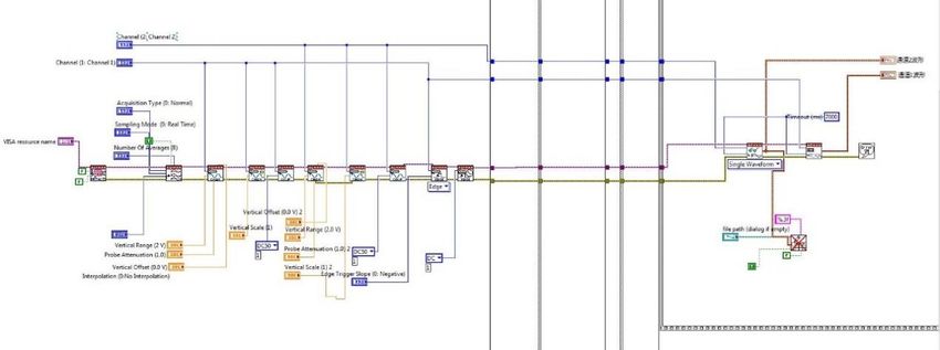
MATEC Web of Conferences 309, 04016 (2020)                         https://doi.org/10.1051/matecconf/202030 904016
CSCNS2019

        program written based on the MATLAB software platform. Among them, each pulsed laser is
        accompanied by a synchronous triggering electrical signal, and each synchronous triggering
        signal is used to trigger a high-definition digital oscilloscope, which will capture the
        photoacoustic signal excited by each laser to the biological tissue. In the experiment, the
        signal acquisition of each point on the sample is called A-Line, and each A-Line signal
        averages 64 times, and 100 sets of data are collected in the X-axis direction and the Y-axis
        direction respectively, which constitutes a photoacoustic microscopy system.
        Two-dimensional data, called raster scanning. Image reconstruction of the raster scan data can
        obtain a two-dimensional image of biological tissue photoacoustic imaging.

        Fig. 2. Photoacoustic imaging system device.

        3 Software design

        3.1 Photoacoustic microscope control program flow chart design

        In order to ensure the continuous operation of the electronically controlled translation stage
        control and realize the parameter configuration function, the core part of the main program is
        implemented by a “for” loop, and the main program is embedded in the sequence structure as
        a whole. Each time the sequence structure is completed, the data acquisition of the XY plane
        is completed. The “for” loop includes five states: Start, Initialize, Acquire, Data Storage, and
        Control Motor. The development process is shown in Figure (3).

        Fig. 3. Development process flow diagram of photoacoustic acquisition system.

                                                        3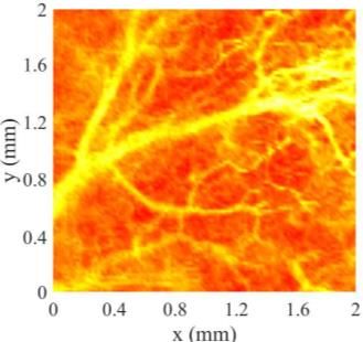
MATEC Web of Conferences 309, 04016 (2020) https://doi.org/10.1051/matecconf/202030 904016
CSCNS2019
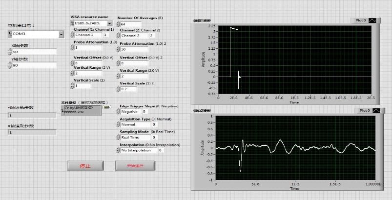
When the sequential structure is running, you need to click the “Start” button on the front
panel to start the acquisition process. The front panel is shown in Figure (4). After starting the
acquisition program, the sequence structure will execute the “Initialize” command to initialize
the configuration of the oscilloscope parameters and read the serial port configuration of the
electronically controlled translation stage. The next step is to execute the “Acquire” and
“Storage” commands, collect and store the data in the front panel waveform graph to the PC,
and then execute the “Move motor” command to control the motor movement to complete a
data acquisition. Then jump to "Acquire" and repeat the command to complete the raster
scanning.
Fig. 4. Front panel photoacoustic signal diagram.
3.2 Photoacoustic microscope control program design
The control program of the photoacoustic microscope consists of two parts: the data
acquisition program and the motor scan control program. The data acquisition program can be
divided into four modules: initial setting, acquisition condition setting, data acquisition and
data storage. The acquisition condition setting module is mainly for setting the parameters of
the high-definition digital oscilloscope, such as selecting the oscilloscope's sampling rate to
be 200MS/s, rising edge triggering, setting the signal acquisition depth, and the average
number of signals. The data acquisition module is shown in Figure 5(a), which is the core of
the data acquisition program. The motor scan control program needs to preset the step speed
and step precision of the motor, and then control the movement of each axis of the motor in the
programmed LabVIEW program. The control program of the motor is shown in Figure 5(b).
4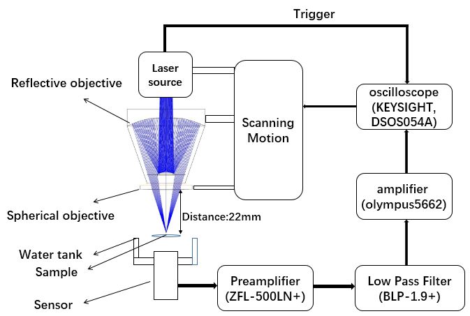
MATEC Web of Conferences 309, 04016 (2020)                           https://doi.org/10.1051/matecconf/202030 904016
CSCNS2019

          (a)

                (b)

        Fig. 5. (a) data acquisition module; (b) motor control program.

        4 Biological experiments
        Blood vessels are an important part of an organism, spread throughout the body, providing
        biological tissue with the nutrients needed to sustain life. In biological organisms, any
        biological tissue lesions can cause changes in vascular structure and intravascular
        composition. Therefore, non-invasive vascular imaging can solve many biomedical problems.
        And has a wide range of research and application value in the field of biomedicine.
            Combined with the data acquisition module developed by LABVIEW software platform,
        the photoacoustic imaging test was performed on the ear blood vessels of mouse, and Fig. 5(a)
        is the photo of the sample. Figure 5(b) is a maximum projection of the photoacoustic
        microscopy of the sample with a scanned imaging area of 2 mm x 2 mm. In the experiment, the
        electronically controlled translation stage drives the laser diode source to have a scanning step
        of 20 μm in the X-Y plane. can be seen from the experimental results that the photoacoustic
        reconstruction images are consistent with the distribution of mouse ear vessels, in which the
        main vessel and microvascular branch structures are clearly imaged, and the surrounding
        subcutaneous capillary network is completely reproduced with an imaging resolution of about
        14 μm. The experiment proved the feasibility of the system to study the biological
        subcutaneous vascular structure and subcutaneous blood vessel distribution imaging.

                                                          5
MATEC Web of Conferences 309, 04016 (2020) https://doi.org/10.1051/matecconf/202030 904016
CSCNS2019
Fig. 5. Photograph of the mouse ear microvasculature and reconstructed images.
5 Conclusion
This paper proposes a small-scale photoacoustic microscopy system based on LabVIEW
2015 virtual instrument development platform. It uses the graphical language LabVIEW
development system software to realize high-resolution mouse ear vascular structure imaging.
The next step is to combine multi-wavelength laser diode excitation, which is expected to
develop into a mark-free, low-cost, miniaturized reflective photoacoustic functional
microscopy technology, providing a real-time monitoring imaging and biometric
identification for subcutaneous microcirculation structures
References
1. Huang Li-na, Yu Xiao-feng, Ding Zhi-hua. Numerical Analysis of Double Pass Rapid
Scanning Optical Delay Line in Optical Coherence Tomography[J]Acta Photonica Sinica,
2005 (11): 1663-1665.
2. Xie Shu-sen Li Hui. Principles and techniques for measuring the optical properties of
biological tissues. [J]. Chinese Journal of Biomedical Engineering, 1997, 16(4):237-332
3. Yang Zi-bin. Review and Prospect on Biomedical engineering [J]. Acta Actademiae
Medicinae Sinicae, 2000, 22(2):280-210.
4. Jackson V P, Hendrick R E, Feig S A, et al. Imaging of the radiographically dense breast.
[J]. Radiology, 1993, 188(2):297-301.
5. Wong T T W, Zhang R, Zhang C, et al. Label-free automated three-dimensional imaging
of whole organs by microtomy-assisted photoacoustic microscopy[J]. Nature
Communications, 2017, 8(1):1386.
6. Murray T W, Haltmeier M, Berer T, et al. Super-resolution photoacoustic microscopy
using blind structured illumination[J]. Optica, 2017, 4(1):17-22.
7. Yao J, Wang L, Yang J M, et al. High-speed label-free functional photoacoustic
microscopy of mouse brain in action[J]. Nature Methods, 2015, 12(5):407-410.
8. Yang X, Jiang B, Song X, et al. Fast axial-scanning photoacoustic microscopy using
tunable acoustic gradient lens[J]. Optics Express, 2017, 25(7):7349-7359.
9. Yang F, Song W, Zhang CL, et al. Broadband graphene-based photoacoustic microscopy
with high sensitivity[J]. Nanoscale, 2018, 10:8606-8614
10. Yang M, Zhao L, He X, et al. Photoacoustic/ultrasound dual imaging of human thyroid
cancers: An initial clinical study[J]. Biomedical Optics Express, 2017, 8(7): 3449-3457.
6MATEC Web of Conferences 309, 04016 (2020) https://doi.org/10.1051/matecconf/202030 904016
CSCNS2019
11. Stylogiannis A, Prade L, Buehler A, et al. Continuous wave laser diodes enable fast
optoacoustic imaging[J]. Photoacoustics, 2018, 9:31-38
12. Liang Y, Jin L, Wang L, et al. Fiber-Laser-Based Ultrasound Sensor for Photoacoustic
Imaging[J]. Scientific Reports, 2017, 7:40849
13. Wang T, Nandy S, Salehi H S, et al. A low-cost photoacoustic microscopy system with a
laser diode excitation[J]. Biomedical Optics Express, 2014, 5(9):3053-3058.
14. Daoudi K, Van d B P J, Rabot O, et al. Handheld probe integrating laser diode and
ultrasound transducer array for ultrasound/photoacoustic dual modality imaging[J].
Optics Express, 2014, 22(21): 26365-26374.
15. Wang P H, Li M L. DVD pickup head based optical resolution photoacoustic
microscopy[J]. Proceedings of SPIE - The International Society for Optical Engineering,
2012, 8223:78.
16. Erfanzadeh M, Kumavor P D, Zhu Q. Laser scanning laser diode photoacoustic
microscopy system[J]. Photoacoustics, 2018, 9:1-9.
17. Beckmann M F, Schwab H M, Schmitz G. Optimized SNR simultaneous multispectral
photoacoustic imaging with laser diodes[J]. Optics Express, 2015, 23(2):1816-1828.
18. Zeng L, Liu G, Yang D, et al. Cost-efficient laser-diode-induced optical-resolution
photoacoustic microscopy for two-dimensional/three-dimensional biomedical
imaging[J]. Journal of Biomedical Optics, 2014, 19(7):076017.
7You can also read