Desmoplastic Spitz nevus - Our Dermatology Online
←
→
Page content transcription
If your browser does not render page correctly, please read the page content below
Our Dermatology Online
Case Report
Desmoplastic Spitz nevus
Vladimír Bartoš
Martin´s Biopsy Center, Ltd., Martin, Slovakia
Corresponding author: Dr. Vladimír Bartoš, PhD., E-mail: vladim.bartos@gmail.com
ABSTRACT
Desmoplastic Spitz nevus (DSN) is an uncommon variant of melanocytic nevus rarely encountered in dermatological
practice. Herein, we describe a 54-year-old male who presented himself with a cutaneous tumor arising from the left
arm. Histology revealed an intradermal proliferation of somewhat pleomorphic, epithelioid, spindled melanocytes in
a background of desmoplastic stroma. A perineural invasion of tumor cells was found. Proliferative and mitotic rates
were minimal. The tumor was diffusely positive for S-100 protein, PNL-2, and SOX-10, and only occasionally reactive
for melan-A and HMB-45. The final diagnosis of DSN was established. Although DSN is a completely benign tumor,
it may result in diagnostic pitfalls. Due to its unusual histopathological features, it may be confused with a malignant
desmoplastic melanoma. A knowledge of the clinicopathological differences between the two prognostically distinct
skin tumor entities is essential for a differential diagnosis.
Key words: Desmoplastic Spitz nevus; Desmoplastic malignant melanoma; Perineural invasion
INTRODUCTION appeared as a light-brown, well-circumscribed, elevated
nodule 6 mm in size. The presumptive clinical diagnosis
Desmoplastic Spitz nevus (DSN) is an uncommon was a benign skin tumor. Total surgical extirpation was
variant of melanocytic nevus characterized by done. Histology revealed an intradermal proliferation
dermal proliferation of large epithelioid or fusiform of somewhat pleomorphic, epithelioid, spindled
melanocytes in a sclerotic stroma [1-5]. It occurs more melanocytes in a background of desmoplastic stroma.
frequently on the limbs of young adults, predominantly No junctional component was present. The lesion was
females, in the third decade of life [1-4]. Compared to symmetrical and had a wedge-shaped configuration,
the classical variants of melanocytic Spitz nevi [6], DSN with the base beneath the epidermis and the apex
exhibits some distinct microscopic features, such as the in the deep dermis. The epithelioid melanocytic
lack of dermoepidermal activity, the absence of Kamino population was mainly located in the superficial portion
bodies, the presence of ganglion-like epithelioid cells, of the tumor mass and exhibited abundant cytoplasm,
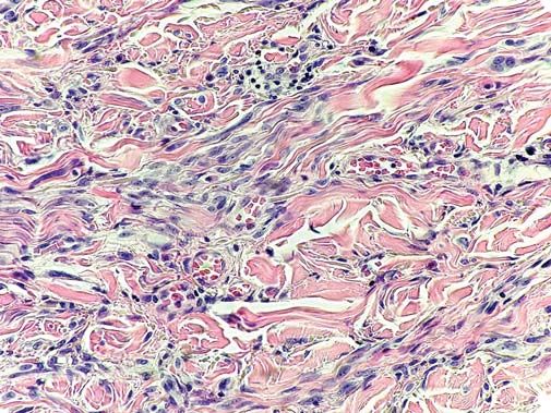
and increased collagen bundles in the dermis [1-5]. large vesicular nuclei with conspicuous nucleoli, and
From a practical point of view, DSN is particularly occasional intranuclear pseudoinclusions (Fig. 1). The
important as it may be confused with a desmoplastic aggregations of melanocytes diminished with the depth
melanoma [1-5]. Therefore, in routine biopsy practice, of the lesion, where clusters of spindled cells were
such cases may be diagnostically challenging. Herein, dispersed among thickened and hyalinized collagen
a case of a patient with DSN is described from bundles (Fig. 2). Interestingly, a perineural propagation
a pathologist’s perspective. was found in the reticular dermis (Fig. 3). Proliferative
activity was minimal (Ki-67 index at ca. 2%) and
CASE REPORT mitoses were only sporadic (in “hot spots,” two mitotic
figures per 1 mm2). Immunohistochemically, the tumor
A 54-year-old male was found to have a cutaneous cell population was diffusely positive for S-100 protein
tumor lesion arising from the left arm. Grossly, it (Fig. 4), PNL-2, and SOX-10, and only occasionally
How to cite this article: Bartoš V. Desmoplastic Spitz nevus. Our Dermatol Online. 2021;12(1):44-46.
Submission: 02.09.2020; Acceptance: 16.11.2020
DOI: 10.7241/ourd.20211.11
© Our Dermatol Online 1.2021 44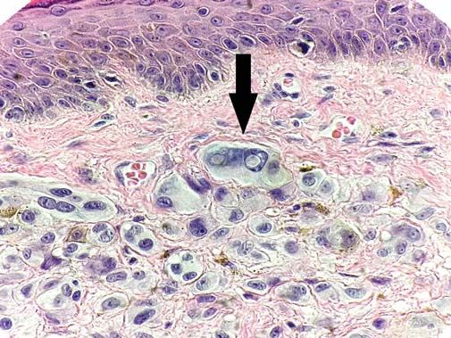
www.odermatol.com
Figure 1: An epithelioid melanocytic population showing nuclear atypia Figure 4: Diffuse immunoreactivity for S-100 protein in the tumor (10×).
with prominent nucleoli and occasional intranuclear pseudoinclusions
(arrow). (H&E, 100×).
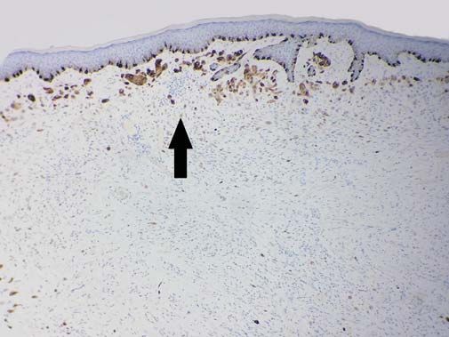
Figure 5: Focal immunoreactivity (arrow) for HMB-45 in the superficial
part of the tumor (40×).
Figure 2: A spindled melanocytic population among thickened collagen
bundles (H&E, 80×). reactive for melan-A and HMB-45 (Fig. 5) in the
superficial part of the lesion. Based on histomorphology
and the immunophenotype, the final diagnosis of
desmoplastic Spitz nevus was established. Resection
margins were free of the tumor.
DISCUSSION
DSN may be a problematic diagnosis in biopsy
practice. As already mentioned, it consists of somewhat
pleomorphic, epithelioid, spindle-shaped melanocytes
distributed among thickened, keloidal-appearing collagen
fibers in the dermis [1-5]. Due to the atypia of the cells,
stromal desmoplasia, and occasional neurotropism, it
may be mistaken for a desmoplastic malignant melanoma
(DMM) and atypical fibrous histiocytoma [1-5,7].
Figure 3: A perineural invasion of tumor cells (arrow) in the deep
Because atypical fibrous histiocytoma is a benign
dermis (H&E, 40×). mesenchymal tumor exhibiting a completely different
© Our Dermatol Online 1.2021 45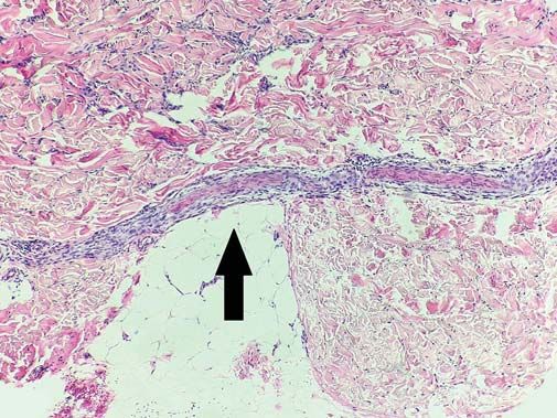
www.odermatol.com
Table 1: Summary of the clinicopathological differences between             CONCLUSION
desmoplastic Spitz nevus and desmoplastic melanoma [1-5,8]
                         Desmoplastic         Desmoplastic
                         Spitz nevus          melanoma                      Desmoplastic Spitz nevus is rarely encountered in
Age                         mean age of 28 yrs    mean age of 65–75         dermatological practice. Although a completely benign
                                                  yrs                       tumor, it may result in diagnostic pitfalls. Due to its
Sex                         female                male predominance
                            predominance
                                                                            unusual histopathological features, it may be confused
Location                    usually extremities   usually head and neck     with a desmoplastic malignant melanoma. A knowledge
Circumscription             usually well-         poorly circumscribed      of the clinicopathological differences between the two
                            circumscribed                                   prognostically distinct skin tumor entities is essential
Symmetry                    present               absent
                                                                            for a differential diagnosis.
Configuration               wedge-shaped          diffusely infiltrative
Junctional component        may be present        may be present
Fascicles of melanocytes    short and discrete    at least some long        Consent
Cytologic atypia            present               present
“Spitzoid” cytomorphology   present               absent                    The examination of the patient was conducted according to the
Intranuclear                common                uncommon                  principles of the Declaration of Helsinki.
pseudoinclusions
Melanocytic maturation      present               absent
Ki-67 index                 low                   usually higher            REFERENCES
Mitotic activity            absent or sporadic    may be low
Solar elastosis             usually absent        common                    1.   Calonje JE, Brenn T, Lazar A, Billings SD (Eds). McKee’s pathology
Lymphocytic aggregations    uncommon              common
                                                                                 of the skin: with clinical correlations. Volume two. 5th edition.
                                                                                 Elsevier; 2020:p.1271-3.
Perineural spreading        may be present        common
                                                                            2.   Massi G, LeBoit PE. Histological diagnosis of nevi and melanoma.
Adnexa involvement          common                uncommon
                                                                                 2nd edition. Springer-Verlag, Berlin, Heidelberg; 2014:p.185-6.
HMB-45 and melan-A          at least focally      often completely
                            positive              negative                  3.   Koc MK, Sudogan S, Kavala M, Kocaturk E, Büyükbabani N,
                                                                                 Altintas S. Desmoplastic spitz naevus can be mistaken for
                                                                                 desmoplastic malignant melanoma and dermatofibroma. Acta Derm
                                                                                 Venereol. 2011;91:74-5.
immunoprofile [7], a strict distinction between DSN and                     4.   Olsen SH, Patel RM, Ma L, Fullen DR. Diffi culties in the
DMM is much more important, as the former represents                             diagnosis of spitzoid melanocytic lesions. Expert Rev Dermatol.
a benign melanocytic lesion, while the latter is an aggressive                   2010;5:549-60.
malignancy with a poor prognosis. Accurate diagnosis of                     5.   Nojavan H, Cribier B, Mehregan DR. Desmoplastic Spitz nevus:
                                                                                 a histopathological review and comparison with desmoplastic
DSN requires an experienced pathologist who will take                            melanoma. Ann Dermatol Venereol. 2009;136:689-95.
into account a combination of clinical, microarchitectural,                 6.   Gundalli S, Kadadavar S, Singhania S, Kolekar R. Histopathological
cytological, and immunohistochemical findings. The                               spectrum of benign melanocytic nevi – our experience in a tertiary
                                                                                 care centre. Our Dermatol Online. 2016;7:21-5.
main clinicopathological differences between DSN                            7.   Dheenadhayalan K, Srinivasan S, Bakthavatsalam S. An unusual
and DMM are summarized in Table 1 [1-5,8]. In our                                misleading multiple nodules on the extremities – a case report. Our
patient, the findings were typical of DSN. Of note                               Dermatol Online. 2016;7:59-61.
                                                                            8.   Adamicova K, Fetisovova Z, Bobrovska M, Homola I. The
was an interesting feature: an apparent perineural                               importance of using SOX10 for exact diagnosis of desmoplastic
spreading of the tumor cells in the deep dermis. This,                           melanoma. Acta Medica Martiniana. 2019;19:111-6.
at first glance worrisome, histopathological finding is                     9.   Xavier-Júnior JCC, Ocanha-Xavier JP, Camilo-Júnior DJ,
                                                                                 et al. Interesting overlooked findings in melanocytic nevi. Surg Exp
generally uncommon in benign tumors. Nevertheless, in                            Pathol. 2019;2:19.
accordance with our observation, some authors [9] have
even described it in benign melanocytic nevi. For this
                                                                             Copyright by Vladimír Bartoš. This is an open access article distributed
reason, it may not necessarily be considered an attribute                    under the terms of the Creative Commons Attribution License, which
of malignancy in atypical melanocytic lesions. In any case,                  permits unrestricted use, distribution, and reproduction in any medium,
this is an adverse prognostic parameter that indicates                       provided the original author and source are credited.
                                                                             Source of Support: Nil, Conflict of Interest: None declared.
a higher risk of local recurrence.

© Our Dermatol Online 1.2021                                                                                                                            46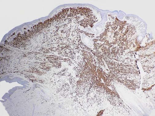
You can also read