Multiple and giant perforating pilomatrixoma: a case report - Our Dermatology Online ...
←
→
Page content transcription
If your browser does not render page correctly, please read the page content below
Our Dermatology Online

                                                                                                                                     Case Report

Multiple and giant perforating pilomatrixoma: a case
report
Sarra Harbaoui, Soumaya Youssef, Imene Ben Lagha, Najla Daadaa, Kahena Jaber,
Mohamed Raouf Dhaoui, Nejib Doss
Department of Dermatology, Military Hospital of Tunis, Tunisia

Corresponding author: Dr. Sarra Harbaoui, E-mail: sarraharbaoui0101@gmail.com

   ABSTRACT

   Pilomatrixoma is benign skin tumor that originates from the pilosebaceous follicle. In most cases it presents as a solitary
   asymptomatic and a firm subcutaneous nodule on the head, neck or upper extremities. Herein, we report a case of
   27-year-old patient who presented with a history of multiple tumors of the upper back, the left arm, the forearms, the
   proximal third of the right thigh and the scalp. The biopsy showed a benign tumoral proliferation on the dermis and
   hypodermis with a transepidermal elimination of shadow cells islands on the perforated lesion. Pilomatrixoma is an
   adnexal skin tumor which may be difficult to diagnose due to different clinical and cytological findings. This diagnosis
   must be evoked in every patient presenting with a firm subcutaneous tumor of the head, neck or upper extremities.
   The histological examination confirms the diagnosis and the treatment is surgical excision.

   Key words: Pilomatrixoma; Multiple; Perforating

INTRODUCTION                                                                    forearms, the proximal third of the right thigh and the
                                                                                scalp. He reported first noticing of these masses on the
Pilomatrixoma, also termed pilomatricoma,                                       left arm, approximately one year earlier, since then, this
trichomatricoma or calcifying epithelioma of Malherbe,                          lesion had rapidly increased in size and other lesions
is a benign skin tumor that originates from the matrix                          had progressively appeared.
of the hair root. It’s a relatively rare condition [1] which
is frequently misdiagnosed clinically and the correct                           He denied any history of trauma, and reported some
diagnosis is only histological [1,2]. This tumor presents                       discomfort in the left arm secondary to the size of
as a solitary asymptomatic, firm, and skin-colored faint                        the lesion in this area. His past medical history was
blue/red nodule in the deep dermis and subcutaneous                             unremarkable. Physical examination revealed a dome-
tissue, with an average size of 0.5 to 3.0 cm [3].                              shaped, polypoïd, stony and movable tumor measuring
However, giant pilomatrixomas (more than 5 cm) have                             10 × 8 × 3 cm, on the distal third of the left arm with
been reported in a few cases [4]. Pilomatrixoma is most                         a blue-red overlying skin showing telangiectatic vessels
commonly seen on the head and neck region followed                              and stretch marks (Fig. 1). Besides, examination showed
by the upper extremities [5]. Although it can be seen                           four painless and freely-movable, firm subcutaneous
in all ages and sexes; it is most often encountered in                          nodules ranging from 0,5 to 2 cm in diameter on
first two decades of life and females [1,4-9].                                  the right upper back, the posterior forearms and
                                                                                the occipital region of the scalp. The overlying skin
CASE REPORT                                                                     was normal. We also found a well-demarcated large
                                                                                area of thick, keloid, blue-red skin, measuring 7cm
A 27-year-old white man, presented with a history of                            in largest diameter and presenting a firm ulcerated,
multiple tumors of the upper back, the left arm, the                            crumbly and spontaneously bleeding tumor on the

How to cite this article: Harbaoui S, Youssef S, Ben Lagha I, Daadaa N, Jaber K, Dhaoui MR, Doss N. Multiple and giant perforating pilomatrixoma: a case
report. Our Dermatol Online. 2018;9(3):299-301.
Submission: 12.11.2017;     Acceptance: 28.01.2018
DOI:10.7241/ourd.20183.18

© Our Dermatol Online 3.2018                                                                                                                               299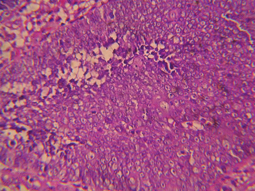
www.odermatol.com
Fig ure 1: Dome-shaped, polypoïd, stony and movable tumor
measuring 10 × 8 × 3 cm, on the distal third of the left arm. Figure 3: Scattered foci and islands of basophilic and shadow cells in
the dermis and hypodermis.
Figure 2: Ulcerated, crumbly and spontaneously bleeding tumor on
the upper back. Figure 4: Trans epidermal elimination of shadow cells islands.
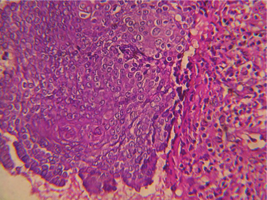
upper back (Fig. 2). Histopathologic examination preponderance in females [1,4,7] and it seems that this
found a benign tumoral proliferation in the dermis tumor occurs usually in Caucasians when compared
and hypodermis which was composed of scattered with Asians and African-Americans [4,8].
foci and islands of basophilic and shadow cells in an
inflammatory stroma with a granulomatous reaction The head and the neck followed by upper extremities are
with foreign body giant cells (Fig. 3). The perforated the most frequent localizations of pilomatrixoma with up
lesion shows a trans epidermal elimination of shadow to 40 % of cases occurring on the head [6,8,10] perhaps
cells islands (Fig. 4). Prior to the study, patient gave due to the higher hair follicle density in the scalp.
written consent to the examination and biopsy after
having been informed about the procedure. Clinically, pilomatrixomas present as solitary, firm
to hard, painless, dermal or subcutaneous nodules
DISCUSSION ranging from 0,5 to 3 cm; although pilomatrixomas
over 10 cm in diameter have rarely been reported in
Pilomatrixoma is a relatively rare benign skin tumor. literature [1,4,10]. Multiple or recurring pilomatrixomas
In fact, it has been reported to be the most common are rarely seen and can be associated with myotonic
cutaneous adnexal tumor in patients younger than dystrophy, Gardner syndrome, Turner syndrome,
20 years [8]. Up to 40% of pilomatrixomas arise before trisomy 9, spina bifida or sarcoidosis [6]. The overlying
the age of 10 years and more than 60% of cases in the skin may be normal or have a reddish or bluish hue in
first two decades [6,8,9]. Most studies report a slight 24% of cases [1,3,8].
© Our Dermatol Online 3.2018 300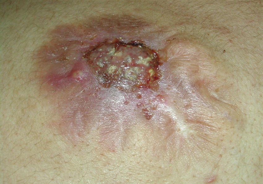
www.odermatol.com

The diagnosis may be reached by physical examination,        REFERENCES
imaging and cytology; however, there is a large number of
                                                             1.  Aydın S, Bilmez ZE, Erdogdu S, Altintoprak N, Kayipmaz Ş.
misdiagnosis [1,3,6]. The differential diagnosis includes        Complicated Giant Pilomatrixoma of the Parotid Region.
dermoid cysts, branchial cleft remnants, pre-auricular           J Maxillofac Oral Surg. 2016;15:111-5.
sinuses, sebaceous cysts, hemangiomas or malignant           2. Piel S, Denisjuk N, Schadendorf D, Dissemond J. Giant
soft tissue tumors [1,3]. Pilomatrixoma do not regress           Pilomatricoma: A Benign Tumor in an Uncommon Presentation.
                                                                 J Pediatr. 2009;154:623.
or disappear spontaneously and transdermal elimination       3. Souto MPA, Matsushita M de M, Matsushita G de M, Souto LRM.
has rarely been reported as perforating or ulcerating            An unusual presentation of giant pilomatrixoma in an adult patient.
pilomatricoma and this was noted in our patient [4].             J Dermatol Case Rep. 2013;7:56-9.
                                                             4. Nadershah M, Alshadwi A, Salama A. Recurrent giant pilomatrixoma
                                                                 of the face: a case report and review of the literature. Case Rep
The present case report is consistent with the published         Dent. 2012;2012:197–273.
literature in terms of incorrect provisional diagnosis,      5. Gupta R, Verma S, Bansal P, Mohta A. Pilomatrixoma of the Arm:
and it’s original in terms of the age of first appearance        A Rare Case with Cytologic Diagnosis. Case Rep Dermatol Med.
                                                                 2012;2012:257405.
of the disease, the number of lesions, their various         6. Schwarz Y, Pitaro J, Waissbluth S, Sam J. Daniel. Review of pediatric
appearance, some atypical localizations [2,3] and the            head and neck pilomatrixoma. Int J Pediatr Otorhinolaryngol.
perforating histological feature.                                2016;85:148-53.
                                                             7. Simi CM, Rajalakshmi T, Correa M. Pilomatricoma: A tumor with
                                                                 hidden depths. Indian J Dermatol Venereol Leprol. 2010;76:543-6.
CONCLUSION                                                   8. Levy J, Ilsar M, Deckel Y, Maly A, Anteby I, Pe’er J. Eyelid
                                                                 pilomatrixoma: A description of 16 cases and a review of the
                                                                 literature. Surv Ophthalmol. 2008;53:526-35.
Pilomatricoma is an adnexal skin tumor which may             9. Topal IO, Singer R, Kocaturk Goncu OE, Topal Y, Yarıkkaya E,
occur at any age especially in the two first decades. This       Sahin IM. An ulcerated giant pilomatricoma mimicking malignancy.
                                                                 J Dtsch Dermatol Ges. 2015;13:329-30.
diagnosis must be evoked in every patient presenting
                                                             10. Kumaran N, Azmy A, Carachi R, Raine PA, Macfarlane JH,
with a firm subcutaneous tumor of the head, neck or              Howatson AG. Pilomatrixoma accuracy of clinical diagnosis.
upper extremities. The histological data confirm the             J Pediatr Surg. 2006;41:1755-8.
diagnosis and the treatment is surgical.

                                                              Copyright by Sarra Harbaoui, et al. This is an open-access article
CONSENT                                                       distributed under the terms of the Creative Commons Attribution License,
                                                              which permits unrestricted use, distribution, and reproduction in any
The examination of the patient was conducted                  medium, provided the original author and source are credited.
                                                              Source of Support: Nil, Conflict of Interest: None declared.
according to the Declaration of Helsinki principles.

© Our Dermatol Online 3.2018                                                                                                         301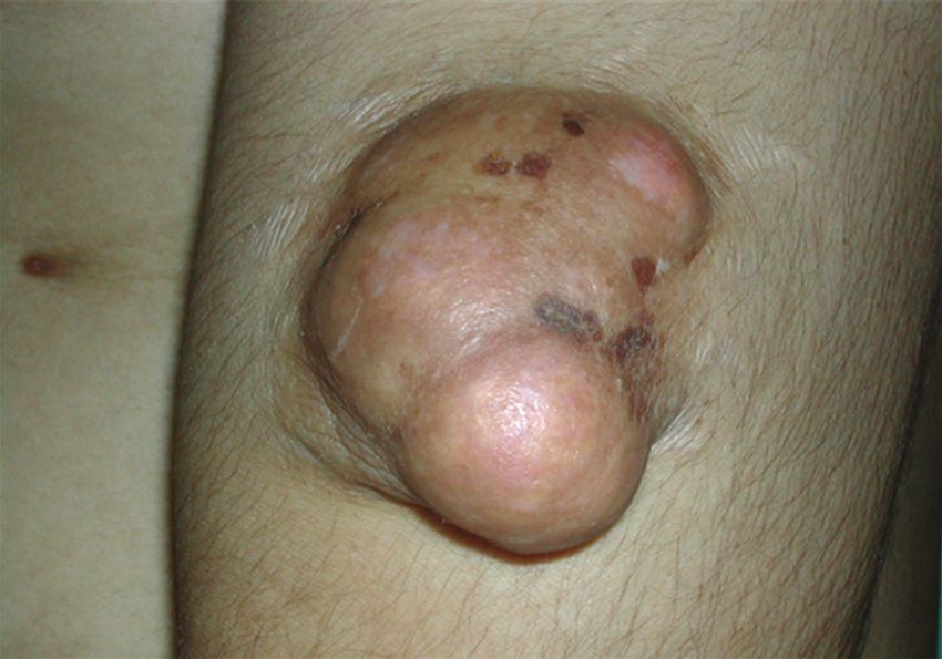
You can also read


