Giant extracranial liposarcoma - Case report
←
→
Page content transcription
If your browser does not render page correctly, please read the page content below
214 V. Pruna et al Giant extracranial liposarcoma
Giant extracranial liposarcoma – Case report
V. Pruna, Narcisa Bucur, Angela Neacsu, A. Giovani,
Anca Buliman, M. Gorgan
First Neurological Clinic, Fourth Department of Neurosurgery
Clinic Emergency Hospital “Bagdasar-Arseni” Bucharest
Abstract series (2). Common sites of occurrence in
Objective: Anaplastic liposarcoma of the the head and neck region include the
head is an extremely rare entity. Seventy- larynx, hypopharynx, oral cavity, orbit, scalp
seven cases of head and neck liposarcomas and soft tissues of the neck. Liposarcomas
have been reported in the world literature rarely arise from a preexisting lipoma.
since 1911. Radical surgery is the form of Liposarcoma tumors are the most
treatment advised. radiosensitive soft-tissue tumors.
Clinical presentation: Authors report the The gross appearance of the tumor
case of a 62 years old female patient depends on the histologic type, degree of
admitted in our institution for a giant vascularity, presence of necrosis, and
extracranial tumor (122/88 mm), developed amount of mature fat and fibrous tissue.
insidious over a period of 3 years and The tumor appears as a smooth, lobulated,
neglected. The patient agreed surgery only or nodular mass, and in most instances, it is
for the epicranial tumor. The lesion was well encapsulated. However, the
completely removed. Postoperatory appearance of an encapsulated tumor may
outcome was excellent concerning this be misleading, because daughter nodules
tumor, although the histopathological result are often present around the main mass.
was not that great: high anaplastic Complete excision is not always possible
liposarcoma. because of the close association of the
Conclusion: Liposarcoma of the scalp is tumor with vital structures; therefore, the
rare. Diagnosis is made histologically. The recurrence rate is high.
histopathologic variant influences clinical
behavior and prognosis. The treatment of Case presentation
choice is wide surgical excision. Authors report the case of a 62 years old
Keywords: giant tumors, anaplastic female patient admitted in our institution
liposarcoma, surgical technique for a giant extracranial tumor, insidiously
developed over a period of 3 years and
Liposarcoma is a malignant neglected. She decided to underwent
mesenchymal neoplasm that arises from surgery for esthetic consideration in first
adipose tissue, most commonly in the place, neurological status being normal.
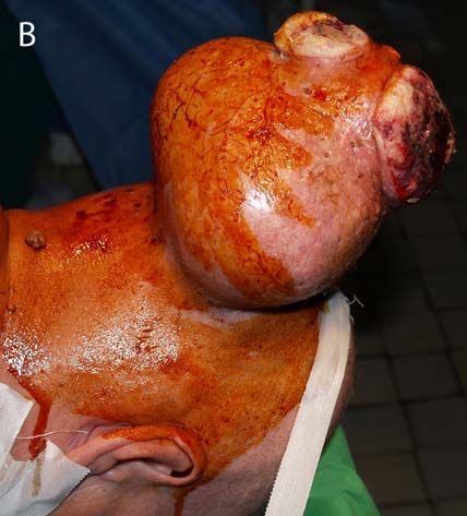
retroperitoneum and lower extremities. A giant epicranial medial-occipital mass
Liposarcoma of the head and neck is rare, (122/88 mm) (Figure 1) and two other
representing 5% to 9% of cases in large masses: left parietal (25/20 mm), and left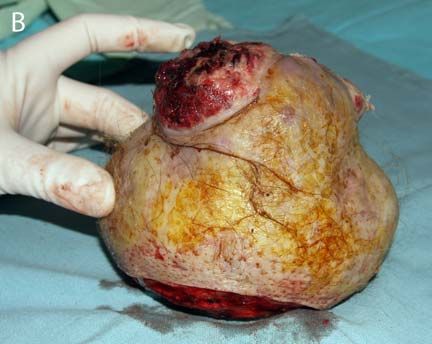
Romanian Neurosurgery (2010) XVII 2: 214 – 218               215

cerebellopontine angle (18/12 mm) was             also    recognizes    a     fifth    variant,
seeing on CT scan (Figure 2). On IRM              dedifferentiated, to describe changes
cerebral the lesions was isodense in T1 and       occurring      within    well-differentiated
T2 weighted (Figure 3).                           liposarcoma that correspond with more
    The patient agreed surgery only for the       aggressive clinical behavior and poor
epicranial tumor, for cosmetic reason. The        outcome (9). Patients with well-
lesion was completely removed; care must          differentiated and myxoid type tumors have
be tacked to avoid excessive scalp removal        higher 5-year survival rates and lower
or potential necrosis, and a good hemostasis      recurrence rates than patients with
was performed for prevent bleeding.               pleomorphic and round-cell types.
    Postoperative outcome was excellent
concerning this tumor, the wound healed
normaly. But the histopathological result
was not that great: high anaplastic
liposarcoma,      with     large    necrotic,
mixomatous and undifferentiated areas
associated with fibrosarcomatous cells.
    The case remains in observation for the
other two intracranial tumors and the
patient referred to oncologist.

Discussion
   Liposarcoma can easily be misdiagnosed
clinically; its relatively indolent course lead                         1A
to often mistakes for lipoma (7), cyst or
benign soft tissue tumors. Nonetheless,
many authors report difficulty in
distinguishing these entities (3) and
therefore histopathology is required for an
appropriate diagnosis (11).
   The histologic characteristics that
distinguish liposarcoma from intramuscular
lipoma include the presence of cellular
pleomorphism, mitotic activity, lipoblasts,
and vascular proliferation (5). Currently,
the      World        Health       Organization
distinguishes the four variants proposed by
Enzinger        and      Weiss     based     on
developmental stage of the lipoblasts and
overall     degree      of    cellularity   and
pleomorphism. These four entities are                                   1B
described as well-differentiated, myxoid,              Figure 1 A, B Preoperative photographs
round-cell and pleomorphic. The WHO
216 V. Pruna et al Giant extracranial liposarcoma
Figure 2 CT brain. A intracranial tumor plated on
the rear face of the left temporal cliff, 18/12 mm; B,
C shows a giant epicranial occipital tumor mass
(122/88 mm), spontaneous homogeneous,
well defined, without reactive
changes of the bone.
2A
3A
2B
3B
2CRomanian Neurosurgery (2010) XVII 2: 214 – 218 217
Figure 3 IRM cerebral shows 2 lesions: left parietal
(25/20 mm) and rear face of the temporal cliff (18/12
mm). A, C, axial T1 weighted image; B, D, axial T2
weighted image; E, sagittal T1 weighted image.
3C 4A
4B
Figure 4 Postoperative pictures. A, CT scan; B,
Postoperative photograph of the lesion.
3D
The incidence of metastasis is also
correlated with histologic type.
Wide surgical excision is the treatment
of choice for liposarcoma. Recurrence rate
increases from 17% to 80% with incomplete
excision (8), as may occur when tumors are
mistakenly believed to be benign lipomas
(4). Although grossly these tumors appear
to be encapsulated, they extend by
infiltration; the likelihood of nearby satellite
nodules necessitates wide excision (1).
Lymph node dissection is not indicated
unless there is concrete evidence of
3E218 V. Pruna et al Giant extracranial liposarcoma
metastasis, since the likelihood of nodal References
metastases in this disease is so rare (9). 1.Collins BT, Gossner G, Martin DS, Boyd JH: Fine
Nonsurgical treatment modalities are of needle aspiration biopsy of well differentiated
liposarcoma of the neck in a young female. A case
limited use in liposarcoma. The use of report. Acta Cytol 43:452-456, 1999.
radiation therapy or chemotherapy remains 2.Enterline HT, Culberson JD, Rochlin DB, Brady LW:
controversial (10). Liposarcoma. A clinical and pathological study of 53
Prognosis of liposarcoma is influenced cases. Cancer 13:932-950, 1960.
3.Evans HL, Soule EH, Winkelmann RK: Atypical
by three factors: histologic variant, lipoma, atypical intramuscular lipoma, and well
adequacy of surgical excision, and location differentiated retroperitoneal liposarcoma: a reappraisal
of the tumor (10). Golledge et al (6) found of 30 cases formerly classified as well differentiated
liposarcoma. Cancer 43:574-584, 1979.
a relatively favorable prognosis for
4.Fusetti M, Silvagni L, Eibenstein A, Chiti-Batelli S,
liposarcoma of the scalp, face and larynx as Hueck S: Myxoid liposarcoma of the oral cavity: case
compared with the oral cavity, pharynx and report and review of the literature. Acta Otolaryngol
neck. The role of tumor size in prognosis is (Stockh) 121:759-762, 2001.
5.Garavaglia J, Gnepp DR: Intramuscular (infiltrating)
unclear. lipoma of the tongue. Oral Surg Oral Med Oral Pathol
63:348-350, 1987.
Conclusion 6.Golledge J, Fisher C, Rhys-Evans PH: Head and neck
liposarcoma. Cancer 76:1051-1058, 1995.
In conclusion, liposarcoma of the scalp is 7.Larson DL, Cohn AM, Estrada RG: Liposarcoma of
rare. Diagnosis is made histologically. The the tongue. J Otolaryngol 5:410-414, 1976.
histopathologic variant influences clinical 8.McCulloch TM, Makielski KH, McNutt MA: Head
and neck liposarcoma. A histopathologic reevaluation of
behavior and prognosis. The treatment of reported cases. Arch Otolaryngol Head Neck Surg
choice is wide surgical excision. The scalp 118:1045-1049, 1992.
region represents a risk factor to the patient 9.Newlands SD, Divi V, Stewart CM: Mixed
myxoid/round cell liposarcoma of the scalp. Am J
because the diagnosis is usually made late.
Otolaryngol 24:121-127, 2003.
10.Pack GT, Pierson JC: Liposarcoma; a study of 105
cases. Surgery 36:687-712, 1954.
11.Wescott WB, Correll RW: Multiple recurrences of a
lesion at the base of the tongue. J Am Dent Assoc
108:231-232, 1984.You can also read