Multiple submucosal lipomas of small intestine: a case report - International ...
←
→
Page content transcription
If your browser does not render page correctly, please read the page content below
International Surgery Journal
Tiwari K et al. Int Surg J. 2020 Sep;7(9):3133-3135
http://www.ijsurgery.com pISSN 2349-3305 | eISSN 2349-2902
DOI: http://dx.doi.org/10.18203/2349-2902.isj20203809
Case Report
Multiple submucosal lipomas of small intestine: a case report
Kritika Tiwari1*, Rhishikesh J. Raghuvanshi1, Anuja Athale1,2, Suresh G. Deshpande1
1
Ruby Hall Clinic, Pune, Maharashtra, India
2
King’s College Hospital, London
Received: 17 June 2020
Accepted: 28 July 2020
*Correspondence:
Dr. Kritika Tiwari,
E-mail: aimgame20@gmail.com
Copyright: © the author(s), publisher and licensee Medip Academy. This is an open-access article distributed under
the terms of the Creative Commons Attribution Non-Commercial License, which permits unrestricted non-commercial
use, distribution, and reproduction in any medium, provided the original work is properly cited.
ABSTRACT
Small bowel obstruction can be due to benign or malignant pathologies. Gastro intestinal lipomas are one of the benign
subepithelial tumours causing obstruction. These are usually detected incidentally if asymptomatic. Adult
intussusception due to intestinal lipoma is a very rare cause. We are presenting a case of male hypertensive patient with
features of multiple subacute obstruction due to multiple submucosal lipomas in ileum. Exploratory laparotomy with
intra-operative enteroscopy was performed and resection-anastomosis of affected segment was done.
Keywords: Submucosal lipoma, Obstruction, Enteroscopy, Multiple submucosal lipoma, Small bowel lipomas
INTRODUCTION diagnosis of multiple submucosal lipomas in resected
segment of ileum.
Submucosal lipomas of small bowel are a rare tumour of
gastrointestinal tract. Gastrointestinal lipomas are benign, CASE REPORT
usually single, slow growing non-epithelial tumors.1 The
common site is the colon, although they may also be found A 54-year-old gentleman presented with features of
in the stomach, oesophagus and small intestine.1,2,3 colicky abdominal pain from last 15 days. Pain was present
in right lower quadrant. It was acute, intermittent, colicky
Submucous lipoma of small intestine which is otherwise and non-radiating with no precipitating factors. It was
silent pathology sometimes clinically manifest as associated with abdominal distention for short period of
gastrointestinal bleeding, intussusception and bowel time. Not associated with fever, melena, vomiting,
obstruction.1,4 jaundice, bleeding per rectum or altered bowel habits.
Asymptomatic lipoma requires only monitoring whereas On examination, vital parameters were found to be in
symptomatic lipoma requires treatment such as endoscopic normal limits. Abdominal examination revealed
or surgical resection. Invasive management is not advised tenderness in right lower quadrant of abdomen with no
unless complications arise such as intussusception, other specific findings. Abdomen was soft with no
obstruction, bleeding or perforation leading to peritonitis.5 organomegaly. Per rectal examination had no obvious
finding.
We present a case of subacute intestinal obstruction with
colicky abdominal pain due to small bowel tumours which On admission, primary investigations were done. X-ray
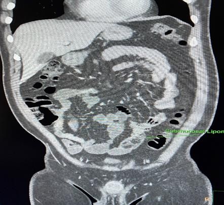
was preoperatively diagnosed by CT scan as submucosal abdomen and ultrasonography were unremarkable.
lipomas. Patient subsequently underwent exploratory However, CT abdomen revealed multiple submucosal
laparotomy with intra-operative enteroscopy for the same. lipomas in ileum, largest measuring 18-19 mm with no
Histopathological examination later confirmed the
International Surgery Journal | September 2020 | Vol 7 | Issue 9 Page 3133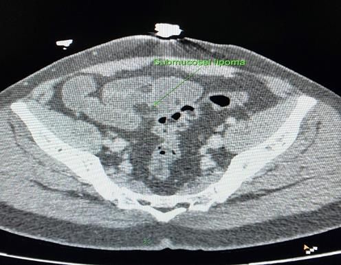
Tiwari K et al. Int Surg J. 2020 Sep;7(9):3133-3135

evidence of intussusception. Rest bowel loops appeared             Resection anastomosis of involved segment of bowel with
unremarkable (Figure 1 and 2).                                     5 cm margin on either side was done. Cut section of
                                                                   resected specimen showed multiple lipomas with intact
                                                                   overlying mucosa, typical of submucosal lesion (Figure 4).

  Figure 1: CT scan showing submucosal lipomas in
                ileum in axial section.
                                                                   Figure 4: Resected segment of ileum cut open to show
                                                                              multiple submucosal lipomas.

                                                                   Histopathological examination of specimen confirmed the
                                                                   diagnosis of benign submucosal lipomas.

                                                                   Post-operative course was uneventful. Patient was
                                                                   discharged and followed up for 3 months and was
                                                                   completely symptom free.

                                                                   DISCUSSION

                                                                   Gastro intestinal lipomas are benign tumours of
                                                                   mesenchymal origin. The incidence of gastro-intestinal
                                                                   lipomas is reported up to 5%. Occurrence is most common
  Figure 2: CT showing multiple lipomas in coronal                 in the colon (65-75%) but they can be found in small
                     section.                                      intestine (20-25%) and very rarely in oesophagus and
                                                                   stomach (less than 5%).6
Patient was planned for exploratory laparotomy with intra-
operative enteroscopy. Intra-operative enteroscopy was             Most lipomas are asymptomatic but sometimes produce
carried out to identify the extent of involved bowel. It           symptoms like gastro-intestinal bleeding, intussusception
showed multiple submucosal lipomas in mid-ileum of                 or obstruction. Large lipomas (more than 2 cm) are most
varying size with largest measuring around 4 cm.                   likely to cause symptoms, so they may be mistaken for
Involvement was limited to mid-ileum and rest of the               malignant lesion. The symptoms of submucosal lipomas
bowel was free from any growth. Mucosal surface was                are not specific and shared with other gastrointestinal
intact and there was no active bleeding (Figure 3).                disease thus the correct diagnosis may be difficult to
                                                                   reach.6

                                                                   Imaging and endoscopic examination contribute to the
                                                                   preoperative diagnosis of intestinal lipomas. CT is the
                                                                   most valuable diagnostic method for intestinal lipomas. It
                                                                   can clearly reveal the typical characteristics of uniform
                                                                   tumour density and fat density, allowing for definite
                                                                   diagnosis. CT scan may also reveal associated
                                                                   intussusception if present.6

                                                                   Asymptomatic lipomas need no treatment. Symptomatic
                                                                   lipoma usually requires surgical intervention. The
                                                                   localised, small, solitary lipoma can be easily and safely
                                                                   removed using endoscope such as endoscopic mucosal
  Figure 3: Intra-operative endoscopy/enteroscopy
                                                                   resection (EMR), unroofing technique, endoscopic
showing multiple submucosal lipomas in small bowel.
                                                                   mucosal resection after pre-cutting (EMR-P), endoscopic

                                                       International Surgery Journal | September 2020 | Vol 7 | Issue 9   Page 3134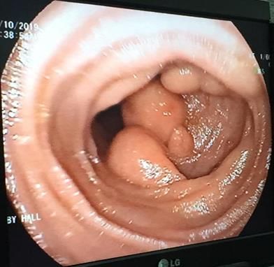
Tiwari K et al. Int Surg J. 2020 Sep;7(9):3133-3135

mucosal dissection (EMD).5 Surgical resection is                   2.     Fernandez MJ, Davis RP, Nora PF. Gastrointestinal
recommended in patients with symptomatic lipoma to                        lipomas. Archives of Surgery. 1983;118(9):1081-3.
relieve symptoms and to exclude malignancy. Surgical               3.     Maderal F, Hunter F, Fuselier G, Gonzales-Rogue P,
treatment has been treatment of choice for large                          Torres O. Gastric lipomas-an update of clinical
submucosal lipomas.                                                       presentation, diagnosis, and treatment. Am J
                                                                          Gastroenterol. 1984;79(12).
CONCLUSION                                                         4.     Botsford TW, Crowe P, Crocker DW. Tumors of the
                                                                          small intestine: a review of experience with 115 cases
Multiple small bowel submucosal lipomas are very rare. It                 including a report of a rare case of malignant
remains undiagnosed if asymptomatic or get diagnosed                      hemangio-endothelioma.           Am        J      Surg.
incidentally. Usually large and multiple lipomas cause                    1962;103(3):358-65.
symptoms. Contrast enhanced CT scan is the investigation           5.     Yoshimoto Y, Yoshida T, Fujikawa T, Shirai Y,
of choice. Although, endoscopy is also very useful.                       Yamamoto T. Novel surgical approach without
Asymptomatic lipomas do not need any intervention                         bowel resection for multiple gastrointestinal
whereas symptomatic submucosal lipomas require                            lipomatosis: a case report. Int J Surg Case Reports.
surgical intervention.                                                    2019;59:54-7.
                                                                   6.     Jiang RD, Zhi XT, Zhang B, Chen ZQ, Li T.
Funding: No funding sources                                               Submucosal lipoma: a rare cause of recurrent
Conflict of interest: None declared                                       intestinal obstruction and intestinal intussusception. J
Ethical approval: Not required                                            Gastrointest Surg. 2015;19(9):1733-5.

REFERENCES                                                              Cite this article as: Tiwari K, Raghuvanshi RJ,
                                                                        Athale A, Deshpande SG. Multiple submucosal
1.   Lee KJ, Kim GH, Shin NR, Lee BE, Ryu DY, Kim                       lipomas of small intestine: a case report. Int Surg J
     DU, Am Song G. Endoscopic resection of                             2020;7(9):3133-5.
     gastrointestinal lipomas: a single-center experience.
     Surgical endoscopy. 2014;28(1):185-92.

                                                       International Surgery Journal | September 2020 | Vol 7 | Issue 9   Page 3135
You can also read