A 70-Year-Old Female with 'Bony' Pain in Her Chest - Journal of Urgent Care Medicine
←
→
Page content transcription
If your browser does not render page correctly, please read the page content below
I N S I G H T S I N I MAG E S
CLINICAL CHALLENGE:
CHALLENGE CASE 1
In each issue, JUCM will challenge your diagnostic acumen with a glimpse of x-rays, electrocardiograms,
and photographs of conditions that real urgent care patients have presented with.
If you would like to submit a case for consideration, please email the relevant materials and
presenting information to editor@jucm.com.
A 70-Year-Old Female with ‘Bony’ Pain
in Her Chest
Figure 1. Figure 2.
Case
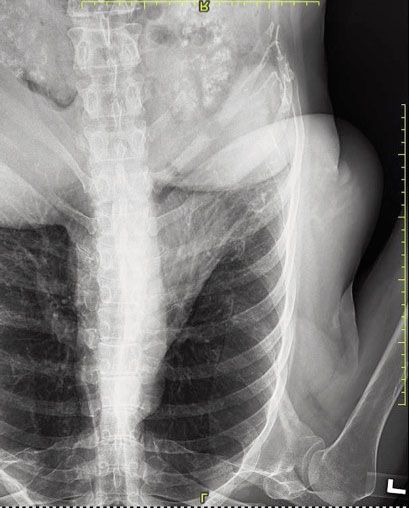
The patient is a 70-year-old female who presents with gradual
onset of constant pain in her left lower chest and back. She
is unsure when she first noticed the pain, but reports that it
has worsened since she experienced a fall several days ago.
She denies shortness of breath, diaphoresis, or an exertional
component.
View the images taken and consider what the diagnosis and
next steps would be. Resolution of the case is described on the
next page.
w w w. j u c m . c o m JUCM T h e J o u r n a l o f U r g e n t C a r e M e d i c i n e | M a y 2 0 1 9 33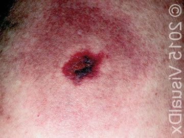
INSIGHTS IN IMAGES: CLINICAL CHALLENGE

 THE RESOLUTION

                                              Figure 3.

     Figure 1.

 Differential Diagnosis                                                                  ! Renal nephrocalcinosis is often asymptomatic but can cause
 !    Bartter syndrome                                                                     renal failure
 !    Dent’s disease                                                                     ! Radiographic findings on plain x-rays reveal abnormal fine
 !    Hyperoxaluria                                                                        granular calcium deposits in the medullary segment of the
 !    Renal manifestation of primary mitochondrial disorders                               kidneys, usually bilateral
 !    Renal nephrocalcinosis                                                             ! Dense deposits can outline the medullary pyramids
                                                                                         ! There may be accompanying renal calculus disease
 Diagnosis                                                                               ! CT findings are more impressive revealing abnormally dense
 The images show bilateral, extensive renal medullary parenchy-                            and calcified medullary segment of the renal parenchyma.
 mal calcifications, which led to a diagnosis of renal nephrocal-                          Ultrasound usually reveals abnormally hyper-echoic medul-
 cinosis—by definition, abnormal deposition of calcium phos-                               lary renal pyramids
 phate and calcium oxalate crystals in the medullary segments
 of the kidneys. This is not a primary disease, but a secondary                          Pearls for Urgent Care Management and
 manifestation of a variety of diseases, many causing hypercal-                          Considerations for Transfer
 cemia. Common primary conditions causing nephrocalcinosis                               ! Treatment for nephrocalcinosis is aimed at alleviating symp-
 include hyperparathyroidism, hypoparathyroidism, hypervita-                               toms and lowering the risk for further build-up of calcium, and
 minosis D, multiple myeloma, prolonged immobilization, sar-                               includes for the primary disease. General management in-
 coidosis, hyper oxaluria, renal tubular acidosis, Alpert syndrome,                        cludes proper hydration, thiazide diuretic and citrate therapy
 Bartter syndrome, sarcoidosis, and other less common causes.
                                                                                         Acknowledgment: Images and case provided by Teleradiology Specialists,
                                                                                         www.teleradiologyspecialists.com.
 Learnings/What to Look for
 ! Renal calculus disease often is concomitantly present and
   may be the presenting feature

34    JUCM T h e J o u r n a l o f U r g e n t C a r e M e d i c i n e | M a y 2 0 1 9                                                                   w w w. j u c m . c o m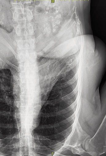
I N S I G H T S             I N     I MAG E S

                         CLINICAL CHALLENGE: CASE 2

   An 18-Year-Old Female with
   Sudden Sharp Chest Pain

   Figure 1.

   Case                                                                       ! Lungs: Clear bilaterally
   The patient is an 18-year-old female who presents to urgent care           ! Cardiovascular: Regular rhythm, without m,r,g
   with 1–2 hours of “sharp” chest pain that worsens with range of            ! Chest: There is point tenderness over the left lower sternal
   motion. She reports it began suddenly while lifting boxes at                  border
   work. Pain is not improved with acetaminophen. She denies                  ! Abdomen: Soft and NT, no distention, without r/r/g
   exertional discomfort, pleuritic pain, and use of hormone therapy.         ! Ext: No edema or asymmetry, pulses are 2+ and equal in
   There is no leg swelling, shortness of breath, or sweating.                   all extremities, no pain with palpation
      Physical exam reveals:                                                  View the ECG and consider what the diagnosis and next
      ! General: Sitting comfortably on the cart, breathing nor-           steps would be. Resolution of the case is described on the
         mally                                                             next page.

w w w. j u c m . c o m                                                  JUCM T h e J o u r n a l o f U r g e n t C a r e M e d i c i n e | M a y 2 0 1 9   35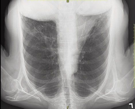
INSIGHTS IN IMAGES: CLINICAL CHALLENGE

 THE RESOLUTION

 Differential Diagnosis                                                                 Learnings/What to Look for
 !   First-degree AV block                                                              ! T wave inversions can be present with ischemia, but manage-
 !   Wolff-Parkinson-White syndrome (WPW)                                                 ment of the patient needs to be based on a clinical history as
 !   Anterior ischemia                                                                    well as the ECG reading
 !   Posterior MI                                                                       ! Juvenile T wave inversions are present with T wave inversion
 !   Persistent juvenile T wave pattern                                                   in leads V1-3 which are asymmetric and less than 3 mm in depth
                                                                                        ! Present commonly in young women of Afro-Caribbean descent
 Diagnosis
 ! This ECG shows normal sinus rhythm with a rate around 80.                            Pearls for Initial Management and Considerations for
   Regarding the possibilities listed in our differential diagnosis                     Transfer
   above, the normal PR interval is 120-200 ms, with first-degree                       ! Perform an ECG in patients suspected of ischemia, syncope,
   AV block being a duration longer than 200 ms (not present on                           and concern for arrhythmia, but not in young patients with an
   this ECG).                                                                             obvious musculoskeletal etiology
 ! WPW is defined by a short PR, a delta wave, and a wide QRS                           ! Compare to an old ECG, if possible
   complex, which is not present here.                                                  ! Young patients can have ischemia, so inquire about exertional
 ! Could this be an anterior ischemia? There is T wave inversion                          pain, vomiting, diaphoresis, and dyspnea as well as risk factors
   anteriorly, but this does not make sense with the clinical picture.                  ! If the T waves are over 3 mm in depth or extend to V4 or fur-
   Additionally, there is no ST elevation.                                                ther, consider ischemia
 ! How about a posterior MI? There is no ST depression in leads                         ! Consider pulmonary embolism in young patients with chest
   V1-3, as well as no clinical story consistent with ischemia or in-                     pain
   farction.                                                                            ! If there is a question about ischemia, err on the side of caution
 ! The correct diagnosis is persistent juvenile T wave pattern,                           and phone consult with cardiology or transfer
   which is a normal variant.                                                           Acknowledgment: Case adapted from Academic Emergency Medicine Education Masters.
                                                                                        MedEDMasters. Persistent juvenile T-waves. Available at: http://www.mededmasters.com/
                                                                                        peristent-juvenile-twi.html. Accessed April 10, 2019.

36   JUCM T h e J o u r n a l o f U r g e n t C a r e M e d i c i n e | M a y 2 0 1 9                                                                    w w w. j u c m . c o m
I N S I G H T S I N I MAG E S
CLINICAL CHALLENGE: CASE 3
A 28-Year-Old Man with Fever,
Diaphoresis, and Nausea
Figure 1.
Case
The patient is a 28-year-old man who presents to urgent care View the photo taken, and consider what your diagnosis
with a single annular ecchymosis lesion on his leg the morning and next steps would be. Resolution of the case is described
after returning home from a trip to visit his family’s cabin in on the next page.
the mountains of North Carolina. He “thought” he noticed a
small insect bite in the vicinity. The lesion has become increas-
ingly painful.
w w w. j u c m . c o m JUCM T h e J o u r n a l o f U r g e n t C a r e M e d i c i n e | M a y 2 0 1 9 37INSIGHTS IN IMAGES: CLINICAL CHALLENGE
THE RESOLUTION
Figure 2.
Differential Diagnosis ! Short of capture or definitive identification, the diagnosis is
! CA-MRSA skin infection clinical
! Brown recluse spider envenomation ! The North American brown recluse (L reclusa) is the most
! Centipede envenoming common species responsible for human injury in the U.S.,
! Coumadin necrosis but deaths are rare
Diagnosis Pearls for Urgent Care Management and
The correct diagnosis is brown recluse spider envenomation. Considerations for Transfer
The brown spiders (Loxosceles species) are found in temperate ! The wound site should be cleaned with soap and water, fol-
and tropical latitudes around the world. They live and build nests lowing by application of a topical antibiotic
in dark areas, either indoors or outdoors. ! Ice may reduce pain and swelling
! Over-the-counter pain relievers may provide further relief
Learnings ! If necrosis develops, patients can later follow up with plastic
! Symptoms of brown recluse spider envenomation include surgery
reddened skin that may be followed by a blister at the site of
the bite Acknowledgment: Images courtesy of VisualDx.
38 JUCM T h e J o u r n a l o f U r g e n t C a r e M e d i c i n e | M a y 2 0 1 9 w w w. j u c m . c o mYou can also read