Detection of Tetracycline tet(k) Gene in Clinical Staphylococcus aureus Isolates - IOPscience
←
→
Page content transcription
If your browser does not render page correctly, please read the page content below

IOP Conference Series: Earth and Environmental Science

PAPER • OPEN ACCESS

Detection of Tetracycline tet(k) Gene in Clinical Staphylococcus aureus
Isolates
To cite this article: K. E. Aziz and Z. F. A. Abdulrahman 2021 IOP Conf. Ser.: Earth Environ. Sci. 761 012128

View the article online for updates and enhancements.

                               This content was downloaded from IP address 46.4.80.155 on 28/07/2021 at 15:06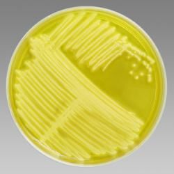
ICCMAT-2021                                                                                                    IOP Publishing
IOP Conf. Series: Earth and Environmental Science 761 (2021) 012128                        doi:10.1088/1755-1315/761/1/012128

Detection of Tetracycline tet(k) Gene in Clinical Staphylococcus
aureus Isolates
                     K. E. Aziz1*and Z. F. A. Abdulrahman2
                     1
                      College of Agricultural Engineering Sciences /Salahaddin University, Iraq
                     2
                      College of Education / Salahaddin University, Iraq
                     *
                     Corresponding author's e-mail: Khalid. Aziz@su.edu.krd.com
                     Abstract. This paper concerned with the identification of S .aureus and detection of tet (k) gene
                     using PCR technique .A total of fifty samples were collected from different clinical sources: 20
                     from burns, (10) surgical wounds, (10) dental carries, and (10) urine samples from patients who
                     were admitted to West Erbil Emergency and Rizgary teaching Hospitals during 1 October to
                     17december 2020. Isolates were identified using conventional method and confirmed by VITEK2
                     compact system. Twenty out of Fifty isolates were identified as Staphylococcus aureus. Disk
                     diffusion method was done for antibiotic resistance, all S. aureus isolates were showed resistance
                     to antibiotic in different percentage: 100% for AMC, AP, AX, and PG, 90% for ME, 65% for
                     CAZ, 65% for TM, 60% for T, 60% for E, 50% for CRO, 45% for CTX, and S, 45% for L, 40%
                     for RA, 40% for DA, 35% for CIP, 35% for C%15 , for KF and 10% for GM while all isolates
                     were sensitive to vancomycin . PCR technique results of partial fragments of tet (K) gene using
                     360bp primer were shown that 7 (35%) of isolates were tetk positive, while 13 (65%) of the
                     isolates were tetk negative.

1. Introduction
  Staphylococcus aureus is a major cause of nosocomial infections and remain a versatile and dangerous
pathogen in human. The fre1quency of both communities acquired and hospital acquired staphylococcal
infections have increased steadily [1]. S. aureus is coagulase-positive, often hemolysis blood and produce
a variety of extracellular enzymes and toxins [2].In humans, infection with S. aureus may cause
suppuration, abscess formation, a variety of pyogenic infections and even fatal septicemia. It can also
cause food intoxication due to elaboration of heat-stable enterotoxin [3;]. Antimicrobial resistance is an
increasing threat afflicting hospitals worldwide [5]. Antimicrobial drug resistance in hospitals is driven by
failures of hospital hygiene, selective pressures created by overuse of antibiotics, and mobile genetic
elements that can encode bacterial resistance mechanisms [6]. Methicillin -resistant S. aureus (MRSA)
isolates were once confined largely to hospitals, other health care environments, and patients frequenting
these facilities [7]. A large number of tetracycline resistance genes have been identified. The main
mechanisms conferring resistance to tetracycline to bacteria are active efflux proteins, ribosomal
protection proteins and enzymatic inactivation. PCR-based molecular methods are often preferred for
determination of antibiotic resistance genes. Therefore, the availability of sensitive and specific methods
for the accurate detection of antibiotic resistance in these multi-drug resistant pathogens has become an
important tool in clinical diagnosis and permits timely implementation of effective antimicrobial therapy,
preventive control strategies, screening of the patient contacts and staff, and appropriate disinfection
measures which in turn reduce the costs [8] .Therefore, this paper concerned with isolation and
identification of S. aureus from different clinical specimen and study both antibiotic resistance pattern
and detection of tet k in S .aureus isolates using PCR technique.

2. Materials and Methods
2.1. Samples collection
Samples were collected from 50 patients who were admitted to West Erbil Emergency, Emergency, and
Rizgary teaching Hospitals during the period from 1 October 2020 to 17december 2020. The age of
patients ranged from one to 45 years. Samples were taken from different sites: burns, surgical wounds,
dental carries and urine. The samples were obtained by rubbing the inflamed or discharged wound, burn,

              Content from this work may be used under the terms of the Creative Commons Attribution 3.0 licence. Any further distribution
              of this work must maintain attribution to the author(s) and the title of the work, journal citation and DOI.
Published under licence by IOP Publishing Ltd                          1
ICCMAT-2021 IOP Publishing
IOP Conf. Series: Earth and Environmental Science 761 (2021) 012128 doi:10.1088/1755-1315/761/1/012128
or decayed teeth by a sterile disposable swabs with normal saline to keep samples fresh while transporting
it to the laboratory for further processing. Urine samples were collected by taking a loop full from the
urine sample and streak it directly on the culture media.
2.2. Identification of the isolates
Identification of these isolates was carried out using microscopically, morphological, biochemical tests
and VITEK2 compact system [ 9]; [10]; [11].
2.3. Antimicrobial susceptibility test (Disk diffusion method)
This test was performed according to Schwalbe et al., [12] Antibiotic impregnated discs with required
concentration were dispensed on the surface of Mueller-Hinton agar medium that has been spread with a
pure bacterial suspension of 105 CFU/ml. After incubation, inhibition zones were measured and translated
into predetermined categories as susceptible, intermediate, or resistant.
2.4. Genomic DNA extraction
A Presto™ Mini gDNA Bacteria Kit was used for genomic DNA extraction from S. aureus isolates. A
loop full of bacteria were incubated over night in a tube containing LB broth. The kit’s instructions was
followed carefully to obtain a good DNA extracts. The purity of extracted DNA was between 1.8-2.0.
2.5.Detection of tet(k) gene in S. aureus clinical isolates
The standard PCR assay was performed using the DNA amplification instrument Master cycler gradient
(Eppendorf, Germany) to detect tetk gene. The tetk - specific primer pairs used for amplification of 360
base pair (bp) fragment are: Forward, 5’-GTAGCGACAATAGGTAATAGT-3’and Reverse, 5’-
GTAGTGACAATAAACCTCCTA-3’ (Bühlmann et al., 2008).A volume of 20µl deionized distilled
water (ddH2O), 1.3 µl Reverse primer, 1.3µl Forward primer and 2.5 µl of extracted DNA (template) was
added to the ready to use PCR reagent tube (Bioneer, South Korea) which contains the following (for the
20µl reaction): 1U Top DNA polymerase, 250 µM of each: dNTP (dATP, dCTP, dGTP, dTTP),10
mMTris-HCl (pH 9.0),30 mMKCl,1.5 mM MgCl2, Stabilizer and tracking dye. The thermal cycling
protocol for PCR was comprised as described by [13]:1.Initial denaturation at 950C for 3 minute. 2. Thirty
three cycles of: Denaturation at 950C for 30sec, annealing at 540C for 30 seconds, elongation at 720C for
30sec and final extension at 720C for 4 minutes.
2.6. Detection of PCR products
About 5µl of the amplified products were visualized by electrophoresis in 1.5% agarose gels stained with
ethidium bromide under UV trans illuminator and photographed. The amplicon (PCR product) generated
from S. aureus gene sequences by this PCR method was a DNA fragment of 360 bp length. Therefore, a
positive PCR test should yield a 360 bp DNA fragment which appeared as an intense band on an ethidium
bromide stained agarose gel. The molecular size of the band was verified by comparing its migration to
that of a DNA marker (100bp DNA ladder) run on the same gel. A negative PCR product did not produce
any visible band on the gel [13].
3. Results and Discussion
3.1. Isolation and Identification of S. aureus
twenty isolates were identified among 50 different clinical specimens including: 20 burn swab, 10 wound
swab, 10 urine and 10 from dental carries. The identification of the isolates were carried out using
conventional method based on cultural characteristics, cell morphology, Gram stain reaction biochemical
properties and VITEK2 compact system. All isolates were able to grow on mannitol salt agar (selective
media for Staphylococcus) (figure 1). S. aureus has the ability to change the color of the media from
Pink-Orange to yellow, because it can ferment the mannitol which present in the medium that leads to
change in the color [14]. circular, smooth, yellow to golden colonies raised on blood agar with various
degrees of hemolysis (mostly beta hemolysis). Prepared smears of S. aureus isolates appeared as purple
single, diplo, and grape like Gram positive cocci under light microscope. All isolates were positive for
catalase, coagulase and DNase. All isolates were identified as S. aureus by VITEK2 compact system with
over 85% probability percentage.
2ICCMAT-2021 IOP Publishing
IOP Conf. Series: Earth and Environmental Science 761 (2021) 012128 doi:10.1088/1755-1315/761/1/012128
Figure 1. Staphylococcus aureus on mannitol salt agar
3.2. Antibiotic resistance pattern of S. aureus isolates
Antibiotic Sensitivity test by disk diffusion method for 50 isolates of S. aureus was done against 20
commonly used antibiotics (AMC, AP, AX, C, CAZ, CIP, CRO, CTX, DA, E, GM, KF, L, ME, PG, RA,
S, T, TM, VA). The resistance percentage of S. aureus isolates varied for different antibiotics used in this
study as shown in table (1).The results revealed that the resistance was 100% for AMC, AP, AX, and PG
and 90% for ME. Resistance percentage for other antibiotics were 65% for CAZ, 65% for TM, 60% for T,
60% for E, 50% for CRO, 45% for CTX, and S, 45% for L, 40% for RA, 40% for DA, 35% for CIP, 35%
for C,15% for KF, and 10 % for GM while all isolates were sensitive for vancomycin. Our results are in
agreement with that of [15] that found that S. aureus isolates from patients with urinary tract infections
were highly resistant to ampicillin and amoxicillin. The results of [16] showed that (77%) of isolates were
oxacillin-resistant Staphylococcus aureus and exhibited multiple resistances to other tested antibiotics
which is close to our results.[17] reported that the resistance patterns of S. aureus were: for levofloxacin
(20 %), for norfloxacin (16 %), for ofloxacin (18 %), for ciprofloxacin (16 %), for levofloxacin (14%)
and for nalidixic acid (50%), while the results of [18] revealed that (8.10%) of S. aureus isolates were
amikacin resistant, (100%) of isolates were amoxicillin resistant ,(86.48 %) of isolates were ampicillin
resistant, (54.05 %) were resistant to (cephotaxim, erythromycin, tetracycline), (21.62%) of isolates were
methecillin resistant (MRSA), and (10.81%) were vancomycin resistant. [19] found that S. aureus isolates
from Africa were: 54% resistant for trimethoprim, 21% for sulfamethoxazole and 19% for
trimethoprim/sulfamethoxazole, while [20] reported that a total of 94 cases from 2010 to 2012 were
diagnosed to have S. aureus infection using conventional bacteriologic methods. From these cases, 38
(40.6%) were identified as MRSA and 37 (39.4%) were inducible clindamycin resistant. In England,
surveillance of surgical site infections has been running since 1997. During the 5 year period between
January 2003 and December 2007, at least one causative microorganism was reported for 77% of surgical
site infections. The most common organism was S. aureus (accounting for 38% of surgical site
infections), of which 64% were MRSA. However, between October 2008 and September 2009, the
proportion of S. aureus isolates (accounting for 31% of surgical site infections) that were methicillin
resistant decreased to 32%. This decrease in surgical site infections due to MRSA in England appears to
mirror the decline in MRSA bacteremia [21]. The antimicrobial agents are losing their efficacy because of
the spread of resistant organisms due to indiscriminate use of antibiotics, lack of awareness, patient
noncompliance and unhygienic condition. It is the need of the time that antibiotic policies should be
formulated and implemented to resist and overcome this emerging problem. Every effort should be made
to prevent spread of resistant organisms. There are multiple factors, which contribute to the global spread
of resistance. Decreasing unnecessary antibiotic use, with narrow spectrum agents, improving compliance
3ICCMAT-2021 IOP Publishing
IOP Conf. Series: Earth and Environmental Science 761 (2021) 012128 doi:10.1088/1755-1315/761/1/012128
with therapy, decrease in use of antibiotic in animal and agriculture, and improving infection control all
have a role in confronting this problem [22].
Table 1. Resistance percentage of S. aureus to antibiotics.
Antimicrobial agent Antibiotics % of resistance No. of resistant isolates
Amoxicillin AMC 100 20
Amoxicillin+ clavulanic acid AP 100 20
Ampicillin AX 100 20
Cefotaxime C 35 7
Ceftazidime CAZ 65 13
Ceftriaxone CIP 35 7
Cephalothin CRO 50 10
Chloramphenicol CTX 45 9
Ciprofloxacin DA 40 8
Clindamycin E 60 12
Erythromycin GM 10 2
Gentamicin KF 15 3
Lincomycin L 45 9
Methicillin ME 90 18
Penicillin G PG 100 20
Rifampin RA 40 8
Streptomycin S 45 9
Tetracyclin T 60 12
Trimethoprim TM 65 13
Vancomycin VA 0 0
3.3. Detection of tet(k) in S.aureus isolates
All isolates were analyzed by PCR to detect the presence of tet(k) gene using forward and reverse primers
described by [13]. The results showed that 7 isolates (35%) were harboring the tet(k) gene (360 bp), while
13 isolate (65%) were lacking the tet(k) gene(figure 1). These results shows contrast with the results of
disc diffusion method for tetracycline, 65% of isolates were resistant to tetracycline while only 35% of it
were harboring tet(k) gene. This may be due to that the resistance may conferred by other class of
tetracycline gene like tet (K), tet(M), tet(O) and tet(L) that not detected in our study. Our results agree
with that obtained by [13] Duran et al.,(2012) who reported 36% of MRSA were positive for tet(k) gene.
In contrast to our results [23] Adwan et al., (2014) reported that 76% isolate of methicillin resistant S.
aureus were tet(k) positive. [24] found that only 58 (44.61%) from total 130 S. aureus isolates showed
tet(k) gene positive.
A large number of tetracycline resistance genes have been identified. There are 38 acquired tetracycline
resistance genes that are known and all use one of three strategies to render the bacteria resistant. These
include (1) efflux proteins, (2) ribosomal protection proteins and (3) enzymatic inactivation of
tetracycline.The majority of these genes (60 %) code for energy-dependent efflux pumps, and different
bacterial genera tend to have the same efflux or ribosomal protection genes [25]. This indicates that
tetracycline resistance genes can be transferred amongst the bacterial population. In fact, resistance to
tetracycline in most bacteria is due to the acquisition of new genes; these genes tend to be associated with
mobile elements such as transposons and plasmids [26].
4ICCMAT-2021 IOP Publishing
IOP Conf. Series: Earth and Environmental Science 761 (2021) 012128 doi:10.1088/1755-1315/761/1/012128
Figure 2: PCR gene product of tetK gene
Tetacycline resistance genes: tetK, tetM, tetO and tetL are four major genes associated with
tetracycline resistance amongst Gram positive bacteria. The tetK and tetL genes code for efflux proteins;
these are energy dependent membrane-associated proteins which prevent tetracycline from accumulating
within the cell [25]. The other two genes, tetM and tetO, code for ribosomal protection proteins, which
reduce the affinity of tetracycline to the ribosome [27].
Our study confirms the usefulness of PCR assay for the detection of antibiotic resistance genes
associated with S. aureus infections. The PCR assay offers a rapid, simple, and accurate identification of
antibiotic resistance profiles and could be used in clinical diagnosis as well as for the surveillance of the
spread of antibiotic resistance determinants in epidemiological studies. Classical methods and molecular
approaches especially PCR based techniques were more effective when used together and could provide
more accurate and reliable information. Laboratory methods used to detect multidrug resistant bacteria
such as MRSA should have high sensitivity and specificity.
4. Conclusion
In this study we can conclude the resistance was 100% for Amoxicillin, Amoxicillin+ clavulanic acid,
Ampicillin and PG, 90% for ME while all isolates were sensitive to vancomycin. Results also indicated
that 7 (35%) of isolates were tet(k) gene positive.
References
[1] Lowy, FD 1998, Staphylococcus aureus infections. The New England Journal of Medicine, 339:
520–532.
[2] Brooks, GF, Butel, JS, Morse, SA and Jawetz, E 2001, Jawetz, Melnick & Adelberg's Medical
Microbiology. 14th ed. McGraw-Hill Companies. USA.
[3] Collee, JG, Fraser, AG, Marmion, BP and Simmons, A 1996, Mackie and McCartney, Practical
Medical Microbiology. 14th ed. Churchil living stone. New York, USA.
5ICCMAT-2021 IOP Publishing
IOP Conf. Series: Earth and Environmental Science 761 (2021) 012128 doi:10.1088/1755-1315/761/1/012128
[4] Kloos, WE and Bannerman, TL 1999, Staphylococcus and micrococcus. in: manual of clinical
microbiology. Murray PR, Baron EJ, Pfaller MA, Tenover FC and Yolken RH (Eds.).
Massachusetts Avenue, Washington, 274- 276.
[5] Graffunder, EM and Venezia, RA 2002, Risk factors associated with nosocomial methicillin –
resistant Staphylococcus aureus (MRSA) infection including previous use of antimicrobials. J.
Antimicrob. Chemother, 49, 999-1005 .
[6] Weinstein, RA 2001, Controlling antimicrobial resistance in hospitals: infection control and use of
antibiotics. Emerg. Infect. Dis, 7(2), 188-192.
[7] David, MZ, and Daum, RS 2010, Community-associated methicillin-resistant staphylococcus
aureus: epidemiology and clinical consequences of an emerging epidemic. Clinical Microbiology
Reviews, 23(3), 616–687.
[8] Woodford, N and Sundsfjord 2005, Molecular detection of antibiotic 9. resistance: when and
where? J Antimicrob Chemother, 56, 259-61.
[9] Forbes, BA, Sahm, DF and Weissfeld, AS 2007, Bailey and Scott’s Diagnostic Microbiology. 12th
ed. Mosby Elsevier. China., 842-855.
[10] Winn, WC and Koneman EW 2006, Koneman's Color Atlas and Textbook of Diagnostic
Microbiology. 6th ed. Lippincott Williams & Wilkins. Washington DC, USA.
[11] Goldman, E and Green, LH 2009, Practical Handbook of Microbiology. 2nd ed. CRC Press,
Taylor and Francis Group. California, USA.
[12] Schwalbe, R, Moore, SL and Goodwin AC 2007, Antimicrobial Susceptibility testing protocols.
CRC Taylor and Francis Group, Boca Raton London. New York.
[13] Duran, N, Burcin, O, Gulay, G., Duran, YO and Cemil, D 2012, Antibiotic resistance genes &
susceptibility patterns in staphylococci. Indian Journal Medical Research, 135, 389-396.
[14] Morello, JA, Granato, PA and Mizer, HE 2003, Laboratory Manual and Workbook in
Microbiology Applications to Patient Care. 7th ed., McGraw-Hill company. New york, USA.
[15] Al-Jebouri, MM and Mdish, SA 2013, Antibiotic resistance pattern of bacteria isolated from
patients of urinary tract infections in Iraq. Open Journal of Urology, 3(2), 8-14.
[16] AL-Marjani, MF, Kadhim, KA, Kadhim, AA, Ibraheem, AS and Kinani, AY 2015, Ciprofloxacin
resistance in Staphylococcus aureus and Pseudomonas aeruginosa isolated from patients in
baghdad. International Journal of Pharma Sciences and Research, 6(2), 10-18.
[17] Al-Ugaili, D, Fadhil, AMA and Wohaieb, SA 2014, Comparison of Oxacillin disc diffusion test
with mecA polymerase chain reaction and cefoxitin disc diffusion test for the detection of oxacillin-
resistant staphylococcus aureus collected from baghdad hospitals. Journal of Al-Nahrain
University, 17(2), 172-180.
[18] Al-Azzawi, HA, Flayyih, MT 2014, Detection of vancomycin-resistance among methicillin-
Resistant Staphylococcus aureus and their effect on autolysis. M.Sc. thesis. College of Science.
University of Baghdad, Iraq.
[19] Nurjadi, D, Olalekan, AO, Layer, F, Shittu, AO and Alabi, A 2014, Emergence of trimethoprim
resistance gene dfrG in Staphylococcus aureus causing human infection and colonization in sub-
saharan africa and its import to europe. Journal Antimicrobial Chemotherapy.,69(9), 2361-8.
[20] Juayang, AC, Reyes, GB, de la Rama, AJ and Gallega, CT 2014, Antibiotic resistance profiling of
Staphylococcus aureus isolated from clinical specimens in a tertiary hospital from 2010 to 2012.
interdisciplinary perspectives on infectious diseases, 2014: 4. Available at:
http://dx.doi.org/10.1155/2014/898457
[21] Johnson, AP 2011, Methicillin-resistant Staphylococcus aureus: the European landscape. Journal
Antimicrobial Chemotherapy, 66 (4), 43–48.
[22] Gupta, M, Gupta, OK, Raduvansh, RK and Vpadhyahy, J 1993, Burn epidemiology: the pink city
scene. Burn, 19, 47-51.
6ICCMAT-2021 IOP Publishing
IOP Conf. Series: Earth and Environmental Science 761 (2021) 012128 doi:10.1088/1755-1315/761/1/012128
[23] Adwan, G, Kamel A, Naser, J and Alaa, A 2014, Molecular detection of nine antibiotic resistance
genes in methicillin resistant Staphylococcus aureus isolates. Romanian Archives of Microbiology
and Immunology, 73(2), 9-18.
[24] Ullah, F, Salman, AM, Jawad, A, Farman U, Syed, MS, Muhammad, A, Sajid, H and Lubna K
2012, Investigation of the Genetic Basis of Tetracycline Resistance in Staphylococcus aureus from
Pakistan. Tropical Journal of Pharmaceutical Research December, 11 (6), 925-931.
[25] Speer, BS, Shoemaker, NB and Salyers, AA 1992, Bacterial resistance to tetracycline:
mechanisms, transfer, and clinical significance. Clinical Microbiology Review, 5(4), 387-399.
[26] Chopra, I and Roberts, M 2001, Tetracycline antibiotics: mode of action, applications, molecular
biology, and epidemiology of bacterial resistance. Microbiology Molecular Biology Review, 65(2),
232-260.
[27] Bismuth, R, Zilhao, R, Sakamoto, H, Guesdon, JL and Courvalin P 1990, Gene heterogeneity for
tetracycline resistance in Staphylococcus spp. Antimicrobial Agents Chemotherapy, 34(8),1611-
1614.
7You can also read