A QIL1 Variant Associated with Ventricular Arrhythmias and Sudden Cardiac Death in the Juvenile Rhodesian Ridgeback Dog - MDPI
←
→
Page content transcription
If your browser does not render page correctly, please read the page content below
G C A T
T A C G
G C A T
           genes
Article
A QIL1 Variant Associated with Ventricular
Arrhythmias and Sudden Cardiac Death in
the Juvenile Rhodesian Ridgeback Dog
Kathryn M. Meurs 1, * , Steven G. Friedenberg 2 , Natasha J. Olby 1 , Julia Condit 1 ,
Jess Weidman 3 , Steve Rosenthal 3 and G. Diane Shelton 4
 1   Department of Clinical Sciences, College of Veterinary Medicine, North Carolina State University,
     Raleigh, NC 27607, USA; njolby@ncsu.edu (N.J.O.); jgcondit@ncsu.edu (J.C.)
 2   Department of Veterinary Clinical Sciences, College of Veterinary Medicine, University of Minnesota,
     Saint Paul, MN 55108, USA; fried255@umn.edu
 3   CVCA at Chesapeake Veterinary Referral Center, 808 Bestgate Road, Annapolis, MD 21401, USA;
     jess.weidman@cvcavets.com (J.W.); Steven.Rosenthal@cvcavets.com (S.R.)
 4   Department of Pathology, University of California San Diego, La Jolla, CA 92093, USA; gshelton@ucsd.edu
 *   Correspondence: kate_meurs@ncsu.edu; Tel.: +919-513-6213

 Received: 21 December 2018; Accepted: 14 February 2019; Published: 21 February 2019

 Abstract: The QIl1 gene produces a component of the Mitochondrial Contact Site and Cristae
 Organizing System that forms and stabilizes mitochondrial cristae junctions and is important
 in cellular energy production. We previously reported a family of Rhodesian Ridgebacks with
 cardiac arrhythmias and sudden cardiac death. Here, we performed whole genome sequencing
 on a trio from the family. Variant calling was performed using a standardized bioinformatics
 approach. Variants were filtered against variants from 247 dogs of 43 different breeds. High impact
 variants were validated against additional affected and unaffected dogs. A single missense G/A
 variant in the QIL1 gene was associated with the cardiac arrhythmia (p < 0.0001). The variant
 was predicted to change the amino acid from conserved Glycine to Serine and to be deleterious.
 Ultrastructural analysis of the biceps femoris muscle from an affected dog revealed hyperplastic
 mitochondria, cristae rearrangement, electron dense inclusions and lipid bodies. We identified
 a variant in the Q1l1 gene resulting in a mitochondrial cardiomyopathy characterized by cristae
 abnormalities and cardiac arrhythmias in a canine model. This natural animal model of mitochondrial
 cardiomyopathy provides a large animal model with which to study the development and progression
 of disease as well as genotypic phenotypic relationships.

 Keywords: QIL1; Rhodesian ridgeback; arrhythmia; mitochondrial cristae

1. Introduction
     The QIl1 gene produces a protein component of the Mitochondrial Contact Site and Cristae
Organizing System (MICOS), a complex made up of seven core subunits that form and stabilize
mitochondrial cristae junctions and determine the placement, distribution and copy number of
the cristae in the mitochondria [1,2]. The mitochondrial cristae contain the respiratory chain complexes
needed for oxidative phosphorylation and the production of a significant amount of cellular ATP [3].
Loss of QIL1 has been associated with the loss of cristae junctions, cristae rearrangement into
stacks of concentric membranes, and reduced cellular respiration [4]. The important role of QIL1 in
cellular energy production would suggest that a dysfunctional protein would have a likely impact
on organs with the highest energy needs, including the liver, brain, skeletal muscle and heart [5].
DNA variants in the QIL1 gene have previously been identified in infants with hepatocellular

Genes 2019, 10, 168; doi:10.3390/genes10020168                                  www.mdpi.com/journal/genes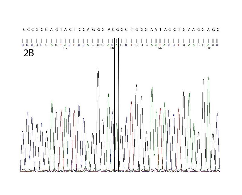
Genes 2019, 10, 168                                                                                          2 of 10

 Genes 2019, 10, x FOR PEER REVIEW                                                                          2 of 9

dysfunction, mitochondrial encephalopathy, and in one patient, hypertrophic cardiomyopathy [1,6,7].
  mitochondrial encephalopathy, and in one patient, hypertrophic cardiomyopathy [1,6,7]. Cardiac
Cardiac arrhythmias in the absence of structural cardiac changes have not yet been reported.
  arrhythmias in the absence of structural cardiac changes have not yet been reported.
     Here, we report the association of a novel variant in the QIL1 gene with familial cardiac
       Here, we report the association of a novel variant in the QIL1 gene with familial cardiac
arrhythmias in the Rhodesian Ridgeback dog. We have previously reported this canine model of
  arrhythmias in the Rhodesian Ridgeback dog. We have previously reported this canine model of
familial arrhythmias and sudden death [8]. Affected dogs had cardiac arrhythmias but had no
  familial arrhythmias and sudden death [8]. Affected dogs had cardiac arrhythmias but had no
evidence of cardiac hypertrophy, myocardial dysfunction or abnormal cardiac histologic findings.
  evidence of cardiac hypertrophy, myocardial dysfunction or abnormal cardiac histologic findings.
Skeletal muscle was found to be consistent with the previously identified mitochondrial abnormalities
  Skeletal muscle was found to be consistent with the previously identified mitochondrial
in human patients with QIl1 variants [6]. We report here the first example of a QIL1 variant associated
  abnormalities in human patients with QIl1 variants [6]. We report here the first example of a QIL1
with a mitochondrial arrhythmic cardiomyopathy.
  variant associated with a mitochondrial arrhythmic cardiomyopathy.
2. Materials and Methods
  2. Materials and Methods
      This study was conducted in accordance with the guidelines of the North Carolina State University
       This study was conducted in accordance with the guidelines of the North Carolina State
Institutional Animal Care and Use Committee (IACUC, 17-168-0).
  University Institutional Animal Care and Use Committee (IACUC, 17-168-0).
      We previously reported an extended family of Rhodesian Ridgebacks with juvenile cardiac
       We previously reported an extended family of Rhodesian Ridgebacks with juvenile cardiac
arrhythmias that occasionally resulted in sudden cardiac death [8]. Affected dogs were noted to
  arrhythmias that occasionally resulted in sudden cardiac death [8]. Affected dogs were noted to have
have ventricular arrhythmias (Figure 1) most commonly between seven and twelve months of age.
  ventricular arrhythmias (Figure 1) most commonly between seven and twelve months of age. Atrial
Atrial premature beats and second-degree atrioventricular block were noted as well, although much
  premature beats and second-degree atrioventricular block were noted as well, although much less
less commonly.
  commonly.

       Figure1.1.Affected
      Figure      AffectedRhodesian
                           RhodesianRidgebacks
                                      Ridgebacks were
                                                  were observed
                                                        observed to
                                                                  to have
                                                                     have three
                                                                          three different
                                                                                different cardiac
                                                                                          cardiacarrhythmias.
                                                                                                  arrhythmias.
       (A)  Second  degree  atrioventricular block. (B) Sinus rhythm  with  supraventricular
      (A) Second degree atrioventricular block. (B) Sinus rhythm with supraventricular        tachycardia. (C)
                                                                                                  tachycardia.
       Sinus  rhythm   with ventricular premature  complexes.
      (C) Sinus rhythm with ventricular premature complexes.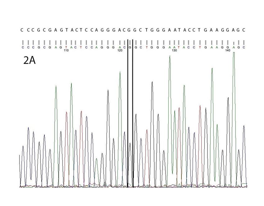
Genes 2019, 10, 168                                                                                 3 of 10

      A trio that included an apparently unaffected 70-month-old sire, an apparently unaffected
58-month-old dam and an affected 13-month-old female offspring was selected from the family
for whole genome sequencing. The affected phenotype was determined by cardiac evaluation
including a 24 h ambulatory electrocardiogram (Holter monitor) with at least 50 ventricular premature
complexes/24 h and echocardiogram by a board-certified veterinary cardiologist that indicated no
structural reason for the arrhythmia [9,10]. The sire and dam were considered to be unaffected
based on ambulatory electrocardiograms with one, and zero ventricular premature beats, respectively,
per 24 h at the time of evaluation. The sire and dam did not have a history of ventricular ectopy or
previous evidence of cardiac disease; however, a previous mating had produced a female offspring
who developed ventricular arrhythmias at 10 months of age. Three male offspring without evidence of
ventricular ectopy were also produced.
      Approximately three milliliters of whole blood was collected in an EDTA tube from each
animal. Genomic DNA was extracted using the QIAmp DNA Blood Kit standard protocol
(Qiagen, Germantown, MD, USA). Three micrograms of DNA from each dog was submitted for library
preparation and whole genome sequencing, using a 150 base pair (bp) paired-end read configuration on
an Illumina HiSeq 4000 high-throughput sequencing system (Genewiz LLC, South Plainfield, NJ, USA).
      Variant calling from next-generation sequencing data was performed using a standardized
bioinformatics pipeline for all samples, as described previously [11]. Sequence reads were trimmed
using Trimmomatic 0.32 to a minimum phred-scaled base quality score of 30 at the start and end
of each read, with a minimum read length of 70 bp, and aligned to the canFam3 reference
sequence using BWA 0.7.13 [12,13]. Aligned reads were prepared for analysis using Picard Tools 2.8
(http://broadinstitute.github.io/picard) and GATK 3.7 following best practices for base quality score
recalibration and indel realignment (Broad Institute, Cambridge, MA, USA) [14–16]. Variant calls
were made using GATK’s HaplotypeCaller walker, and variant quality score recalibration (VQSR)
was performed using sites from dbSNP 146 and the Illumina 174K CanineHD BeadChip as training
resources. A VQSR tranche sensitivity cutoff of 99.9% to SNPs and 99% to indels was used for
downstream analyses; genotype calls with a phred-scaled quality score < 20 were flagged but not
removed from the variant callset.
      Variants in the trio of dogs were filtered for polymorphisms consistent with both an autosomal
dominant and recessive inheritance pattern. The resulting variants were then filtered against
a previously established database of variants from 247 non-Rhodesian Ridgeback dogs of 43 different
dog breeds. Any variants with a minor allele frequency greater than 1% in the non-Rhodesian
Ridgebacks were removed. The remaining variants were categorized by Variant Effect Predictor 91
(https://useast.ensembl.org/info/docs/tools/vep/index.html) and prioritized by their functional
impact (e.g., stop codon, frameshift, change in amino acid, etc.) [17]. They were manually curated
for potential cardiac involvement of the gene (cardiac expression, encoding for cardiac channel proteins,
sarcomeric proteins, cytoskeletal or mitochondrial proteins, previous association with cardiomyopathy
or arrhythmic disease). Missense variants were evaluated for genomic functional significance with
Polyphen (http://genetics.bwh.harvard.edu/pph2/), SIFT (http://sift.jcvi.org/) and Provean (http:
//provean.jcvi.org/index.php).
      High impact variants (missense, stop/start gained or lost, inframe deletion, frameshift)
with potential cardiac involvement were evaluated for previous identification in the canine population
in the DogSD (http://bigd.big.ac.cn/dogsdv2/) SNP database. DNA SNPs that were not previously
reported were pursued with Sanger Sequencing in five affected and five apparently unaffected dogs,
and assessed for statistical association to the arrhythmia with a Fisher’s exact test. Variants that
were significantly associated with disease (p-value of
Genes 2019, 10, 168 4 of 10
To determine the impact of the mutation at the skeletal muscular level, a biopsy of the biceps
femoris muscle was performed under general inhalational anesthesia on a 15-month-old affected
female Rhodesian Ridgeback dog homozygous for a QIL1 variant. Following collection, the samples
were immersion-fixed in Karnovsky’s fixative. Samples were evaluated with electron microscopy.
For comparison, archived control muscle from a large mixed breed dog were similarly processed.
3. Results
Affected dogs demonstrated ventricular and/or supraventricular tachycardia and occasional
atrioventricular block that developed between 7–12 months of age (Figure 1). Analysis of the whole
genome sequences identified 271,877 variants consistent with an autosomal recessive pattern.
Variants were the filtered to identify those that were in the affected Rhodesian Ridgeback, sire and dam,
and not in >1% of the alleles in the non-Rhodesian Ridgeback dog database. This reduced the number
of variants to 32,599 that would be consistent with an autosomal recessive pattern. Similarly, analysis
identified 1,080,041 variants consistent with an autosomal dominant pattern, and 239,780 remained
after filtering.
The majority of the variants were predicted to be of low or moderate impact (synonymous, 30 or 50
untranslated regions, upstream or downstream of a gene, intronic), and were not pursued for additional
evaluation. One hundred and seven of the variants were predicted to be of higher impact, including
five variants predicted to create a frameshift, 10 predicted to create either an inframe deletion or insertion,
twenty splice site variants and seventy-two missense mutations. Thirteen of these higher impact variants
were predicted to have cardiac involvement, including variants identified in the ADCY3, AGRN, BSCL2,
FASTKD3, FAT1, HCN4, LAMA4, MYO9B, PIEZO2, PRDM8, QIL1, SMTNL1 and SORBS2 genes. Each of
these variants was evaluated by Sanger Sequencing of ten (five affected, five unaffected) additional dogs,
and a Fisher’s Exact test was performed to test for association of the variant to the arrhythmia.
Only one variant, a single missense variant ENSCAFG00000018796 g.54343438 G>A in exon four of
the C19orf70 (QIL1) gene, had a statistical association with the arrhythmia (p = 0.04) (Table 1) (Figure 2).
Table 1. Variants evaluated by Sanger sequencing. Gene, variant location, effect and statistical
association by Fisher’s exact test are provided.
Gene Variant Effect p Value
ENSCAFG00000004090
ADCY3 Missense/Splice site NP
g.19164291 G>A
ENSCAFG00000019342
AGRN Missense 0.25
g.56260122 C>A
ENSCAFG00000023629 Initiator codon
BSCL2 0.62
g.53960802 A>G variant
ENSCAFG00000010129
FASTKD3 Missense 0.63
g.6105469 C>T
ENSCAFG00000007273
FAT1 Missense 0.16
g.44199325 G>T
ENSCAFG00000031809
HCN4 Frameshift NP
g.36680881_36680885del
ENSCAFG00000004043
LAMA4 In frame deletion NP
g.68553193_68553195del
ENSCAFG00000015532
MYO9B Missense 0.35
g.45524791 C>TGenes 2019, 10, 168 5 of 10
Table 1. Cont.
Gene Variant Effect p Value
ENSCAFG00000018761
PIEZO2 Frameshift NP
g.76508892_76508892insG
ENSCAFG00000008881
PRDM8 Inframe insertion NP
g.4450426_4450427insG
ENSCAFG00000018796
C19orf70 Missense 0.04
g.54343438 G>A
ENSCAFG00000007843
SMTNL1 Missense >0.99
g.38627683 C>T
ENSCAFG00000007475
SORBS2 Missense 0.035
g.45035993 G>A
Additionally, the variant was significantly associated with the arrhythmia in the Rhodesian
Ridgeback family (p = 0.001) and was identified as homozygous in the affected offspring
and heterozygous in both of the parents. The variant was strongly associated with the arrhythmic
disease (Fisher’s exact p < 0.0001) in the population of 106 affected Rhodesian Ridgebacks
compared to the control population of non-Ridgeback dogs. The SNP was not identified in DogSD
(http://bigd.big.ac.cn/dogsdv2/) as a known canine SNP.
The QIL1 variant was predicted to change the amino acid produced at this location from a highly
conserved Glycine to Serine, and was predicted to be a deleterious change by all three variant prediction
algorithms. Polyphen predicted the variant to be likely damaging (score of 1; scores of 0.85–1 predicted
to be deleterious); SIFT predicted it to be deleterious (score of 0; scores of 0–0.05 predicted to be
deleterious) and Provean predicted it to be a deleterious change (score of −3.5; scored of −2.5 or less
predicted to be deleterious).
Ultrastructural analysis of the biceps femoris muscle from an affected homozygous Ridgeback
revealed hyperplastic mitochondria, cristae rearrangement including irregular membranous swirls,
and electron dense inclusions and lipid bodies (Figure 3A,B and Figure 4) consistent with pathologic
changes described in the similar human disorder [6] and mouse model [4]. Large mitochondria
spanned over 3 sarcomeres (Figure 3B). In contrast, mitochondria from control muscle were variable in
size, and the largest spanned up to 1 sarcomere.ENSCAFG00000007843
SMTNL1 Missense >0.99
g.38627683 C>T
ENSCAFG00000007475
SORBS2 Missense 0.035
g.45035993 G>A
Genes 2019, 10, 168 6 of 10
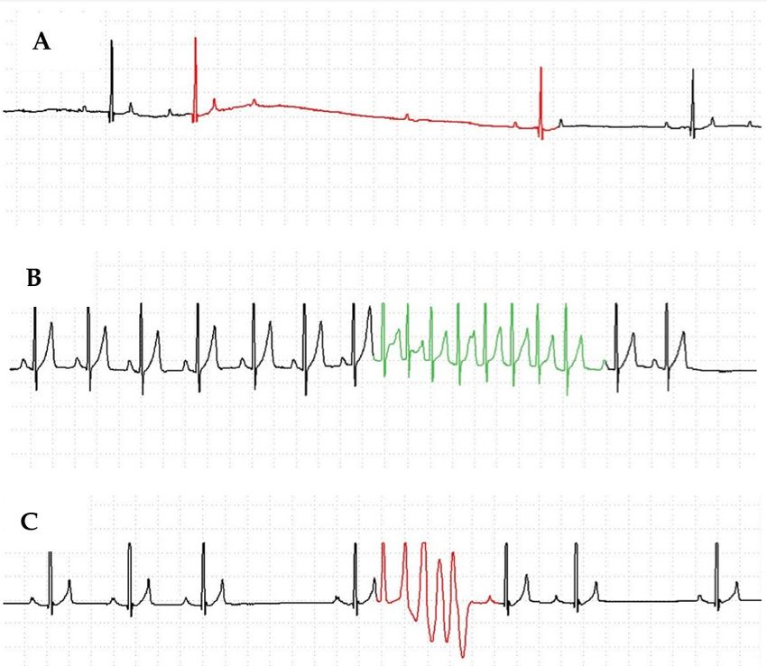
Figure 2. (A) Normal genetic sequence. (B) Sequence of a Rhodesian Ridgeback with a homozygous
variant (G/A) outlined by black box. Reference sequence (ENSCAFG00000018796) alignment is above
Figure
the DNA2. (A) Normal genetic sequence. (B) Sequence of a Rhodesian Ridgeback with a homozygous
chromatogram.
variant (G/A) outlined by black box. Reference sequence (ENSCAFG00000018796) alignment is
above the DNA chromatogram.
Additionally, the variant was significantly associated with the arrhythmia in the Rhodesian
Ridgeback family (p = 0.001) and was identified as homozygous in the affected offspring and
heterozygous in both of the parents. The variant was strongly associated with the arrhythmic disease
(Fisher’s exact p < 0.0001) in the population of 106 affected Rhodesian Ridgebacks compared to theUltrastructural analysis of the biceps femoris muscle from an affected homozygous Ridgeback
revealed hyperplastic mitochondria, cristae rearrangement including irregular membranous swirls,
and electron dense inclusions and lipid bodies (Figures 3A, 3B, 4) consistent with pathologic changes
described in the similar human disorder [6] and mouse model [4]. Large mitochondria spanned over
3Genes
sarcomeres (Figure 3B). In contrast, mitochondria from control muscle were variable in size,7 and
2019, 10, 168 of 10
the largest spanned up to 1 sarcomere.
Figure
Figure 3.3. (A)
(A)Electron
Electronmicrograph
micrographshowing
showinghyperplastic
hyperplasticmitochondria
mitochondria with
with irregular
irregular cristae
cristae (*).
(*).
Arrows highlightlipid
Arrows highlight lipid bodies.
bodies. (B)
(B) A A mitochondrion
large large mitochondrion spans
spans over over 3 sarcomeres.
3 sarcomeres. Membrane
Membrane proliferation
proliferation in swirlsdense
in swirls and electron and electron
inclusionsdense inclusions
(arrow) (arrow)
were also noted. were
Z = zalso
lines.noted. Z = z lines.Genes 2019, 10, 168 8 of 10
Genes 2019, 10, x FOR PEER REVIEW 7 of 9
Figure 4.
Figure 4. For
For comparison
comparison toto Figure
Figure 3,
3, an
an electron
electron micrograph
micrograph from
from archived
archived control dog muscle
control dog shows
muscle shows
variability in the size of mitochondria with the largest mitochondrion spanning 1 sarcomere Z = zz line,
variability in the size of mitochondria with the largest mitochondrion spanning 1 sarcomere Z = line,
m=
m = mitochondria.
mitochondria.
4. Discussion
4. Discussion
In
In the
the study
study presented
presented here,here, we
we report
report aa mitochondrial
mitochondrial arrhythmic
arrhythmic cardiomyopathy
cardiomyopathy associated
associated
with
with aa DNA
DNA variant
variant in the QIL1
in the QIL1 gene
gene in in aa spontaneous
spontaneous canine
canine model.
model. We We have
have previously
previously reported
reported
that
that this cardiomyopathy was characterized by familial ventricular arrhythmias and sudden cardiac
this cardiomyopathy was characterized by familial ventricular arrhythmias and sudden cardiac
death
death in in aapopulation
populationofofyoung young Rhodesian
Rhodesian Ridgeback
Ridgeback dogsdogs [8]. Here
[8]. Here we report
we report the association
the association of this
of this cardiomyopathy
cardiomyopathy and characteristic
and characteristic mitochondrialmitochondrial abnormalities
abnormalities in skeletalinmuscle
skeletalwith
muscle with
the QIL1
the QIL1
variant. variant.
Mitochondrial cardiomyopathiescan
Mitochondrial cardiomyopathies can be associated
be associated withwith
eithereither
nuclearnuclear or mitochondrial
or mitochondrial variants
variants
[18]. QIL1 [18].
is a QIL1
nuclearis aprotein
nuclearthatprotein that is into
is imported imported into the mitochondria
the mitochondria and is for
and is important important
proper
for proper assembly of the MICOS, which stabilizes mitochondrial
assembly of the MICOS, which stabilizes mitochondrial cristae junctions and determines the cristae junctions and determines
the placement
placement andand distribution
distribution of of mitochondrial
mitochondrial cristae[1,2].
cristae [1,2].QILI
QILIdepletion
depletionhas hasbeen
been associated
associated withwith
enlarged mitochondria, increased lipid droplets, cristae morphologic defects
enlarged mitochondria, increased lipid droplets, cristae morphologic defects including a curvilinear including a curvilinear
pattern
pattern andand concentric
concentric stacking
stacking of of the
the inner
inner mitochondrial
mitochondrial membrane,
membrane, and and reduced
reduced mitochondrial
mitochondrial
respiration
respiration [1,2,4,7]. Since mitochondrial
[1,2,4,7]. Since mitochondrial respiration
respiration is is critical
critical for
for the
the generation
generation of of ATP
ATP via
via electron
electron
transport
transport and oxidative phosphorylation systems, organ systems that have particularly high energy
and oxidative phosphorylation systems, organ systems that have particularly high energy
demands,
demands, including
including thethe brain, liver, skeletal
brain, liver, skeletal muscle
muscle andand the
the heart,
heart, are
are most
most likely
likely to
to be
be impacted
impacted by by
mitochondrial
mitochondrial dysfunction
dysfunction[5]. [5].QILI
QILIvariants
variantshavehavebeen
beenpreviously
previously associated
associated with
withthethe
development
development of
infantile encephalopathy, liver dysfunction and in one patient, hypertrophic
of infantile encephalopathy, liver dysfunction and in one patient, hypertrophic cardiomyopathy cardiomyopathy [1,6,7].
Since
[1,6,7].the
Sinceheart is oneis of
the heart onethe mostmost
of the energyenergydemanding
demanding organs,
organs,mitochondrial
mitochondrialdiseases
diseases often
preferentially impact the heart [18], and and it has been estimated that cardiac involvement including
it has been estimated that cardiac involvement including
structural and/or arrhythmic abnormalities
and/or arrhythmic abnormalities can can occur
occur in 20–40% of children with with mitochondrial
mitochondrial
disease [5,19,20]. We We report here on young young Rhodesian
Rhodesian Ridgeback dogs with familial familial arrhythmias
arrhythmias
including
including supraventricular and ventricular tachycardia and atrioventricular block. previously
supraventricular and ventricular tachycardia and atrioventricular block. We have We have
reported
previously thereported
absence ofthestructural
absencemyocardial
of structural involvement
myocardial in this model [8]. These
involvement in thisarrhythmic
model [8]. findings
These
are consistentfindings
arrhythmic with thoseare previously
consistent reported
within those
mitochondrial
previouslycardiomyopathies
reported in [18]. It has been
mitochondrial
hypothesized
cardiomyopathies that the development
[18]. It has beenofhypothesized
these arrhythmias that in
themitochondrial
developmentcardiomyopathies
of these arrhythmias may be in
mitochondrial cardiomyopathies may be associated with dysfunctional mitochondrial respirationGenes 2019, 10, 168 9 of 10
associated with dysfunctional mitochondrial respiration and decreased ATP synthesis and its impact
on cardiovascular action potential development, myocardial conduction [21] and electrical stability.
5. Conclusions
In conclusion, we identify here a variant in the Q1l1 gene resulting in a mitochondrial
cardiomyopathy characterized by cristae abnormalities and cardiac arrhythmias in a canine model.
This natural animal model of mitochondrial cardiomyopathy provides a large animal model with
which to study the development and progression of this disease as well as our understanding of
genotypic phenotypic relationships. Additionally, it serves as model with which to study the impact of
medical management on mitochondrial dysfunction.
Author Contributions: Conceptualization, K.M., J.W. and S.R.; Data curation, S.F.; For mal analysis, K.M. and S.F.;
Investigation, N.O., J.C., J.W., S.R. and D.S.; Methodology, K.M., S.F., N.O. and J.C.; Validation, S.F. and J.C.;
Writing—original draft, K.M.; Writing—review & editing, K.M., S.F., N.O., J.W., S.R. and D.S.
Funding: This research received no external funding
Conflicts of Interest: The authors declare no conflict of interest. North Carolina State University offers the canine
variant test as a risk assessment tool for dogs.
References
1. Zeharia, A.; Friedman, J.R.; Tobar, A.; Saada, A.; Konen, O.; Fellig, Y.; Shaag, A.; Nunnari, J.; Elpeleg, O.
Mitochondrial hepato-encephalopathy due to deficiency of QIL1/MIC13 (C19orf70), a MICOS complex
subunit. Eur. J. Hum. Genet. 2016, 24, 1778–1782. [CrossRef] [PubMed]
2. Zerbes, R.M.; Höß, P.; Pfanner, N.; Van Der Laan, M.; Bohnert, M. Distinct Roles of Mic12 and Mic27 in
the Mitochondrial Contact Site and Cristae Organizing System. J. Mol. Biol. 2016, 428, 1485–1492. [CrossRef]
[PubMed]
3. Vogel, F.; Bornhövd, C.; Neupert, W.; Reichert, A.S. Dynamic subcompartmentalization of the mitochondrial
inner membrane. J. Cell Biol. 2006, 175, 237–247. [CrossRef] [PubMed]
4. Guarani, V.; McNeill, E.M.; Paulo, J.A.; Huttlin, E.L.; Fröhlich, F.; Gygi, S.P.; Van Vactor, D.; Wade Harper, J.
QIL1 is a novel mitochondrial protein required for MICOS complex stability and cristae morphology.
Elife 2015, 4, 1–23. [CrossRef] [PubMed]
5. El-Hattab, A.W.; Scaglia, F. Mitochondrial Cardiomyopathies. Front. Cardiovasc. Med. 2016, 3, 1–9. [CrossRef]
[PubMed]
6. Guarani, V.; Jardel, C.; Chretien, D.; Lombes, A.; Benit, P.; Labasse, C.; Lacene, E.; Bourillon, A.; Imbard, A.;
Benoist, J.F.; et al. QIL1 mutation causes MICOS disassembly and early onset fatal mitochondrial
encephalopathy with liver disease. Elife 2016, 5, 1–18. [CrossRef] [PubMed]
7. Godiker, J.; Gruneberg, M.; DuChesne, I.; Reunert, J.; Rust, S.; Westermann, C.; Wada, Y.; Classen, G.;
Langhans, C.D.; Schlingmann, K.; et al. QIL1-dependent assembly of MICOS complex-lethal mutation in
C19ORF70 resulting in liver disease and severe neurological retardation. J. Hum. Genet. 2018, 63, 707–716.
[CrossRef] [PubMed]
8. Meurs, K.M.; Weidman, J.A.; Rosenthal, S.L.; Lahmers, K.K.; Friedenberg, S.G. Ventricular arrhythmias in
Rhodesian Ridgebacks with a family history of sudden death and results of a pedigree analysis for potential
inheritance patterns. J. Am. Vet. Med. Assoc. 2016, 248, 1135–1138. [CrossRef] [PubMed]
9. Meurs, K.M.; Spier, A.W.; Wright, N.A.; Hamlin, R.L. Use of ambulatory electrocardiography for detection of
ventricular premature complexes in healthy dogs. J. Am. Vet. Med. Assoc. 2001, 218, 1291–1292. [CrossRef]
10. Thomas, W.P.; Gaber, C.E.; Jacobs, G.J.; Kaplan, P.M.; Lombard, C.W.; Sydney Moise, N.; Moses, B.L.
Recommendations for standards in transthoracic Two-Dimensional echocardiography in the dog and cat.
Vet. Radiol. Ultrasound 1994, 35, 173–178. [CrossRef]
11. Friedenberg, S.G.; Meurs, K.M. Genotype imputation in the domestic dog. Mamm. Genome 2016, 27, 485–494.
[CrossRef] [PubMed]
12. Bolger, A.M.; Lohse, M.; Usadel, B. Trimmomatic: A flexible trimmer for Illumina sequence data.
Bioinformatics 2014, 30, 2114–2120. [CrossRef] [PubMed]Genes 2019, 10, 168 10 of 10
13. Li, H.; Durbin, R. Fast and accurate short read alignment with Burrows-Wheeler transform.
Bioinformatics 2009, 25, 1754–1760. [CrossRef] [PubMed]
14. DePristo, M.A.; Banks, E.; Poplin, R.E.; Garimella, K.V.; Maguire, J.R.; Hartl, C.; Philippakis, A.A.;
del Angel, G.; Rivas, M.A.; Hanna, M.; et al. A framework for variation discovery and genotyping using
next-generation DNA sequencing data. Nat. Genet. 2011, 43, 491–498. [CrossRef]
15. McKenna, A.; Hanna, M.; Banks, E.; Sivachenko, A.; Cibulskis, K.; Kernytsky, A.; Garimella, K.; Altshuler, D.;
Gabriel, S.; Daly, M.; et al. The Genome Analysis Toolkit: A MapReduce framework for analyzing
next-generation DNA sequencing data Aaron. Genome Res. 2010, 20, 1297–1303. [CrossRef] [PubMed]
16. Van Der Auwera, G.; Carneiro, M.O.; Hartl, C.; Poplin, R.; del Angel, G.; Levy-Moonshine, A.; Jordan, T.;
Shakir, K.; Roazen, D.; Thibault, J.; et al. From FastQ data to high confidence varant calls: The Genonme
Analysis Toolkit best practices pipeline. Curr. Protoc. Bioinform. 2014, 11, 1–33.
17. Mclaren, W.; Gil, L.; Hunt, S.E.; Riat, H.S.; Ritchie, G.R.S.; Thormann, A.; Flicek, P.; Cunningham, F.
The Ensembl Variant Effect Predictor. Genome Biol. 2016, 17, 1–14. [CrossRef] [PubMed]
18. Lee, S.R.; Han, J. Mitochondrial mutations in cardiac disorders. Adv. Exp. Med. Biol. 2017, 982, 81–111.
19. Scaglia, F.; Towbin, J.A.; Craigen, W.J.; Belmont, J.W.; Smith, E.O.; Neish, S.R.; Ware, S.M.; Hunter, J.V.;
Fernbach, S.D.; Vladutiu, G.D.; et al. Clinical spectrum, morbidity, and mortality in 113 pediatric patients
With mitochondrial disease. Pediatrics 2004, 114, 925–931. [CrossRef]
20. Finsterer, J.; Kothari, S. Cardiac manifestations of primary mitochondrial disorders. Int. J. Cardiol. 2014,
177, 754–763. [CrossRef]
21. Montaigne, D.; Pentiah, A.D. Mitochondrial cardiomyopathy and related arrhythmias. Card. Electrophysiol. Clin.
2015, 7, 293–301. [CrossRef] [PubMed]
© 2019 by the authors. Licensee MDPI, Basel, Switzerland. This article is an open access
article distributed under the terms and conditions of the Creative Commons Attribution
(CC BY) license (http://creativecommons.org/licenses/by/4.0/).You can also read