A Stop-Gain Mutation within MLPH Is Responsible for the Lilac Dilution Observed in Jacob Sheep
←
→
Page content transcription
If your browser does not render page correctly, please read the page content below
G C A T
T A C G
G C A T
           genes
Communication
A Stop-Gain Mutation within MLPH Is Responsible
for the Lilac Dilution Observed in Jacob Sheep
Christian J. Posbergh 1, * , Elizabeth A. Staiger 1,2        and Heather J. Huson 1, *
 1   Department of Animal Science, Cornell University, Ithaca, NY 14853, USA; eas0115@auburn.edu
 2   Department of Animal Sciences, Auburn University, Auburn, AL 36849, USA
 *   Correspondence: cjp98@cornell.edu (C.J.P.); hjh3@cornell.edu (H.J.H.)

 Received: 7 May 2020; Accepted: 2 June 2020; Published: 4 June 2020

 Abstract: A coat color dilution, called lilac, was observed within the Jacob sheep breed. This dilution
 results in sheep appearing gray, where black would normally occur. Pedigree analysis suggested
 an autosomal recessive inheritance. Whole-genome sequencing of a dilute case, a known carrier,
 and sixteen non-dilute sheep was used to identify the molecular variant responsible for the coat
 color change. Through investigation of the genes MLPH, MYO5A, and RAB27A, we discovered
 a nonsynonymous mutation within MLPH, which appeared to match the reported autosomal recessive
 nature of the lilac dilution. This mutation (NC_019458.2:g.3451931C>A) results in a premature stop
 codon being introduced early in the protein (NP_001139743.1:p.Glu14*), likely losing its function.
 Validation testing of additional lilac Jacob sheep and known carriers, unrelated to the original case,
 showed a complete concordance between the mutation and the dilution. This stop-gain mutation is
 likely the causative mutation for dilution within Jacob sheep.

 Keywords: Ovis aries; coat color; whole-genome sequencing; genomics

1. Introduction
     Coat color is suspected to be one of the first traits selected for in livestock species after domestication.
Historically, selection in sheep has been for white wool, due to its ability to be dyed, as opposed to
nonwhite wool. While white wool remains the dominant product in the commercial wool market,
nonwhite wool can bring higher prices in niche markets. Nonwhite wool comes in a variety of patterns
and colors, which breeders can select to increase the revenue from wool sales. One such nonwhite coat
color variation is dilution, which is commonly represented by lighter shades of color pigmentation.
A dilute phenotype has been observed within the Jacob breed, often called lilac. This dilution results in
the nonwhite portions of the wool appearing gray, rather than the traditional black. Based on pedigree
analysis of the Jacob Sheep Breeders Association registry, the dilution is inherited and expressed in
an autosomal recessive pattern [1].
     Dilute coat color phenotypes are commonly the result of impaired melanosome transport, leading
to an irregular clustering of pigment. This irregular clustering of melanosomes results in decreased
light absorption in the fiber, resulting in black hair or wool that appears grey. Melanophilin, together
with myosin Va and Rab27a, form a protein complex that is responsible for transporting melanosomes
to the cytoskeleton of melanocytes [2]. This complex has been shown to be required for proper
melanosome transport [3]. Defects in any of these three genes, melanophilin (MLPH), myosin Va
(MYO5A), and Rab27a (RAB27A), have been linked with several dilute phenotypes and the autosomal
recessive Griscelli syndromes in humans (OMIM #214450, 607624, 609227) [4,5]. Of the Griscelli
syndromes, type 3 (OMIM #609227) is linked to mutations within MLPH and is the only one of the three
types to exhibit hypopigmentation in the absence of neurological or immunological abnormalities [5].

Genes 2020, 11, 618; doi:10.3390/genes11060618                                       www.mdpi.com/journal/genes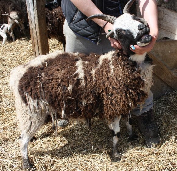
Genes 2020, 11, x FOR PEER REVIEW 2 of 8
Genes 2020, 11, 618 2 of 8
Until now, no dilute phenotypes in sheep or goats have been associated with any specific
molecular variants. Dilute phenotypes observed in chickens (OMIA #001445-9031) [6], dogs (OMIA
Until now, no dilute phenotypes in sheep or goats have been associated with any specific molecular
#000031-9615) [7–9], rabbits (OMIA #000031-9986) [10–12], cats (OMIA #000031-9685) [13], American
variants. Dilute phenotypes observed in chickens (OMIA #001445-9031) [6], dogs (OMIA #000031-9615) [7–9],
minks (000031-452646) [14,15], and Belgian Blue cattle (OMIA #000031-9913) [16] have all been linked
rabbits (OMIA #000031-9986) [10–12], cats (OMIA #000031-9685) [13], American minks (000031-452646) [14,15],
to variants within the melanophilin gene (MLPH). Therefore, MLPH was the most promising
and Belgian Blue cattle (OMIA #000031-9913) [16] have all been linked to variants within the melanophilin
candidate gene to investigate for variants contributing to the dilute phenotype in Jacob sheep. The
gene (MLPH).
purpose of this Therefore,
study was to MLPH wasthe
identify thegenomic
most promising
variant(s) candidate
responsiblegene to investigate
for the for variants
lilac color seen in
contributing
Jacob to the dilute
sheep, using phenotype
whole-genome in Jacob sheep.
sequencing and aThe purposegene
candidate of this study was
approach. Bytoutilizing
identifywhole-
the genomic
variant(s)sequencing
genome responsible of forathe lilac color
known seencase
dilute in Jacob
and sheep,
a known usingcarrier,
whole-genome sequencing
a premature and apoint
stop-gain candidate
gene approach.
mutation By utilizing
was identified within whole-genome
MLPH. This work sequencing
adds to of our
a known dilute of
knowledge case
MLPHand amutations,
known carrier,
a premature
leading stop-gain
to dilute point mutation
phenotypes in domestic was identified within MLPH. This work adds to our knowledge of
species.
MLPH mutations, leading to dilute phenotypes in domestic species.
2. Materials and Methods
2. Materials and Methods
2.1. Sample Collection
2.1. Sample Collection
All sheep were sampled in accordance with the Cornell University Institutional Animal Care &
All sheep were
Use Committee sampled
(Protocol in accordance
#2014-0121). Ownerwith the Cornell
consent was givenUniversity
prior toInstitutional Animalfor
sample collection Care &
Use Committee
privately owned(Protocol #2014-0121).
sheep. Whole blood wasOwner consent
collected wasthe
from given priorvein
jugular to sample collection
via 10-mL for privately
vacutainers
owned sheep.
containing Whole
K2EDTA, andblood was collected
genomic DNA wasfrom the jugular
extracted veinthe
following viaQiagen
10-mL Puregene
vacutainers containing
Protocol
(Gentra
K2 EDTA, Systems, Inc. Minneapolis,
and genomic DNA was MN, USA).following
extracted The genomic DNA was
the Qiagen stored at
Puregene −80 ℃ (Gentra
Protocol until it was
Systems,
Inc. Minneapolis, MN, USA). The genomic DNA was stored at −80 ◦ C until it was sequenced.
sequenced.
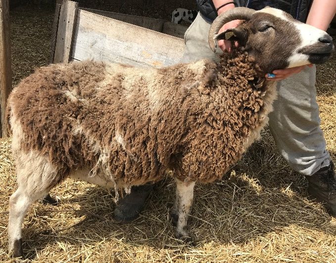
The
Thedilute
dilutephenotype
phenotypewas wasvisually
visuallycharacterized
characterizedbyby diluted
diluted pigment
pigment in in
thethenonwhite
nonwhite portions
portions of
of the fleece. An example of dilute and non-dilute Jacobs can be seen in Figure
the fleece. An example of dilute and non-dilute Jacobs can be seen in Figure 1. The two Jacobs 1. The two Jacobs used
used for
for whole-genome
whole-genome sequencing
sequencing werewere unrelated
unrelated within
within fivefive generations.
generations. Additional
Additional dilute
dilute Jacobs
Jacobs were
were sourced
sourced from an unrelated flock. In total, 22 dilute Jacobs, 13 known carriers, and
from an unrelated flock. In total, 22 dilute Jacobs, 13 known carriers, and 26 non-dilute Jacobs were 26 non-dilute Jacobs
were available
available for testing.
for testing. The carriers
The carriers werewere determined
determined via pedigree
via pedigree analysis,
analysis, usingusing the Jacob
the Jacob Sheep Sheep
Breeders
Breeders Association pedigree database [17]. An additional 163 sheep that were not dilute,
Association pedigree database [17]. An additional 163 sheep that were not dilute, representing the Icelandic,
representing the Icelandic, Karakul, California Red, Romeldale, Romney, Finnsheep, Lincoln, and
Karakul, California Red, Romeldale, Romney, Finnsheep, Lincoln, and Shetland breeds, were used for
Shetland breeds, were used for validation testing.
validation testing.
(a) (b)
Figure 1. Cont.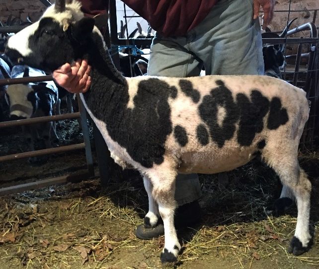
Genes 2020, 11, 618
Genes 2020, 11, x FOR PEER REVIEW 3 of 8
3 of 8
(c)
(d)
Figure
Figure 1.1. Photos
Photosofofdilute
dilute(lilac) andand
(lilac) non-dilute Jacob
non-dilute sheep
Jacob that were
sheep used for
that were whole-genome
used sequencing
for whole-genome
are in panelsare
sequencing (a) and (b): (a)
in panels (a)and
and(c)(b):
are(a)
examples
and (c) of
arethe lilac dilution
examples inlilac
of the Jacobdilution
sheep; (b) and (d)
in Jacob are examples
sheep; (b)
of non-dilute Jacob sheep.
and (d) are examples of non-dilute Jacob sheep.
2.2. Whole Genome Sequencing
2.2. Whole Genome Sequencing
TruSeq PCR-Free libraries were prepared for a known dilute case, a known dilute carrier,
TruSeq PCR-Free libraries were prepared for a known dilute case, a known dilute carrier, and
and sixteen additional
sixteen additional non-dilutenon-dilute
controlcontrol
samples samples from
from other other The
breeds. breeds. The were
libraries libraries were sequenced
sequenced using
using
150 bp paired-end reads on an Illumina HiSeq X Ten platform to generate approximately 20x 20x
150 bp paired-end reads on an Illumina HiSeq X Ten platform to generate approximately
coverage
coverage perperindividual.
individual. These
These sequences
sequences have
havebeenbeendeposited
depositedininNCBI’s
NCBI’ssequence
sequenceread readarchive
archive(SRA)
and can be found under the BioProject accession: PRJNA480684. Reads
(SRA) and can be found under the BioProject accession: PRJNA480684. Reads were aligned to were aligned to Oar_v4.0
using
Oar_v4.0the using
Burrows–Wheeler aligner mem
the Burrows–Wheeler aligneralgorithm, versionversion
mem algorithm, 0.7.12-r1039 [18]. [18].
0.7.12-r1039 Average genome
Average
coverage was calculated
genome coverage using goleft
was calculated usingcovstats
goleft [19]. The[19].
covstats alignments were locally
The alignments wererealigned, and variants
locally realigned,
were called and
and variants werefiltered following
called and filteredthe Genome
following theAnalysis
Genome Toolkit’s best practices,
Analysis Toolkit’s versionversion
best practices, 4.0.3.0 [20].
4.0.3.0nucleotide
Small [20]. Smallvariants
nucleotide
werevariants were called
called using using the HaploytpeCaller,
the HaploytpeCaller, within the Genomewithin the Genome
Analysis Toolkit.
Analysis were
Variants Toolkit. Variants
filtered fromwere
thefiltered from
analysis the analysis
using the Genome using the Genome
Analysis Analysis
Toolkit, Toolkit,
with with
the following
the followingquality
thresholds: thresholds:
depth < 2.0,
quality depth < 2.0, Phred-scaled
Phred-scaled strand biasstrand > 60.0,> 60.0,
bias p-value
p-value mapping
mapping < 40.0,
quality
quality
MQRankSum < −12.5, and ReadPosRankSum < −8.0. We also evaluated the predictedimpact
< 40.0, MQRankSum < −12.5, and ReadPosRankSum < −8.0. We also evaluated the predicted impact of
of these
these variants
variants usingSNPEff
using SNPEffandandthethe NCBI
NCBI Ovis
Ovisaries
ariesannotation
annotation release
release102102
[21]. Based
[21]. on previous
Based on previous
work in other domestic species [6–16], we focused our investigation
work in other domestic species [6–16], we focused our investigation on variants within on variants within MLPH. MLPH.
Variants within and surrounding MLPH (Oar_v4.0: OAR1:3,383,028-3,478,858) were filtered to be
Variants within and surrounding MLPH (Oar_v4.0: OAR1:3,383,028-3,478,858) were filtered to be
homozygous alternate in the dilute case, heterozygous in the known carrier, and homozygous
homozygous alternate in the dilute case, heterozygous in the known carrier, and homozygous reference
reference in the sixteen non-dilute non-Jacob samples. Due to the nature of the melanosome transport
in the sixteen non-dilute non-Jacob samples. Due to the nature of the melanosome transport and the
and the known influence of MYO5A and RAB27A, we performed the same approach on the identified
known influence of MYO5A and RAB27A, we performed the same approach on the identified variants
variants within MYO5A and RAB27A.
within MYO5A and RAB27A.
2.3. Candidate Variant Validation
2.3. Candidate Variant Validation
To validate the candidate variant, a PCR was designed to take advantage of the RFLP removed
To validate the candidate variant, a PCR was designed to take advantage of the RFLP removed by
by the NC_019458.2:g.3451931C>A mutation. The following forward and reverse primers were
the NC_019458.2:g.3451931C>A
designed to capture the first mutation.
exon The
of following
MLPH, using forwardPrimer3
and reverse primers[22]:
software were designed
F: 5’- to
capture the first exon of MLPH, using Primer3 software [22]: F: 5 0 -GTCCCGCCACACACACTTAC-30 ; R:
GTCCCGCCACACACACTTAC-3’; R: 5’-TCGGTGTTTTCTGCATTGTC-3’. PCR amplification was
0 -TCGGTGTTTTCTGCATTGTC-30 . PCR amplification was performed in a 20 µL volume, containing
5performed in a 20 µL volume, containing 2 µL of DNA (diluted to a concentration of 25 ng/µL), 2 µL
2ofµL of DNA (diluted
10× PCR reaction to awith
buffer concentration
20 mM MgCl2of 25(Roche
ng/µL), 2 µL of 10×0.2
Diagnostics), PCR reaction
µL of Taq DNAbuffer with 20 mM
Polymerase
MgCl2 (Roche Diagnostics), 0.2 µL of Taq DNA Polymerase [23], 2 µL of 2 mM dNTPs,
[23], 2 µL of 2 mM dNTPs, 2 µL of forward and reverse 5 µM primers, and 9.8 µL PCR-grade water. 2 µL of forward
and
The reverse
PCR was5 carried
µM primers,
out in and 9.8 µL
a BioRad PCR-grade
T100 water.(BioRad
thermal cycler The PCR was carriedwith
Laboratories), out the
in afollowing
BioRad T100
◦ C followed by
thermal cycler
conditions: 3 min at 95 ℃Laboratories),
(BioRad followed by 40with
cyclesthe s at 95 ℃,
following
of 30 30 s at 59 ℃,
conditions: 3 and
min30ats95
at 72 ℃, and a
40 cycles of 30 s at 95 ◦ C, 30 s at 59 ◦ C, and 30 s at 72 ◦ C, and a final extension time of 3 min. The PCR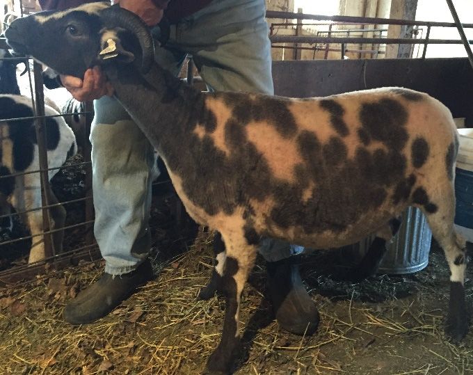
Genes 2020, 11, 618                                                                                 4 of 8

product was Sanger sequenced to validate the candidate variant, using one case, one carrier, and one
wild-type individual. To genotype additional animals, the restriction digest used 10 µL of the 245 bp
PCR product, 0.1 µL of EarI restriction enzyme [1.0 U per reaction, New England Biolabs (NEB),
Ipswich, MA], 1 µL of 10× NEB CutSmart buffer, and 8.9 µL MilliQ of water to bring the reaction
volume to 20 µL and was incubated for 16 h at 37 ◦ C. The resulting products were visualized by agarose
gel electrophoresis, using a 3% agarose gel, run for 40 min at 150 V, and a 100-bp standard ladder
(New England BioLabs) as a reference. The normal allele (C) resulted in four expected fragments of
121, 57, 38, and 29 bp, while the dilute allele (A) resulted in only three expected fragments of 178, 38,
and 29 bp.

3. Results

3.1. Whole-Genome Sequencing
      Whole-genome sequencing generated 220,771,257 raw paired reads for the dilute case, 205,778,310
raw reads for the known carrier, and an average of 230,685,048 raw reads for the remaining sixteen
individuals. After aligning the reads to the Oar_v4.0 reference genome, the dilute individual had
an average genome coverage of 17.34×, the carrier 15.88×, while the remaining sixteen samples
averaged 20.41×.
      We identified 2572 small nucleotide variants within and surrounding MLPH (NC_019458.2:
g.3383028-3478858). After 254 SNPs and 3 indels did not pass our variant filtering criteria, we were
left with 2315 variants. Of these, only 37 SNPs and 4 indels were predicted to have an impact on the
resulting MLPH protein. Within the SNPs, 15 were predicted to have a low impact, 21 a moderate
impact, and only 1 was predicted to have a high impact. Of the indels, two were classified as low
impact, and two were classified as high impact. The two high impact indels appear in the alternate
state in every individual, which we suspect is the result of a reference assembly error, encompassing the
homopolymer that these indels are located near. After filtering the impactful variants for variants that
were homozygous alternate in the dilute animal, heterozygous in the known carrier, and homozygous
reference in the sixteen other individuals, only the single nonsynonymous high impact SNP remained
(NC_019458.2:g.3451931C>A). This SNP is located within exon 1, and results in a premature stop codon
early in the protein (NP_001139743.1: p.Glu14*).
      We identified 2994 small nucleotide variants within MYO5A, and 1173 small nucleotide variants
within RAB27A. However, none of the identified variants exhibited the pattern of being a homozygous
alternate in the dilute sheep, heterozygous in the known carriers, and a homozygous reference in the
sixteen other individuals. They were not investigated further in relation to the dilution phenotype.
A full list of variants discovered within these three genes can be found in Table S1.

3.2. RFLP Validation
     Using the RFLP genotyping test validation of the NC_019458.2:g.3451931C>A mutation within
unrelated lilac cases and additional Jacob and non-Jacob non-dilute sheep, we found a complete
concordance of the phenotype with the dilute genotype. See Figure 2 for the band sizes seen across the
different genotypes. See Table 1 for MLPH NC_019458.2:g.3451931C>A genotype counts.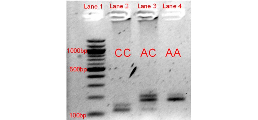
Genes2020,
Genes 11,x 618
2020,11, FOR PEER REVIEW 5 5ofof8 8
Gelimage
Figure2.2.Gel
Figure imageofofthe
theRFLP
RFLPgel gelelectrophoresis
electrophoresisvalidation
validationofofthe
the NC_019458.2:g.3451931C>A
NC_019458.2:g.3451931C>A
mutation. Lane 1 represents the 100-bp reference ladder, with the 1000,
mutation. Lane 1 represents the 100-bp reference ladder, with the 1000, 500, and 500, and100100
bpbp bands
bands labeled.
labeled.
Lanes 2 through 4 represent the three genotypes at this variant. The normal allele (C) results ininfour
Lanes 2 through 4 represent the three genotypes at this variant. The normal allele (C) results four
expectedfragments
expected fragmentsofof121,
121,57,
57,38,
38,and
and2929bp
bpwhile
whilethethedilute
diluteallele
allele(A)
(A)results
resultsininonly
onlythree
threeexpected
expected
fragments of 178, 38, and 29 bp. The 178- and 121-bp fragments were the
fragments of 178, 38, and 29 bp. The 178- and 121-bp fragments were the primary ones used primary ones usedfor
for
genotyping, as the smaller bands were more difficult to identify. The bands of approximately
genotyping, as the smaller bands were more difficult to identify. The bands of approximately 150 bp 150 bp
and 200 bp are likely to be the result of an incomplete digestion of the last EarI recognition
and 200 bp are likely to be the result of an incomplete digestion of the last EarI recognition site within site within
thePCR
the PCRproduct.
product.
Table 1. MLPH (NC_019458.2:g.3451931C>A) genotypes by dilute status.
Table 1. MLPH (NC_019458.2:g.3451931C>A) genotypes by dilute status.
Breed Dilute Status N NC_019458.2:g.3451931C>A Genotype
NC_019458.2:g.3451931C>A
Breed Dilute Status N AA AC CC
Genotype
Jacob Non-dilute 39 0 13 26
AA AC CC
Dilute 22 22 0 0
JacobNon-Jacob Non-dilute
Non-dilute 16339 0 0 0 13 163 26
Total Dilute 22422 22 22 13 0 189 0
Non-Jacob Non-dilute 163 0 0 163
Total
4. Discussion 224 22 13 189
Prior studies in dogs [7], cats [13], rabbits [12], and other domestic species have identified the
4. Discussion
mutations within MLPH as responsible for dilute coat colors. While dilution can impact both eumelanin
andPrior studies in dogs
phaeomelanin, there[7],
havecatsbeen
[13],no
rabbits
reports [12],
of and other
dilute domestic species
phaeomelanin have identified
phenotypes the
within sheep.
mutations
The presentwithin
studyMLPH as responsible
has likely identified theforcausative
dilute coat colors.forWhile
mutation dilutedilution
coat colorcan impact
within the both
Jacob
eumelanin
breed. Thisand phaeomelanin,
is the there have
first report linking been nowithin
mutations reports of dilute
MLPH to a phaeomelanin phenotypes
coat color phenotype within
within sheep.
sheep. The to
This adds present study knowledge
the limited has likely identified the causative
in sheep coat mutation
color molecular for dilute
genetics, coat color
as recent within
reports the
on causal
Jacob breed.
variants This
have is the
been first to
limited report linking[24],
the Agouti mutations within
Extension MLPH
[25,26], andtoBrown
a coat Loci
color[27,28].
phenotype within
sheep. This adds to the limited knowledge in sheep coat color molecular genetics, as recent reports
on causal variants have been limited to the Agouti [24], Extension [25,26], and Brown Loci [27,28].Genes 2020, 11, 618 6 of 8
The mechanism of dilution in sheep appears to be similar to mutations previously described as
responsible for dilution in several species. In cats, the dilution is the result of a premature stop codon
caused by a single base-pair deletion within the second exon of MLPH [13]. Within Belgian Blue cattle,
a 10-bp deletion in the first MLPH exon results in a premature stop codon being introduced [16], differing
from the point mutation described in the present study but still causing a loss-of-function mutation
early in the protein. Exon-skipping within rabbits, caused by a frameshift mutation, also introduces
a premature stop codon, which leads to a dilute phenotype [10]. These reported mutations, occurring
early in the protein, fall within the R27BD domain. This is necessary for targeting MLPH to RAB27A,
without which melanosomes are unable to be transported along actin filaments [29]. This is likely to
result in the irregular clustering of melanosomes that cause the change in color.
Despite the limited molecular knowledge of coat color regulation in sheep, there are various shades
of black and brown observed within other breeds, such as the Shetland, Icelandic, and Romeldale.
Further study is needed to identify whether other mutations in MLPH, MYO5A, or RAB27A exist and
contribute to the various shades seen in these breeds. Future work should investigate multiple species
protein alignments, to extensively characterize the protein changes required for dilute coat color across
species. The discovered variant within MLPH will be useful for Jacob breeders wishing to increase the
frequency of the lilac color within their flocks through marker-assisted selection.
In conclusion, we have identified a stop-gain mutation (NC_019458.2:g.3451931C>A) within MLPH,
which appears to be the causative mutation for the coat color dilution, called lilac, within Jacob sheep.
Supplementary Materials: The following are available online at http://www.mdpi.com/2073-4425/11/6/618/s1,
Table S1. SNVs discovered from WGS near MLPH, RAB27A, and MYO5A.
Author Contributions: Conceptualization, C.J.P.; methodology, C.J.P. and E.A.S.; formal analysis, C.J.P.; funding
acquisition, C.J.P.; investigation, C.J.P. and E.A.S.; resources, C.J.P. and H.J.H.; supervision, H.J.H.; writing—original
draft preparation, C.J.P.; writing—review and editing, C.J.P., E.A.S., and H.J.H.; All authors have read and agreed
to the published version of the manuscript.
Funding: This research was funded by an Engaged Graduate Student Grant from the Office of Engagement
Initiatives (engaged.cornell.edu). The APC was funded by the Engaged Graduate Student Grant.
Acknowledgments: We thank the shepherds which provided us samples for this study. We thank Asha Miles and
Niko Kochendoerfer for their sampling assistance.
Conflicts of Interest: The authors declare no conflict of interest. The funders had no role in the design of the
study; in the collection, analyses, or interpretation of data; in the writing of the manuscript, or in the decision to
publish the results.
References
1. Anjola, O.; McEwan, N. Inheritance patterns of coat colouration and horn number in Jacob sheep. Open Agric.
2018, 3, 363–367.
2. Nascimento, A.A.; Roland, J.T.; Gelfand, V.I. Pigment cells: A model for the study of organelle transport.
Annu. Rev. Cell Dev. Biol. 2003, 19, 469–491. [CrossRef]
3. Fukuda, M.; Kuroda, T.S.; Mikoshiba, K. Slac2-a/Melanophilin, the Missing Link between Rab27 and Myosin
Va IMPLICations Of A Tripartite Protein Complex For Melanosome Transport. J. Biol. Chem. 2002, 277,
12432–12436. [CrossRef]
4. Hume, A.N.; Ushakov, D.S.; Tarafder, A.K.; Ferenczi, M.A.; Seabra, M.C. Rab27a and MyoVa are the
primary Mlph interactors regulating melanosome transport in melanocytes. J. Cell Sci. 2007, 120, 3111–3122.
[CrossRef] [PubMed]
5. Van Gele, M.; Dynoodt, P.; Lambert, J. Griscelli syndrome: A model system to study vesicular trafficking.
Pigment Cell Melanoma Res. 2009, 22, 268–282. [CrossRef] [PubMed]
6. Vaez, M.; Follett, S.A.; Bed’Hom, B.; Gourichon, D.; Tixier-Boichard, M.; Burke, T. A single point-mutation
within the melanophilin gene causes the lavender plumage colour dilution phenotype in the chicken.
BMC Genet. 2008, 9, 7. [CrossRef]Genes 2020, 11, 618 7 of 8
7. Philipp, U.; Hamann, H.; Mecklenburg, L.; Nishino, S.; Mignot, E.; Günzel-Apel, A.-R.; Schmutz, S.M.; Leeb, T.
Polymorphisms within the canine MLPH gene are associated with dilute coat color in dogs. BMC Genet.
2005, 6, 34. [CrossRef]
8. Drögemüller, C.; Philipp, U.; Haase, B.; Günzel-Apel, A.-R.; Leeb, T. A noncoding melanophilin gene (MLPH)
SNP at the splice donor of exon 1 represents a candidate causal mutation for coat color dilution in dogs.
J. Hered. 2007, 98, 468–473. [CrossRef]
9. Bauer, A.; Kehl, A.; Jagannathan, V.; Leeb, T. A novel MLPH variant in dogs with coat colour dilution.
Anim. Genet. 2018, 49, 94–97. [CrossRef]
10. Lehner, S.; Gähle, M.; Dierks, C.; Stelter, R.; Gerber, J.; Brehm, R.; Distl, O. Two-exon skipping within MLPH
is associated with coat color dilution in rabbits. PLoS ONE 2013, 8. [CrossRef]
11. Fontanesi, L.; Scotti, E.; Allain, D.; Dall’olio, S. A frameshift mutation in the melanophilin gene causes the
dilute coat colour in rabbit (Oryctolagus cuniculus) breeds. Anim. Genet. 2014, 45, 248–255. [CrossRef]
[PubMed]
12. Demars, J.; Iannuccelli, N.; Utzeri, V.J.; Auvinet, G.; Riquet, J.; Fontanesi, L.; Allain, D. New Insights into
the Melanophilin (MLPH) Gene Affecting Coat Color Dilution in Rabbits. Genes 2018, 9, 430. [CrossRef]
[PubMed]
13. Ishida, Y.; David, V.A.; Eizirik, E.; Schäffer, A.A.; Neelam, B.A.; Roelke, M.E.; Hannah, S.S.; O’Brien, S.J.;
Menotti-Raymond, M. A homozygous single-base deletion in MLPH causes the dilute coat color phenotype
in the domestic cat. Genomics 2006, 88, 698–705. [CrossRef] [PubMed]
14. Cirera, S.; Markakis, M.N.; Christensen, K.; Anistoroaei, R. New insights into the melanophilin (MLPH) gene
controlling coat color phenotypes in American mink. Gene 2013, 527, 48–54. [CrossRef]
15. Manakhov, A.D.; Andreeva, T.V.; Trapezov, O.V.; Kolchanov, N.A.; Rogaev, E.I. Genome analysis identifies
the mutant genes for common industrial Silverblue and Hedlund white coat colours in American mink.
Sci. Rep. 2019, 9, 1–8. [CrossRef]
16. Li, W.; Sartelet, A.; Tamma, N.; Coppieters, W.; Georges, M.; Charlier, C. Reverse genetic screen for
loss-of-function mutations uncovers a frameshifting deletion in the melanophilin gene accountable for
a distinctive coat color in Belgian Blue cattle. Anim. Genet. 2016, 47, 110–113. [CrossRef]
17. Jacob Sheep Pedigree Search. Available online: https://jsba.org/search.html (accessed on 20 April 2018).
18. Li, H.; Durbin, R. Fast and accurate short read alignment with Burrows–Wheeler transform. Bioinformatics
2009, 25, 1754–1760. [CrossRef]
19. Goleft Github Repository. Available online: https://github.com/brentp/goleft (accessed on 1 February 2020).
20. McKenna, A.; Hanna, M.; Banks, E.; Sivachenko, A.; Cibulskis, K.; Kernytsky, A.; Garimella, K.; Altshuler, D.;
Gabriel, S.; Daly, M.; et al. The Genome Analysis Toolkit: A MapReduce framework for analyzing
next-generation DNA sequencing data. Genome Res. 2010, 20, 1297–1303. [CrossRef]
21. Cingolani, P.; Platts, A.; Wang, L.L.; Coon, M.; Nguyen, T.; Wang, L.; Land, S.J.; Lu, X.; Ruden, D.M. A program
for annotating and predicting the effects of single nucleotide polymorphisms, SnpEff: SNPs in the genome of
Drosophila melanogaster strain w1118; iso-2; iso-3. Fly 2012, 6, 80–92. [CrossRef]
22. Untergasser, A.; Cutcutache, I.; Koressaar, T.; Ye, J.; Faircloth, B.C.; Remm, M.; Rozen, S.G. Primer3—
New capabilities and interfaces. Nucleic. Acids. Res. 2012, 40, e115. [CrossRef]
23. Desai, U.J.; Pfaffle, P.K. Single-step purification of a thermostable DNA polymerase expressed in Escherichia
coli. BioTechniques 1995, 19, 780–782, 784.
24. Norris, B.J.; Whan, V.A. A gene duplication affecting expression of the ovine ASIP gene is responsible for
white and black sheep. Genome Res. 2008, 18, 1282–1293. [CrossRef]
25. Våge, D.I.; Klungland, H.; Lu, D.; Cone, R.D. Molecular and pharmacological characterization of dominant
black coat color in sheep. Mammalian Genome Off. J. Int. Mamm. Genome Soc. 1999, 10, 39–43. [CrossRef]
26. Våge, D.I.; Fleet, M.R.; Ponz, R.; Olsen, R.T.; Monteagudo, L.V.; Tejedor, M.T.; Arruga, M.V.; Gagliardi, R.;
Postiglioni, A.; Nattrass, G.S.; et al. Mapping and characterization of the dominant black colour locus in
sheep. Pigment cell Res./Spons. Eur. Soc. Pigment Cell Res. Int. Pigment Cell Soc. 2003, 16, 693–697. [CrossRef]
27. Gratten, J.; Beraldi, D.; Lowder, B.V.; McRae, A.F.; Visscher, P.M.; Pemberton, J.M.; Slate, J. Compelling
evidence that a single nucleotide substitution in TYRP1 is responsible for coat-colour polymorphism in
a free-living population of Soay sheep. Proc. R. Soc. Lond. B Biol. Sci. 2007, 274, 619–626. [CrossRef]Genes 2020, 11, 618 8 of 8
28. Posbergh, C.; Huson, H. Making Moorit: Mutations in TYRP1 are responsible for brown coat color in different
United States sheep breeds. In Proceedings of the 11th World Congress on Genetics Applied to Livestock
Production, Auckland, New Zealand, 11–16 February 2018.
29. Ramalho, J.S.; Lopes, V.S.; Tarafder, A.K.; Seabra, M.C.; Hume, A.N. Myrip uses distinct domains in the
cellular activation of myosin VA and myosin VIIA in melanosome transport. Pigment Cell Melanoma Res.
2009, 22, 461–473. [CrossRef]
© 2020 by the authors. Licensee MDPI, Basel, Switzerland. This article is an open access
article distributed under the terms and conditions of the Creative Commons Attribution
(CC BY) license (http://creativecommons.org/licenses/by/4.0/).You can also read