Beginner's guide to producing super-resolved images on a widefield fluorescence microscope
←
→
Page content transcription
If your browser does not render page correctly, please read the page content below
Beginner's Guide
Beginner's guide to producing
super-resolved images on a
widefield fluorescence
microscope
Downloaded from http://portlandpress.com/biochemist/article-pdf/42/4/52/890972/bio20200045.pdf by guest on 10 November 2020
Ilijana Vojnovic and The development of super-resolution microscopy techniques, which are able to achieve resolutions
Ulrike Endesfelder (Max in the nanometre range and as such allow the visualization of subcellular structures and dynamics,
Planck Institute for has considerably expanded the possibilities of fluorescence microscopy in the life sciences. While
Terrestrial Microbiology a majority of these techniques require highly specialized hardware, single-molecule localization
and LOEWE Center for microscopy (SMLM) can be implemented on conventional widefield fluorescence microscopes. Here,
Synthetic Microbiology we describe what technical upgrades are necessary and discuss some of the difficulties that can be
(SYNMIKRO), Germany) encountered during sample preparation and imaging.
Visualizing life with fluorescence fluorescence spots on the detector whose centres can be
microscopy localized with high precision.
As a consequence, SMLM, in its simplest form, can be
Fluorescence microscopy sheds light on all areas of implemented on a widely available widefield fluorescence
life. It visualizes the embryonic development of large microscope. This makes it ideal for anyone wishing to
multicellular organisms as well as the molecular explore the possibilities of super-resolution microscopy
organization within tiny microbes. The molecules of at a relatively low cost. In the following sections, we
describe how you can equip your conventional widefield
interest are labeled with fluorophores, which reveal their
microscope for SMLM and what needs to be considered
positions through their emitted fluorescence, producing
during sample preparation and imaging.
colourful and contrast-rich images.
The development of super-resolution techniques
has expanded the possibilities of fluorescence
Preparing your microscope
microscopy even further. These techniques are not
restricted by the diffraction limit of light, and thus are
The basic design of a widefield fluorescence microscope
able to attain resolutions of only a few nanometres. This
is depicted in Figure 1a. The excitation light, depicted in
has in many cases revealed unexpected, spectacular green, is reflected by a dichroic mirror onto the objective
new biology. The demand for super-resolution setups, to illuminate the sample. The emitted fluorescence,
access to imaging facilities and collaborations with depicted in orange, is collected by the same objective
research groups operating such systems is, therefore, and now passes the dichroic mirror due to its Stokes shift
high. to longer wavelengths. The fluorescence image is then
Whilst some super- resolution techniques work recorded by a light-sensitive detector.
with complex illumination patterns and require highly Unfortunately, most conventional widefield
specialized hardware, the class of single-molecule microscopes are not sensitive enough to detect the signal
localization microscopy (SMLM) techniques relies on of single fluorophores, which is essential for SMLM.
special fluorophores that can be photoswitched between However, this can be remedied by upgrading three core
a non-emitting ‘dark state’ and a fluorescent ‘on-state’ to components – the light sources, the objective and the
achieve sub-diffraction resolution. They are modulated detector.
in such a way that only few of them fluoresce at the Conventional fluorescence microscopes typically
same time. Fluorophores, therefore, appear as distinct employ gas discharge lamps or an array of light-emitting
52 August 2020 © The Authors. Published by Portland Press Limited under the Creative Commons Attribution License 4.0 (CC BY-NC-ND)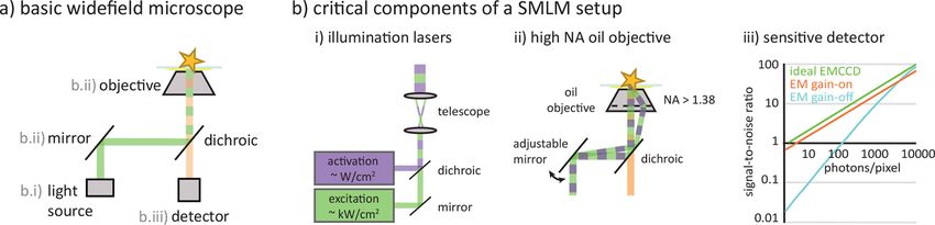
Beginner's Guide
Figure 1. Adapting a widefield microscope for SMLM imaging. In order to adapt an epifluorescence widefield microscope system as depicted in (a) for SMLM
imaging, three components are necessary: illumination lasers (b.i), an immersion oil objective with high numeric aperture (b.ii) and a sensitive detector such
Downloaded from http://portlandpress.com/biochemist/article-pdf/42/4/52/890972/bio20200045.pdf by guest on 10 November 2020
as an EMCCD camera (b.iii). Together, these allow for single-molecule sensitivity.
diodes (LEDs) as light sources, thereby accommodating to reduce phototoxicity and (in combination with
a wide range of fluorophores with diverse excitation a near- UV switchable fluorophore) for dual colour
spectra and allow for straightforward multicolour imaging. Another very convenient add-on is the fast
imaging. For SMLM imaging, these light sources need mechanical shutters or an acousto- optic tunable filter
to be replaced by lasers. Unlike ensemble fluorescence (AOTF) to modulate the activation and excitation lasers
microscopy, which records the joint signal of all for fast temporal illumination control.
fluorescent molecules, SMLM relies on the ability to The second crucial prerequisite for the detection
detect individual fluorophores. This is only possible if of single fluorophores is an objective that collects as
a minimum of several hundred to a few thousands of much fluorescence as possible from the sample (Figure
photons per fluorophore reach the detector in a single 1b.ii). As the fluorescence emitted by a fluorophore
imaging frame. As each emission of a photon requires the is typically isotropic, an objective with a large light
absorption of an excitation photon, fluorophores have collection angle (also called aperture angle) is essential.
to be excited as efficiently as possible to maximize the However, light hitting the glass surface of the objective
emitted fluorescence per time. Lasers provide the strong, at a sharp angle will simply be reflected away if it comes
coherent illumination to achieve this (Figure 1b.i). from a less-dense medium. The space between cover
Typically, small diode lasers or diode-pumped solid slip and objective, therefore, needs to be filled with an
state lasers with wavelengths matching the absorption immersion oil with a refractive index matching that of
spectra of common SMLM fluorophores are used. By glass (about 1.51). This also prevents photon loss due to
means of a telescope, the laser beams' diameters are refraction at the boundary of cover slip and immersion
adjusted in such a way that the illuminated area within medium. Aperture angle and the refractive index of the
the sample covers the field of view of the detector. In immersion liquid are, therefore, the key performance
an efficient setup, lasers with powers of 100–500 mW indicators of any objective. Together, they determine
are sufficient to achieve an illumination intensity of the so-called numerical aperture (NA) of an objective,
0.5–2 kW/cm2 that is needed for SMLM imaging. This which is defined as the product of the refractive index
requires that the expansion of the laser beams is not and the sinus of the half aperture angle. The higher the
larger than necessary and that about 50% of the lasers’ NA, the better the ability of the objective to collect light.
original power reaches the sample (this is typically only For SMLM imaging, an objective with an NA of 1.45 or
possible with open laser paths). higher is typically necessary.
In addition to the excitation lasers, a so- called High NA objectives also come with a second
activation laser is needed to modulate the photoswitching advantage: they allow for objective-based total internal
fluorophores. Most fluorophores suitable for SMLM reflection fluorescence (TIRF) microscopy. The TIRF
are photoswitched by near- UV laser illumination of geometry creates an evanescent light field of only
~400 nm wavelength. Typically, intensities of a few 100–200 nm width at the cover slip surface. It only
W/cm² of a 405- nm diode laser are sufficient excites molecules close to the surface, and as such
(Figure 1b.i). Additionally, variants exist of many drastically improves the signal- to-
noise ratio when
commonly used fluorescent proteins that can be imaging small objects. An adjustable mirror can be
photoswitched via so- called primed photoconversion, installed in the optical path of the illumination lasers
which uses a combination of 488 nm and near-infrared to easily switch between widefield and TIRF imaging
illumination. Primed photoconversion can be useful modes (Figure 1b.ii).
August 2020 © The Authors. Published by Portland Press Limited under the Creative Commons Attribution License 4.0 (CC BY-NC-ND) 53
Beginner's Guide
The last critical component is a detector sensitive quality. While the labeling efficiency is still acceptable
enough to detect the weak signal of single fluorophores. for widefield resolution (Figure 2b.i), prominent gaps
As a widefield microscopy technique, SMLM records are visible in the vimentin structures in the SMLM
images with a two- dimensional array detector, e.g., image (Figure 2b.ii). In general, low labeling efficiencies
a charge-coupled device (CCD) sensor. CCD chips can have many different causes: for affinity tags such as
consist of an array of photodiodes that detect incident antibodies, their affinity could just not be sufficiently
fluorescence photons by means of the photoelectric high or the targeted epitopes cannot be reliably reached
effect. To attain single-molecule sensitivity, CCDs are (e.g., labels physically too big, epitopes partially buried
enhanced by electron multipliers (EMCCDs), which in the structure). For transient expressions of genetic
cause photoelectrons to trigger an avalanche of secondary labels, the filaments simply could be a mixture of native
electrons through impact ionization. The strength of this and labeled proteins of interest. It is also possible for the
amplification can be adjusted through the EM gain of the sample preparation protocols to be too harsh, quenching
Downloaded from http://portlandpress.com/biochemist/article-pdf/42/4/52/890972/bio20200045.pdf by guest on 10 November 2020
detector, which can make even the signal of only a few fluorescence or destroying the epitopes of extrinsic
photons detectable (Figure 1b.iii). Apart from EMCCD labels.
cameras, the new generation of scientific Complementary The quality of SMLM images can also be impacted
Metal Oxide Semiconductor (sCMOS) sensors are by false-positive signals caused by unspecific staining
sensitive enough for SMLM imaging. or autofluorescence. This is especially critical if the
native abundance of the molecules of interest is low.
We simulate unspecific, false-positive signals in the
Labelling and imaging performance at data set for our example by distributing random data
single-molecule fidelity points throughout the field of view (Figure 2c). Even
though vimentin is a highly structural, filamentous
The right hardware, however, is only part of the story. protein, the interpretation of the image becomes
Due to the delicate nature of nanoscale imaging, special ambiguous. It is difficult to evaluate if a registered
care needs to be taken during sample preparation and signal results from specific staining, e.g., a pool of
imaging. Single-molecule–sensitive microscopes provide vimentin monomers, or from an unspecific, non–
single-molecule resolution. This means that factors that target-bound fluorophore (Figure 2c.ii). In general,
are hardly noticeable in conventional widefield images autofluorescence can be mostly attributed to colourful
can have a large impact on the quality of an SMLM metabolites or pigments within the specific organism,
image. or to uptakes from the growth media. Unspecific
One important aspect of fluorescence microscopy staining originates from non–target-bound labels that
is the notion that one always only visualizes the were not washed out during sample preparation, e.g.,
fluorophores, not the molecules of interest themselves. sticking by charge. Genetic labels can be troublesome
As such, fluorescence microscopy techniques strongly as well: overexpression can produce a large pool of
depend on the fidelity of the chosen fluorescence label. non-physiological monomers which are not integrated
Good labeling will have a high efficiency, i.e., a large into native structures, or tags can disturb the biological
part of the target molecules carry a label, and a high function of the protein of interest.
specificity, i.e., most other things do not. It is easy to imagine how the quantitative evaluation
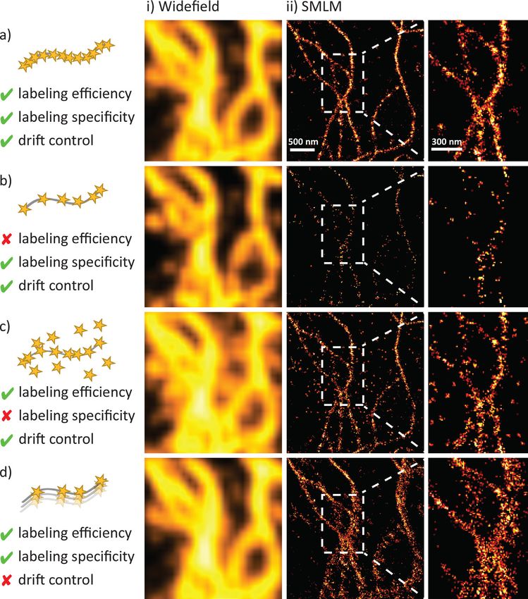
In Figure 2a, we showcase a high-quality image of of SMLM data quickly becomes unfeasible for less-
vimentin filaments in a mammalian HeLa cell at widefield structured or low copy number targets if labeling
(left), as well as SMLM resolution (right). In this specific efficiency and specificity are low. For example, a staining
example, the vimentin structures are labeled by a small, efficiency of 75% for a homotrimeric structure will, on
only a few nanometres, sized single-chain antibody, a average, result in only about 50% of labeled structures
so-called nanobody, which yields high labeling efficiency actually appearing as trimers – under otherwise
and specificity. The labels densely populate the filaments ideal imaging conditions. In practice, the underlying
which are thoroughly stained. The nanobody carries structure would be further obscured by random
an organic dye, Alexa Fluor 647, in a quantitative one- noise and signals lost due to insufficient brightness,
to-one labeling ratio, which is one of the best performing premature bleaching or improper photoswitching
fluorophores in the SMLM field. It is a bright probe, its control. All in all, ensuring high labeling efficiency
photoswitching is well-controlled and reliable, and its and specificity is essential in every SMLM project. It
bleaching rate in SMLM buffers is low. will often be necessary to modify or completely replace
Using this SMLM data set, we simulate typical the chosen labeling strategy if, after some test runs, no
problems of labeling and imaging procedures. satisfactory results are obtained. It is not uncommon
In Figure 2b, we use only a fraction of our data set to pursue two, three or even four different approaches
to visualize how low labeling efficiencies impact image before being successful.
54 August 2020 © The Authors. Published by Portland Press Limited under the Creative Commons Attribution License 4.0 (CC BY-NC-ND)
Beginner's Guide
Downloaded from http://portlandpress.com/biochemist/article-pdf/42/4/52/890972/bio20200045.pdf by guest on 10 November 2020
Figure 2. Labeling and imaging performance at single-molecule fidelity. (a) SMLM image of vimentin structures in a HeLa
cell. Cells were stained using nanobodies carrying the SMLM-suitable photoswitching fluorescent dye, Alexa Fluor 647. This
original recording (taken from Virant et al., Nat Commun, 2018. 9(1)) illustrates a good practice example for SMLM imaging
with high labeling efficiency and specificity and robust drift control. In (b)–(d), we used this data set to simulate typical
problems in labeling and imaging performance. These include low labeling efficiency (b), unspecific signal (c) and drift during
image acquisition (d). In all cases, shortcomings in fluorescence labeling and imaging performance are clearly visible in the
SMLM images (i), and as such limit their quality. At the same time, they are hardly noticeable in the corresponding widefield
fluorescence images (ii).
Last but not least, a robust drift correction is needed. typically post-processed to remove residual drift, e.g.,
Recording SMLM images takes time, which increases by means of trajectories of fiducial markers measured
the chance for substantial sample drift accumulating with the sample or by calculating spatiotemporal cross-
over minutes of imaging (Figure 2d). Most microscope correlations using the sample data itself. Most SMLM
setups are built in a heavy, mechanically stable fashion software supports such corrections.
which passively prevents large drift. Additionally, most To illustrate the severity of residual drift in SMLM
commercial systems actively stabilize the focal plane to imaging, we added an artificial linear shift of 0.02
suppress axial drift, detecting shifts via the reflection nm per imaging frame to our data set. This small
of infrared lasers from the cover slip on quadrant drift sums up to 200 nm of total drift over the 10,000
photodiodes. However, these measures usually do not imaging frames – hard to spot in the widefield image
suffice to achieve nanometre precise drift control as is (Figure 2d.i) but clearly visible in the SMLM image
required for SMLM imaging. Thus, SMLM images are (Figure 2d.ii).
August 2020 © The Authors. Published by Portland Press Limited under the Creative Commons Attribution License 4.0 (CC BY-NC-ND) 55Beginner's Guide
To gather some initial experience with SMLM and can serve as a good control for successful staining
imaging and to test the setup, it is a good idea to first and imaging.
image a well-known test sample. Cytoskeletal structures
such as microtubules, actin or vimentin filaments are Conclusion
often used as visualized standards. For all of them,
well-tested commercial antibodies – pre-labeled with We hope that this beginners’ guide has been able to
Alexa Fluor 647 – are available. Microtubules have the convince you that the first steps towards SMLM imaging
additional advantage that they are hollow, have a well- are not too difficult. Requirements are a widefield setup
defined diameter of 25 nm and do not form higher- equipped for single- molecule sensitivity and careful
order filaments bundling together several fibres. In sample preparation and imaging routines. For further
high-quality SMLM images, a cross-section through a steps, e.g., towards quantitative or live cell SMLM
microtubule should, therefore, show two clear peaks imaging, please have a look at the (open access) overview
■
Downloaded from http://portlandpress.com/biochemist/article-pdf/42/4/52/890972/bio20200045.pdf by guest on 10 November 2020
separated by 25 nm plus two times the size of the label articles listed below. Happy imaging.
Further reading
Introduction into the technical details of light microscopy
• Wegerhoff, R., Weidlich, O., Kässens, M. (2006) Basics of Light Microscopy & Imaging, GIT Verlag GmbH & Co. https://
analyticalscience.wiley.com/do/10.1002/imaging.1654
Introduction into suitable fluorophores and their photoswitching. This review includes several overview tables.
• Turkowyd, B., Virant, D. and Endesfelder, U. (2016) From single molecules to life: microscopy at the nanoscale. Anal.
Bioanal. Chem. 408, 6885–6911 10.1007/s00216-016-9781-8
Strategies for multicolor labeling in SMLM imaging. This review includes an experimental design guideline and a
troubleshooting table.
• Vojnovic, I., Winkelmeier, J. and Endesfelder, U. (2019) Visualizing the inner life of microbes: practices of multi-color
single-molecule localization microscopy in microbiology. Biochem. Soc. Trans. 47, 1041–1065. 10.1042/bst20180399
The example vimentin data set of Figure 2 is taken from this publication. It introduces a new nanobody label for
SMLM imaging.
• Virant, D., Traenkle, B., Maier, J., et al. (2018) A peptide tag-specific nanobody enables high-quality labeling for
dSTORM imaging. Nat. Commun. 9, 930 10.1038/s41467-018-03191-2
Ilijana Vojnovic studied chemistry at the Goethe University in Frankfurt, Germany, and is now a PhD student
at the Department for Systems and Synthetic Microbiology at the Max Planck Institute for Terrestrial
Microbiology in Marburg, Germany. She applies super-resolution methods for structural studies in microbes,
investigating multi-protein complexes. Email: ilijana.vojnovic@synmikro.mpi-marburg.mpg.de
Ulrike Endesfelder studied physics and has been a group leader at the Max Planck Institute for Terrestrial
Microbiology in Marburg since 2014. She is a member of the German Young Academy. In summer 2020, she
and her group will move to Carnegie Mellon University in Pittsburgh, USA, where she accepted a professorship
position for Experimental Biophysics. Email: ulrike.endesfelder@synmikro.mpi-marburg.mpg.de
56 August 2020 © The Authors. Published by Portland Press Limited under the Creative Commons Attribution License 4.0 (CC BY-NC-ND)You can also read