The Topical Tranexamic Acid Have Potential Hazard of Promoting Biofilm Formation of Staphylococcus aureus in Microenvironment of the Prosthetic Joint
←
→
Page content transcription
If your browser does not render page correctly, please read the page content below
Hindawi
BioMed Research International
Volume 2021, Article ID 5748069, 8 pages
https://doi.org/10.1155/2021/5748069

Research Article
The Topical Tranexamic Acid Have Potential Hazard of
Promoting Biofilm Formation of Staphylococcus aureus in
Microenvironment of the Prosthetic Joint

          Feiyang Zhang ,1 Wenjun Dong ,1 Fengyan Wang ,1 Jinlong Yu ,1 Feng Jiang ,1
          Jin Tang ,2 Yan Qian ,1 and Hao Shen 1
          1
              Department of Orthopaedics, Shanghai Jiao Tong University Affiliated Sixth People’s Hospital, #600, Yishan Rd, Shanghai, China
          2
              Department of Clinical Laboratory, Shanghai Jiao Tong University Affiliated Sixth People’s Hospital, #600, Yishan Rd,
              Shanghai, China

          Correspondence should be addressed to Jin Tang; tangjin6ph@163.com, Yan Qian; yanzilaoma@126.com,
          and Hao Shen; shenhao7212@sina.com

          Received 11 August 2020; Revised 1 February 2021; Accepted 4 March 2021; Published 12 March 2021

          Academic Editor: Yu Sheng Li

          Copyright © 2021 Feiyang Zhang et al. This is an open access article distributed under the Creative Commons Attribution License,
          which permits unrestricted use, distribution, and reproduction in any medium, provided the original work is properly cited.

          Background. Perioperative topical tranexamic acid as antifibrinolytic agent is often used for total joint replacement to reduce
          bleeding currently. Staphylococcus aureus was the most common isolates from perioperative infection of prosthetic joint. The
          influence of topical application with tranexamic acid on the incidence of acute prosthetic joint infection of Staphylococcus
          aureus has not been clarified. Methods. Mouse model of Staphylococcus aureus knee prosthesis infection was constructed.
          Tranexamic acid was intra-articular injected during the perioperative period. CFU counting from tissue and implant sample was
          evaluated 3 days and 7 days after inoculating of Staphylococcus aureus. Bacterial growth curve, biofilm formation, aggregation,
          and plasmin inhibition of Staphylococcus aureus were tested with tranexamic acid added to the synovial culture medium.
          Results. There were no significant differences of CFU counting from tissue and implant samples in knee prosthesis infection
          after a single local injection of tranexamic acid at the postoperative 3 or 7 days. The amount of bacterial colonization on the
          surface of implant increased after 3 days’ continuous local injection of tranexamic acid. Tranexamic acid has no effect on
          bacterial growth at the concentration (10 mg/ml) of clinical application, but it can inhibit bacterial aggregation and mildly
          inhibit biofilm formation. Plasmin can significantly inhibit biofilm formation which can be revised by adding tranexamic acid.
          Conclusion. Although continuous local injection of tranexamic acid can promote the biofilm formation of Staphylococcus aureus
          on the surface of articular implant, it has clinical safety for using one single local injection of tranexamic acid during the
          perioperative period.

1. Introduction                                                          ber of clinical studies showed that tranexamic acid can
                                                                         significantly reduce the incidence of postoperative adverse
Tranexamic acid can competitively inhibit the activation of              events [4, 8].
plasminogen and the binding of plasmin to fibrin as synthetic                 Prosthetic joint infection is considered to be the most
analogues of the amino acid lysine, thus inhibiting fibrin deg-           severe complication related to total joint replacement [9,
radation [1, 2]. A large number of studies have confirmed                 10]. Staphylococcus aureus was predominant pathogen in
that tranexamic acid can significantly reduce the amount of               postoperative and acute prosthetic joint infection [10–12].
blood loss during surgery without increasing the risk of                 Because of the numerous virulence factors and the ability to
thromboembolism [3–6]. Intravenous administration and                    form biofilm on the surfaces of implant, Staphylococcus
local injection of tranexamic acid are usually used in the peri-         aureus leads to more severe clinical presentation and worse
operative period of total joint replacement [7]. A large num-            prognosis [12].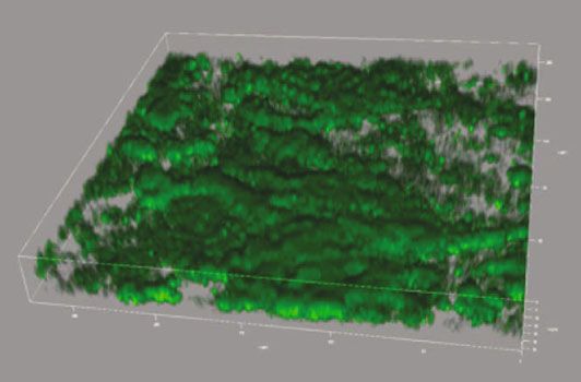
2                                                                                                  BioMed Research International

     Some studies has found that application of tranexamic           of tranexamic acid (i.e., first and second groups) were eutha-
acid can exacerbate infections of Staphylococcus aureus in           nized, and the mice with continuous local injection of
mice model [13, 14].                                                 tranexamic acid were euthanized in 3 days after surgery.
     But in clinical practice, the topical application of tranexa-   Peri-implant tissues and implant in tibia were harvested for
mic acid has been used more and more widely and has not              CFU counting.
shown increasing of infection rate [15–19]. The influence of
topical tranexamic acid on Staphylococcus aureus infection           2.3. Bacteria CFU Counting. The peri-implant tissues were
is still controversial.                                              weighed and homogenized in tubes with 1 ml normal of
     Therefore, it needs to be clarified whether local injection      saline (NS) by high-speed homogenizer (Jingxin Industrial
of tranexamic acid promotes the formation of biofilm and              Limited Company, Shanghai, China). The implant were
leads to the increased risk of Staphylococcus aureus infection.      washed gently 3 times with NS to remove the planktonic bac-
                                                                     teria and placed in tube containing 1 ml of NS, sonicated, and
                                                                     homogenized to detach the adherent bacteria. The homoge-
2. Materials and Methods                                             nates and suspensions were serially diluted in NS and spread
2.1. Bacterial Strains, Reagents, and Ethics Statement. The          on sheep blood agar (SBA). The plates were cultured over-
strains used in this study were methicillin-susceptible Staph-       night at 37°C. The colonies were counted and expressed by
ylococcus aureus (MSSA) ST1792 isolated from PJI prosthe-            log10CFU/g of peri-implant tissues and log10CFU/per
sis. The fluorescently label Staphylococcus aureus ST1792-            implanted rods.
sfGFP which was preserved in our laboratory were used for
confocal microscopic assay. The tranexamic acid (Aladdin,            2.4. The Effect Test of TXA In Vitro. The ST1792 were cultured
Shanghai, China) was used in in vivo and in vitro study of           overnight at 37°C on SBA plates before each experiment. Sin-
bacterial growth, aggregation, and biofilm formation. The             gle colony of each strain was collected and cultured overnight
plasmin (PLM, Sigma Aldrich, St Louis, MO, USA) was used             in TSB at 37°C. The ST1792-sfGFP were cultured on Tryptic
in in vitro study of biofilm formation was purchased from             Soy Agar (TSA) plates with 10 μg/ml chloramphenicol and
and used at final concentrations of 100 μg/ml. Synovial fluid          cultured overnight in TSB containing 10 μg/ml chloramphen-
(SF) was added to the medium and used in in vitro experi-            icol at 37°C.
ment. Collection of human synovial fluid from patients with                The preconditioning method of medium was divided into
osteoarthritis was approved by the Institutional Review              six groups: the first group containing 10% SF, the second
Board of Shanghai Jiao Tong University Affiliated Sixth Peo-           group containing 10%SF and 10 mg/ml TXA, the third group
ple’s Hospital. Because synovial fluid was aspirated as part of       containing 10%SF and 50 mg/ml TXA, the fourth group con-
the routine procedure before injection of hyaluronic acid and        taining 10%SF and 100 μg/ml PLM, the fifth group contain-
would have been discarded otherwise, and patient informa-            ing 10%SF and 100 μg/ml PLM and 10 mg/ml TXA, and the
tion not collected, a waiver for informed written consent            sixth group containing 10%SF and 100 μg/ml PLM and
was granted by the IRB.                                              50 mg/ml TXA.

                                                                     2.4.1. Staphylococcus aureus Growth Curve Assay. Overnight
2.2. Staphylococcus aureus Prosthetic Joint Infection Mouse
                                                                     culture of ST1792 was serially diluted 1 : 1000 in groups 1, 2,
Model. All procedures of animal experiment had been
                                                                     and 3 with TSB and cultured at 37°C. 100 μl of solution were
reviewed and approved by the Animal Care and Experiment
                                                                     aspirated at 0-2-4-6-8-12 hours and transferred to the wells
Committee of Shanghai Jiao Tong University affiliated Sixth
                                                                     of a 96-well tissue culture plate (Corning Co., NY, USA).
People’s Hospital.
                                                                     The absorbance was measured by a microplate reader
     Six-week-old and weighing (20 ± 5) g male C57BL/6 mice
                                                                     (BIO-TEK, ELX 800) at a wavelength of 600 nm.
were used to construct Staphylococcus aureus prosthetic joint
infection model. The mice were anesthetized intraperitone-           2.4.2. Staphylococcus aureus Aggregation Test. Overnight cul-
ally with 3% pentobarbital sodium (0.1 ml per 10 g body              ture of ST1792 was serially diluted to 1 : 1000 in groups 1, 2,
weight) and then shaving the both knees. A medial parapatel-         and 3 with TSB and three additional control groups without
lar incision was made at the knee joint of mice after sterilized     10%SF and cultured at 37°C. Strain clumped together and
with 75% alcohol. Proximal tibia was exposed, and the                sank to the bottom of the tube after 8 h; the turbidity of
medullary cavity was inserted with sterile stainless steel           medium supernatant was significantly decreased. 100 μl of
implant rods. After closing the wounds by sutures, tranexa-          culture medium supernatant were aspirated and transferred
mic acid was injected into the joint cavity of one side at a dose    to the wells of a 96-well tissue culture plate, measuring the
of 10 mg/kg; the other side was injected with an equal volume        absorbance at a wavelength of 490 nm by a microplate reader.
of saline as control. Two hours later, both knee joints were
inoculated with ST1792 solution of ~ 5 ∗ 106 . And then,             2.4.3. Biofilm Formation and Biomass. Overnight culture of
the mice were randomly divided into three groups                     ST1792 was serially diluted to 1 : 1000 in TSB supplemented
(Table 1). The first group was housed for 3 days, and the sec-        with 0.5% glucose (TSBG). Diluted bacteria, with supple-
ond group was housed for 7 days. The third group was con-            mentation from group 1 to group 6, were used for assessment
tinuous local injected with tranexamic acid at a dose of             of biofilm formation on (1) the bottom of 96-well polystyrene
10 mg/kg every 24 hours and housed for 3 days. Three days            microtiter plate (Corning Co., NY, USA) or (2) Ultra High
and 7 days after surgery, the mice with a single local injection     Molecular Weight Polyethylene (UHMWPE) washers that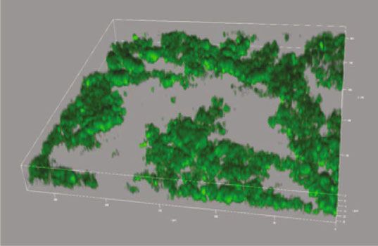
BioMed Research International 3
Table 1: The grouping of mouse.
Treatment
Group Time (day)
Left knee Right knee
1 A single local injection of TXA A single local injection of NS 3
2 A single local injection of TXA A single local injection of NS 7
3 Continuous local injection of TXA Continuous local injection of NS 3
Periprosthetic tissue
10 Implant
10
8
8
Log10 CFU/g
6
Log10 CFU
6
4
4
2 2
0 0
TXA (3 d)
TXA (7 d)
Control (3 d)
Control (7 d)
Control (3 d)
TXA (3 d)
Control (7 d)
TXA (7 d)
Single local injection Single local injection
(a) (b)
Periprosthetic tissue Implant
10 10
⁎
8 8
Log10 CFU/g
6 6
Log10 CFU
4 4
2 2
0 0
TXA (3 d)
Control (3 d)
Control (3 d)
TXA (3 d)
Continuous local injection Continuous local injection
(c) (d)
Figure 1: CFU counting after local injection of tranexamic acid on Staphylococcus aureus PJI mouse model. (a, b) The bacterial CFU of PJI
mice with a single local injection of tranexamic acid in periprosthetic tissue (Figure 2(a)) and implants (Figure 2(b)) after 3 and 7 days. (c) The
bacterial CFU of periprosthetic tissue after continuous local injection of tranexamic acid for 3 days. (d) The bacterial CFU of implants after
continuous local injection of tranexamic acid for 3 days. ∗ P < 0:05; error bars represent standard deviations.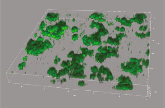
4                                                                                                       BioMed Research International

were affixed to bottom of 24-well polystyrene plates (Corning                             0.4
Co., NY, USA) with Lubriseal grease (Thomas Scientific) and
sterilized by UV irradiation.                                                           0.3

                                                                              OD600nm
     96-well plate were incubated overnight at 37°C.
                                                                                        0.2
Completely aspiring the supernatant from each well and
washing gently 3 times with 200 μl NS to remove the plank-                              0.1
tonic bacteria. Then, 100 μl methanol was used to fix the bio-
film for 30 min and then dried. After that, biofilm was stained                           0.0
with 100 μl of 0.1% crystal violet for 15 min. The biofilm bio-                                0   2    4      6       8   10   12
mass on the bottom of the well was dissolved in 200 μl 33%
                                                                                                           Time (h)
acetic acid after the unbound crystal violet was rinsed by
NS for 3 times. 100 μl solution of each well were transferred                                     Control
into a new 96-well tissue culture plate. The absorbance was                                       TXA (10 mg/ml)
measured by a microplate reader at a wavelength of 590 nm.                                        TXA (50 mg/ml)
     UHMWPE washer inoculated with ST1792 in groups 1,
2, 3, 4, 5, and 6 with TSBG was incubated overnight. The            Figure 2: Bacterial growth curve of Staphylococcus aureus ST1792
UHMWPE washers were washed 3 times with saline to                   cultured separately in TSB medium containing 10% SF
                                                                    (Blank,10 mg/ml TXA and 50 mg/ml TXA).
remove nonadherent cells. The washed UHMWPE washers
were transferred into a new 24-well polystyrene plate. Adher-
ent biofilms were fixed with methanol and stained with crys-
tal violet and washed 3 times with sterile water. Biomass on        3.3. The Influence of Tranexamic Acid on the Bacterial
the surfaces of UHMWPE washers was determined by solu-              Aggregation in Different Media. The aggregation of Staphy-
bilizing crystal violet with 33% acetic acid as described above.    lococcus aureus inhibited by tranexamic acid with clinically
                                                                    used concentration(10 mg/ml) in TSB medium. The inhib-
                                                                    itory effect was attenuated in tranexamic acid with high
2.4.4. Confocal Laser Scanning Microscopy (CLSM). Over-
                                                                    concentration(50 mg/ml) (Figure 3(a)). However, the anti-
night culture of ST1792-sfGFP was serially diluted 1 : 1000
                                                                    aggregation capacity of tranexamic acid disappears when
in groups 1, 2, 3, 4, 5, and 6 with TSBG. 500 μl of bacteria sus-
                                                                    cultured in TSB medium containing 10% SF(Figure 3(b)).
pension were incubated with cover glass overnight in 24-well
                                                                    The results suggested that inhibitory effect of tranexamic
tissue culture plate at 37°C. The cover glasses were washed
                                                                    acid on bacterial aggregation attenuated in the microenvi-
gently 3 times with NS and imaged by CLSM. The ST1792-
                                                                    ronment of the prosthetic joint.
sfGFP showed green fluorescence.

                                                                    3.4. The Individual and Mutual Inhibition for Biofilm
3. Results                                                          Formation of Staphylococcus aureus by Tranexamic Acid
3.1. The Colonization on the Surface of Implant Were                and Plasmin. Whether on 96-well plates or UHMWPE
Increased by Continuous Use with TXA In Vivo. Mice with             washer, the biofilm formation of Staphylococcus aureus was
a single local injection of tranexamic acid during the periop-      slight inhibited by tranexamic acid (Figures 4(a) and 4(b)).
erative period showed no significant difference in Staphylo-          Biofilm formation were inhibited more significantly by tra-
coccus aureus colonization of tissue and implant sample             nexamic acid in high concentration (50 mg/ml) with the
after 3 and 7 days (Figures 1(a) and 1(b)). Although mice           inhibitory ability of bacterial growth. When plasmin was
with continuous local injection of tranexamic acid for 3 days       added into the culture medium, the biofilm formation was
showed no significant increase of bacterial colonization in          significantly inhibited. However, the antibiofilm formation
tissue (Figure 1(c)), there was significant higher CFU count-        ability of plasmin inhibited by tranexamic acid; thus, the cul-
ing from implant (Figure 1(d)). The experimental results sug-       ture medium exhibited a weak antibiofilm formation effect.
gested that the continuous local injection of tranexamic acid
can promote biofilm formation.                                       3.5. Tranexamic Acid Offset the Inhibition of Biofilm
                                                                    Formation with Plasmin via CLSM. Biofilm formed obviously
3.2. Influence of Tranexamic Acid on the Growth of                   in TSBG with 10%SF by ST1792 and slight inhibited with
Staphylococcus aureus. For analysis of the influence of tra-         TXA(10 mg/ml). The inhibitory effect of TXA at high con-
nexamic acid on bacterial growth curve, tranexamic acid             centration (50 mg/ml) was more obvious. However, the bio-
with clinically used concentration(10 mg/ml) and high con-          film disappeared after PLM was added, and it can be found
centration (50 mg/ml) was used. Compared to the medium              that antibiofilm formation function of plasmin much more
without tranexamic acid, the medium containing tranexamic           strong than TXA in TSBG with 10%SF (Figure 5). The weak
acid with clinically used concentration (10 mg/ml) has no           antibiofilm formation function exhibited in medium when
influence on bacterial growth. Bacterial growth was inhibited        acting simultaneously with tranexamic acid and plasmin.
by tranexamic acid with high concentration (50 mg/ml)               The results of CLSM provide further confirmation that tra-
(Figure 2). It showed that tranexamic acid has no antibacte-        nexamic acid offset the inhibition of biofilm formation with
rial capacity in concentration of clinical use.                     plasmin.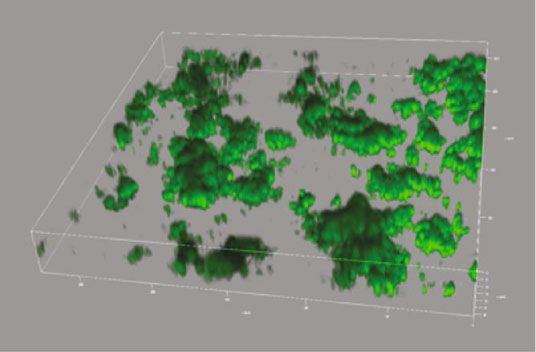
BioMed Research International 5
TSB
⁎⁎
⁎⁎⁎
0.15
TSB +10%SF
Turbidity (OD490 )
0.10 0.15
Turbidity (OD490 )
0.10
0.05
0.05
0.00 0.00
Control
TXA (10 mg/ml)
TXA (50 mg/ml)
Control
TXA (10 mg/ml)
TXA (50 mg/ml)
(a) (b)
Figure 3: Tranexamic acid shows difference of inhibiting bacterial aggregation in TSB with or without SF. (a) The turbidity of medium
supernatant increased in TSB medium containing TXA (n = 6). (b) No significant difference of the turbidity was observed in TSB medium
containing SF and TXA. ∗∗ P < 0:01, ∗∗∗ P < 0:001. Error bars represent standard deviations.
2.5 2.5
⁎ 2.0
2.0 ⁎
⁎⁎
OD590nm
⁎⁎⁎
OD590nm
1.5 1.5 ⁎⁎⁎
⁎⁎⁎
1.0 1.0 ⁎⁎⁎
0.5 0.5 ⁎⁎⁎
⁎⁎⁎
⁎⁎⁎
0.0 0.0
Control
TXA (10 mg/ml)
TXA (50 mg/ml)
PLM
PLM+TXA (10 mg/ml)
PLM+TXA (50 mg/ml)
Control
TXA (10 mg/ml)
TXA (50 mg/ml)
PLM
PLM+TXA (10 mg/ml)
PLM+TXA (50 mg/ml)
(a) (b)
Figure 4: Inhibitory effect of biofilm formation with tranexamic acid were observed in 96-well tissue culture plate (Figure 4(a)) and on the
surface of UHMWPE washer (Figure 5(b)). Biofilm formation inhibited more obviously by plasmin and the inhibition of plasmin were
reversed by tranexamic acid. ∗ P < 0:05, ∗∗ P < 0:01, ∗∗∗ P < 0:001. Error bars represent standard deviations.
4. Discussion infused with tranexamic acid at a dose of 700-800 mg/kg
every 8 hours [14] which was much higher dosage than that
Mice studies with staphylococcal sepsis or septic arthritis for clinical use [14], and previous study had proposed that
showed increasing severity and mortality of infection by high-dose tranexamic acid caused significant cytotoxicity
treatment with tranexamic acid; however, the mice were [20]. In present study, we constructed Staphylococcus aureus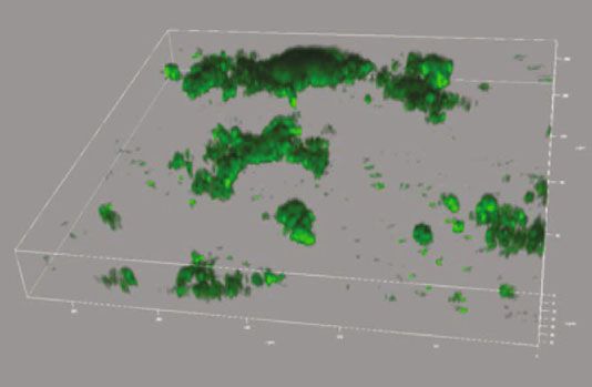
6 BioMed Research International
TSBG+10%SF TSBG+10%SF+TXA (10 mg/ml)
(a) (b)
TSBG+10%SF+TXA (50 mg/ml) TSBG+10%SF+PLM
(c) (d)
TSBG+10%SF+TXA (10 mg/ml)+PLM TSBG+10%SF+TXA (50 mg/ml)+PLM
(e) (f)
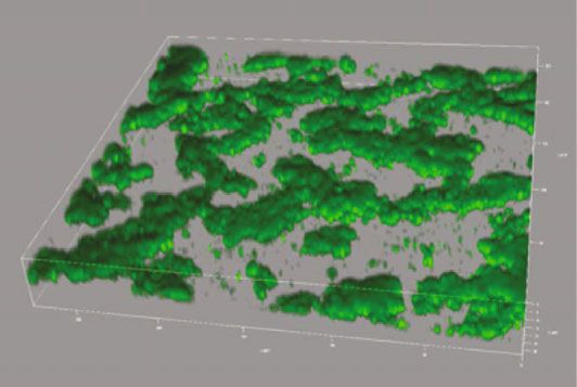
Figure 5: CLSM results of biofilm forming with ST1792-sfGFP. (a) Biofilm formation of Staphylococcus aureus in TSBG medium containing
10% SF. (b) Biofilm formation was inhibited by tranexamic acid (10 mg/ml). (c) Biofilm formation was significantly inhibited by tranexamic
acid (50 mg/ml). (d) Biofilm formation was significantly inhibited by plasmin. (e, f) Tranexamic acid offset the inhibition of biofilm formation
with plasmin.
prosthetic joint infection model with topical tranexamic acid growth. However, tranexamic acid showed inhibitory func-
at 10 mg/kg which dose was similar with that using clinically tion on aggregation and biofilm formation of Staphylococcus
and found that continuous local injection of tranexamic acid aureus. The function of antibacterial aggregation disap-
promoted biofilm formation in vivo. peared in microenvironment containing synovial fluid. High
Clinically, a single local injection of tranexamic acid is concentration of TXA (50 mg/ml) showed inhibition of bac-
commonly used in joint replacement surgery, and the safety terial growth and relatively stronger function of antibiofilm
of this method is well documented [15, 16, 18]. In our formation, but the significant cytotoxicity exhibited in the
in vivo experiments, it was observed that a single local injec- high concentration of TXA which was not used in clinically
tion of tranexamic acid did not aggravate Staphylococcus cases [20].
aureus infection. However, for patients bleeding after joint We can observe some special phenomenon in bacte-
implanted surgery, we hope to explore the feasibility of con- ria aggregation experiments. In the infected setting,
tinuous local injection of tranexamic acid. With the help of polysaccharide-based aggregates and biofilms can be
mice model, continuous local injection of tranexamic acid regarded as different phases of the same process. Aggregate
was observed to promote the formation of S. aureus biofilm, seeding biofilms, while biofilms dispersing into free floating
which is the potential risk of using tranexamic acid. Contin- aggregates [21]. The aggregates can enhance the protection
uous local injection of tranexamic acid is not recommended from phagocytosis and be more tolerant to antibiotic treat-
for clinical treatment. ment [22]. Therefore, the supernatant of bacteria in TSB with
In order to clarify action of tranexamic acid against TXA had higher turbidity, which proves that the clinical con-
Staphylococcus aureus, bacterial growth, aggregation, and centration of TXA can inhibit bacteria aggregation, and the
biofilm formation has been studied in vitro experiment. high concentration of TXA can inhibit the growth of Staphy-
Results indicated no influence of tranexamic acid with clini- lococcus aureus, resulting in lower turbidity. However, tra-
cal concentration (10 mg/ml) on Staphylococcus aureus nexamic acid’s inhibitory ability on bacterial aggregationBioMed Research International 7
disappeared in synovial fluid environment, which may be Conflicts of Interest
related to the complex joint microenvironment, and further
research is needed. The authors have no conflicts of interest.
The primary target of action for tranexamic acid was
plasmin [2]. The double-sided effect of plasmin in the process Authors’ Contributions
of Staphylococcus aureus infection has been shown in previ-
ous studies. On one hand, plasmin can act in a proinflamma- Zhang, Feiyang, Dong, Wenjun, and Wang, Fengyan contrib-
tory manner via triggering chemotaxis and cytokine release uted equally to this work.
[23], and the histones in neutrophil extracellular traps
(NETs) cleaved by plasmin, which lead to protection of Acknowledgments
Staphylococcus aureus in vivo [24]. Therefore, plasmin
showed the function of promoting infection. On the other This study was supported by National Natural Science Foun-
hand, fibrin-containing bacterial biofilms were decomposed dation of China (Grant no: 81772364) and Medical Guidance
by the specifically targeting fibrin function of plasmin, and Scientific Research Support Project of Shanghai Science and
the antibiotic efficiency were greatly improved [22, 25]. Thus, Technology Commission (Grant no: 19411962600).
plasmin showed the function of inhibiting infection.
In this study, the significant antibiofilm function of plas- References
min was found in experiments compared with tranexamic
acid. The obvious inhibitory effect of plasmin on Staphylo- [1] C. Longstaff, “Studies on the mechanism of action of aprotinin
coccus aureus biofilm formation was mentioned in previous and tranexamic acid as plasmin inhibitors and antifibrinolytic
studies [22], but this significant antibiofilm function of plas- agents,” Blood Coagulation & Fibrinolysis: an International
min inhibited by tranexamic acid. Therefore, the increased Journal in Haemostasis and Thrombosis, vol. 5, pp. 537–542,
colonization of Staphylococcus aureus made it easier to form 1994.
biofilms by tranexamic acid on the surface of implant. While [2] P. L. McCormack, “Tranexamic acid: a review of its use in the
tranexamic acid promoted biofilm by inhibiting plasmin, it treatment of hyperfibrinolysis,” Drugs, vol. 72, no. 5, pp. 585–
617, 2012.
also inhibited the degradation of histones in NETs. There-
fore, one single local injection has not shown to promote [3] R. Gandhi, H. M. Evans, S. R. Mahomed, and N. N. Mahomed,
“Tranexamic acid and the reduction of blood loss in total knee
infection in vivo, but continuous use of tranexamic acid
and hip arthroplasty: a meta-analysis,” BMC Research Notes,
in vivo has shown increased Staphylococcus aureus coloniza- vol. 6, no. 1, p. 184, 2013.
tion, which suggested the potential risk of topical tranexamic
[4] J. Poeran, R. Rasul, S. Suzuki et al., “Tranexamic acid use and
acid. postoperative outcomes in patients undergoing total hip or
There are still some limitations in this study. Although knee arthroplasty in the United States: retrospective analysis
the clinical used concentration of tranexamic acid has been of effectiveness and safety,” BMJ, vol. 349, no. aug12 8, article
applied to avoid the cytotoxicity caused by high concentra- g4829, 2014.
tion, the viewing time of mouse model was relatively short [5] J. T. Moskal and S. G. Capps, “Intra-articular tranexamic acid
and the effect of continuous topical tranexamic acid for a lon- in primary total knee arthroplasty: meta-analysis,” The Journal
ger time has not been tested. The influence of tranexamic of Knee Surgery, vol. 31, no. 1, pp. 056–067, 2017.
acid on antibiotic sensitivity of Staphylococcus aureus has [6] Z. Wang and X. Shen, “The efficacy of combined intra-
not been explored in the present study. Although Staphylo- articular and intravenous tranexamic acid for blood loss in
coccus aureus ST1792 was used in terms of strains, the primary total knee arthroplasty: a meta-analysis,” Medicine,
response of various Staphylococcus aureus subtypes to tra- vol. 96, no. 42, article e8123, 2017.
nexamic acid has not been verified. [7] D. Gulabi, Y. Yuce, K. H. Erkal, N. Saglam, and S. Camur, “The
combined administration of systemic and topical tranexamic
acid for total hip arthroplasty: is it better than systemic?,” Acta
5. Conclusions Orthopaedica et Traumatologica Turcica, vol. 53, no. 4,
pp. 297–300, 2019.
In summary, here, we clarified the function of topical tra-
nexamic acid use with Staphylococcus aureus. Continuous [8] H. Yazdi, M. R. Klement, M. Hammad et al., “Tranexamic acid
is associated with reduced periprosthetic joint infection after
topical injection of tranexamic acid promoted biofilm forma-
primary total joint arthroplasty,” The Journal of Arthroplasty,
tion of Staphylococcus aureus which has potential risks to vol. 35, no. 3, pp. 840–844, 2020.
patients undergoing total joint replacement. Although it has
[9] S. M. Kurtz, E. Lau, H. Watson, J. K. Schmier, and J. Parvizi,
clinical safety for using one single local injection of tranexa- “Economic burden of periprosthetic joint infection in the
mic acid, it is recommended to use antibiotics in combina- United States,” The Journal of Arthroplasty, vol. 27, no. 8,
tion with topical using of tranexamic acid in clinic to pp. 61–65.e1, 2012.
minimizing the rate of infection. [10] J. L. Del Pozo and R. Patel, “Clinical practice. Infection
associated with prosthetic joints,” The New England Journal
Data Availability of Medicine, vol. 361, no. 8, pp. 787–794, 2009.
[11] V. K. Aggarwal, H. Bakhshi, N. U. Ecker, J. Parvizi, T. Gehrke,
The data used to support the findings of this study are and D. Kendoff, “Organism profile in periprosthetic joint
available from the corresponding author upon request. infection: pathogens differ at two arthroplasty infection8 BioMed Research International
referral centers in Europe and in the United States,” The Jour-
nal of Knee Surgery, vol. 27, no. 5, pp. 399–406, 2014.
[12] M. Titécat, E. Senneville, F. Wallet et al., “Bacterial epidemiol-
ogy of osteoarticular infections in a referent center: 10-year
study,” Orthopaedics & Traumatology, Surgery & Research,
vol. 99, no. 6, pp. 653–658, 2013.
[13] D. F. Draxler, M. M. Awad, G. Hanafi et al., “Tranexamic acid
influences the immune response, but not bacterial clearance in
a model of post-traumatic brain injury pneumonia,” Journal of
Neurotrauma, vol. 36, no. 23, pp. 3297–3308, 2019.
[14] M. Kłak, N. Anäkkälä, W. Wang et al., “Tranexamic acid, an
inhibitor of plasminogen activation, aggravates staphylococcal
septic arthritis and sepsis,” Scandinavian Journal of Infectious
Diseases, vol. 42, no. 5, pp. 351–358, 2010.
[15] J. Y. Chen, N. N. Lo, D. K. Tay, P. L. Chin, S. L. Chia, and S. J.
Yeo, “Intra-articular administration of tranexamic acid in total
hip arthroplasty,” Journal of Orthopaedic Surgery (Hong
Kong), vol. 23, no. 2, pp. 213–217, 2015.
[16] J. S. Kang, K. H. Moon, B. S. Kim, and S. J. Yang, “Topical
administration of tranexamic acid in hip arthroplasty,” Inter-
national Orthopaedics, vol. 41, no. 2, pp. 259–263, 2017.
[17] C. Wang, G. J. Xu, Z. Han et al., “Topical application of
tranexamic acid in primary total hip arthroplasty: a systemic
review and meta-analysis,” International Journal of Surgery,
vol. 15, pp. 134–139, 2015.
[18] B. S. Waddell, T. Zahoor, M. Meyer, L. Ochsner, and
G. Chimento, “Topical tranexamic acid use in knee peripros-
thetic joint infection is safe and effective,” The Journal of Knee
Surgery, vol. 29, no. 5, pp. 423–429, 2016.
[19] F. Reichel, C. Peter, V. Ewerbeck, and M. Egermann,
“Reducing blood loss in revision total hip and knee arthro-
plasty: tranexamic acid is effective in aseptic revisions and
in second-stage reimplantations for periprosthetic infection,”
BioMed Research International, vol. 2018, Article ID
3891870, 9 pages, 2018.
[20] M. McLean, K. McCall, I. D. M. Smith et al., “Tranexamic acid
toxicity in human periarticular tissues,” Bone & Joint Research,
vol. 8, no. 1, pp. 11–18, 2019.
[21] H. A. Crosby, J. Kwiecinski, and A. R. Horswill, “_Staphylo-
coccus aureus_ aggregation and coagulation mechanisms,
and their function in host-pathogen interactions,” Advances
in Applied Microbiology, vol. 96, pp. 1–41, 2016.
[22] S. Dastgheyb, J. Parvizi, I. M. Shapiro, N. J. Hickok, and
M. Otto, “Effect of biofilms on recalcitrance of staphylococcal
joint infection to antibiotic treatment,” The Journal of Infec-
tious Diseases, vol. 211, no. 4, pp. 641–650, 2015.
[23] D. F. Draxler and R. L. Medcalf, “The fibrinolytic system–more
than fibrinolysis?,” Transfusion Medicine Reviews, vol. 29,
no. 2, pp. 102–109, 2015.
[24] G. Pietrocola, G. Nobile, M. J. Alfeo et al., “Fibronectin-bind-
ing protein B (FnBPB) from _Staphylococcus aureus_ protects
against the antimicrobial activity of histones,” The Journal of
Biological Chemistry, vol. 294, no. 10, pp. 3588–3602, 2019.
[25] Y. Guo, J. Li, E. Hagström, and T. Ny, “Protective effects of
plasmin (ogen) in a mouse model of Staphylococcus aureus-
induced arthritis,” Arthritis and Rheumatism, vol. 58, no. 3,
pp. 764–772, 2008.You can also read