DICODerma: A practical approach for metadata management of images
←
→
Page content transcription
If your browser does not render page correctly, please read the page content below
DICODerma: A practical approach for metadata management of images
                                                                  in dermatology
                                                                                         Preprint, compiled February 18, 2021

                                                                      Bell Raj Eapen1 , Feroze Kaliyadan2 , and Ashique Karalikkattil T3
                                                                              1
                                                                                McMaster University, Hamilton, Ontario, L8S 4L8, Canada
                                                                          2
                                                                              Faculty of Dermatology, King Faisal University, Saudi Arabia
                                                                                  3
                                                                                    Amanza Skin Clinic, Perinthalmanna, Kerala, India

                                                                                                      Abstract
arXiv:2102.08673v1 [cs.IR] 17 Feb 2021

                                                  Clinical images are vital for diagnosing and monitoring skin diseases, and their importance has increased with
                                                  the growing popularity of machine learning. Lack of standards has stifled innovation in dermatological imaging,
                                                  unlike other image-intensive specialties such as radiology. We investigate the meta-requirements for utilizing
                                                  the popular DICOM standard for metadata management of images in dermatology. We propose practical design
                                                  solutions and provide open-source tools to integrate dermatologists’ workflow with enterprise imaging systems.
                                                  Using the tool, dermatologists can tag, search, organize and convert clinical images to the DICOM format. We
                                                  believe that our less disruptive approach will improve the adoption of standards in the specialty.

                                         1   Introduction                                                    the patient demographics along with the image. Traditionally
                                                                                                             dermatologists rely on auxiliary systems such as the electronic
                                                                                                             medical records (EMRs) for the clinical metadata.
                                         Dermatology being a visual specialty, dermatologists rely on
                                         images for documenting and evaluating patient outcomes. How-        DICODerma is a tool and a preliminary standard to reconcile the
                                         ever, unlike radiology that relies widely on accepted standards     best of both worlds — the simplicity of consumer image tools
                                         for imaging, dermatologists lack standardized methods for ac-       and the DICOM and PACS based enterprise imaging infrastruc-
                                         quisition, transfer and archival of clinical images [1]. The lack   ture. DICODerma can encode some of the relevant DICOM tags
                                         of standardisation has been a major drawback when it comes to       in the EXIF (Exchangeable Image File Format) header space of
                                         large-scale imaging and documentation in dermatology. With          ordinary digital images. Using DICODerma we built a plugin for
                                         machine learning (ML) gaining momentum and popularity in the        the popular open-source image viewer for healthcare — ImageJ
                                         recent times, the need for standardised digital imaging has also    — to manage these metadata in digital images in dermatology.
                                         increased. Many of these emerging ML methods need efficient         ImageJ has been used previously in dermatological applications
                                         and effective management of images for training, testing and        such as constructing three-dimensional images from optical co-
                                         validating models.                                                  herence tomography [4] and quantifying allergic and irritant
                                                                                                             patch test reactions [5]. Using our plugin called DIT4IJ, meta-
                                         The lack of a well-established standard has an impact on patient
                                                                                                             data can be added to any digital image, search images based
                                         privacy as well [2]. Dermatologists do not have standards-based
                                                                                                             on the metadata and convert ordinary digital images to the DI-
                                         solutions such as the Picture Archival and Retrieval System
                                                                                                             COM format. DIT4IJ stands for Dermatology Image Tagger for
                                         (PACS) to rely on for sharing images among them and peers.
                                                                                                             ImageJ.
                                         Hence, they are often compelled to resort to less secure methods
                                         such as email and social media platforms. Most dermatologists       DIR4IJ allows dermatologists to use the existing tools that they
                                         rely on their own personal methods for image archival. Hence,       are familiar with, and at the same time leverage some of the ad-
                                         they find it difficult to compile or retrieve images belonging to   vantages of an enterprise imaging infrastructure such as greater
                                         a specific category (example: images of mucosal lesions) for        patient privacy, patient safety, and better compliance with leg-
                                         discussions, presentations or any academic activity, a task which   islative requirements for image retention.
                                         is very easily done by their radiology colleagues.
                                                                                                             The rest of the article is structured as follows. First, we briefly
                                         Digital Imaging and Communications in Medicine (DICOM)              describe the DICOM specifications and the associated terminolo-
                                         is a widely accepted and comprehensive standard for image           gies and how they pertain to dermatology. Then we systemati-
                                         acquisition, transmission and storage in radiology and related      cally explore the meta-requirements for extending the DICOM
                                         specialties. Most devices for image acquisition and display         standard to dermatology based on our personal experience. Next,
                                         support the DICOM standard. Much work has been done to              we describe our meta-design — a java library for storing and
                                         port the DICOM standard to dermatology, but the efforts so far      retrieving patient metadata as EXIF tags called DICODerma.
                                         have been largely unsuccessful [3]. The consistent display of an    Then we describe how we used DICODerma to build an ImageJ
                                         image is less critical in dermatology for diagnosis and the imag-   plugin for dermatologists (DIT4IJ) to tag and organize images
                                         ing needs are (or traditionally were) less intensive compared to    and to convert them to the DICOM format. Finally, we discuss
                                         radiology. This led to the resistance in adopting DICOM -- a        some of the advantages and limitations of our approach.
                                         comprehensive and complex standard for image management.
                                         Unlike standard consumer image file formats such as JPEG and
                                         BMP, DICOM supports the storage of clinical metadata such as

                                                                                          *correspondence: eapenbp@mcmaster.ca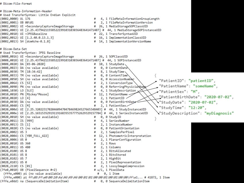
Preprint – DICODerma: A practical approach for metadata management of images in dermatology                                     2

2   The DICOM Standard                                             The workgroup 19 (WG19) of the DICOM consortium has ex-
                                                                   plored ways in which DICOM can be extended to dermatological
DICOM is one of the most widely used standards in healthcare       applications though the group did not propose a complete final
defining formats for images and structured data, workflow man-     standard [3]. The existing IODs such as the Visible Light (VL)
agement and network protocols [6]. The National Electrical         and the Standard Capture (SC) can be used for dermatological
Manufacturers Association (NEMA) foresees the administration       applications with little modifications. Device and acquisition-
of the standard but has no license requirement for use. Some       related metadata are captured by consumer-devices and encoded
of the common terms associated with DICOM are the service          in the EXIF header supported by many digital image storage
object pair (SOP) and the image object definition (IOD). Though    formats. There is some overlap between EXIF and DICOM
IODs are generic classes, most IODs represent individual real-     header tags.
world entities such as X-rays and MRI along with the associated
metadata. The combination of an IOD with a service such as
storage, print or query, is the SOP.                              3.1 The Machine Learning revolution
The various metadata associated with the images includes pa- The growing popularity of machine learning (ML) and artifi-
tient demographics, series (a group of closely related images), cial intelligence (AI) applications in dermatology has brought
study (all series associated with one procedure) and the acquired new requirements for image management [1]. The need for
binary image data. The metadata has a numerical key called standardized images, labelled with appropriate metadata, is an
the tag, data type called the value representation (VR) and the enabler for AI applications. The digital revolution encourages
value multiplicity (VM) count. The metadata is organized into sharing of images with peers and experts from other disciplines
logical groups such as the patient module. The list of these for opinion and as such being part of the wider institutional
specifications that a product supports is called the conformance image management infrastructure such as the picture archiv-
statement. In short, DICOM specifies storage for storing, pro- ing and communication system (PACS). Adoption of Electronic
cessing, transmitting and displaying imaging data. The DICOM Health Record (EHR) systems made it necessary to have a com-
header is seen in Figure 1.                                       plete digital longitudinal patient record that includes clinical
                                                                  images captured during a dermatology encounter. The need for
                                                                  adopting enterprise-imaging standards is becoming increasingly
3 Imaging standards in Dermatology                                important in dermatology.
Imaging standards have a crucial role in the clinical image
management in dermatology owing to its highly visual nature.       4   Our Approach
Dermatologists use different types of images ranging from der-
moscopy to total-body maps. Sophisticated methods such as         Guided by the design science research methodology [7], we
reflectance confocal microscopy are also becoming increasingly    systematically investigated the solution space for the problem
popular. In this article, we give emphasis to the common digital  of standardizing the digital image workflow for dermatology.
photographs, but some of the discussions may apply to other       Our aim was to find generalizable design knowledge that can
modalities as well.                                               guide system designers and policymakers. Though specific
                                                                  requirements vary among different user groups of an information
Image metadata is important in dermatology as in other do-
                                                                  system, they follow generic laws called meta-requirements [8].
mains. The useful metadata includes demographic details, clini-
                                                                  We identified some of the meta-requirements as below:
cal findings, device settings and image characteristics. Accurate
rendering of images and acquisition context is important in der-        1. The existing DICOM standard should be leveraged as
matology as well [3]. Dermatology has a distinct ontology that             much as possible so that existing solutions such as
is used for an accurate textual description of lesions. The meta-          PACS can be directly used in dermatology.
data standards should support the domain-specific ontology of
dermatology and support the emerging modalities.                        2. The users should be able to enter the DICOM ecosys-
                                                                           tem without adopting the entire standard, ideally using
Though dermatology is highly visual, dermatologists do not com-            simple tools that are already in use.
pletely rely on the captured images for diagnostic, prognostic
and therapeutic decision making, and as such accuracy of colour         3. The solution should be usable even with no vendor
and resolution is not very crucial. Images are mainly used for             adoption, but vendors who adopt the standard should
documentation, but with the increasing popularity of telederma-            have an incentive to do so.
tology, parameters like resolution and color accuracy have also         4. The solution should support improved patient privacy.
become more important. Dermatologists, especially those work-           5. Search Engine Optimisation [SEO]: Search engines
ing in the community and those in limited resource settings, rely          and social media platforms have an increasingly im-
on consumer devices such as digital cameras and smartphones                portant role in knowledge dissemination in a privacy-
for image capture and documentation. Image capture mostly                  preserving manner. Potential solutions should address
happens during a face-to-face consultation and routine physical            the needs of these platforms [9].
examinations. Hence, though the DICOM standard can be used
as it is in dermatology, its overall adoption by vendors as well        6. The standard should support emerging techniques such
as practitioners has not been very encouraging as of now. The              as machine learning and artificial intelligence.
lack of adoption is mostly due to the large overhead required for       7. The meta-design should be sufficiently abstract so that
the implementation and adoption of a comprehensive standard                it can be easily implemented by vendors and users to
such as DICOM.                                                             support new needs.
Preprint – DICODerma: A practical approach for metadata management of images in dermatology 3
8. The standard should be simple and easy to adopt and EXIF enabling the inclusion of patient metadata in consumer im-
adapt to, leveraging existing tools. age files. DICODerma uses popular and open-source dcm4che
java library [14] for writing DICOM (dcm) files from JPEG file
format, a popular format supported by most capture devices and
5 Design image editing software. These converted DICOM files can be
used in any system that supports these standards.
As potential users of DICODerma, we adopt a meta-design
approach to translate the generalizable meta-requirements as
described above into a prototype that can be extended. We 5.3 DIT4IJ
created two software artifacts (meta-design) in the solution space
that aligns with the above meta-requirements. One is a java ImageJ has several plugins that can display, edit, save and pro-
library called DICODerma, to encode some of the important cess digital images in various formats including DICOM. Owing
DICOM tags as EXIF tags. The other is a plugin called DIT4IJ to the extensible, plugin architecture of ImageJ, advanced uses
for the popular open-source biomedical image management not natively supported by ImageJ can be added. The modules
software — ImageJ. Both are open-source available from the are typically written in Java and can be installed from the ImageJ
GitHub repository [10]. Before we describe our meta-design user interface or manually copied to the plugins folder in the
in detail, we will briefly introduce the EXIF standard and the ImageJ folder structure. The additional functions introduced by
ImageJ platforms that form the building blocks for our meta- the plugins can be easily integrated into the ImageJ graphical
design. user interface (GUI). The plugins, depending on their type and
functions, implement certain abstract base classes in the ImageJ
5.1 EXIF Tags core and provide implementations for methods such as run and
setup.
EXIF tags (hereafter EXIF) are metadata tags added by con-
sumer devices such as digital cameras to digital images captured DIT4IJ is an ImageJ plugin that adds the following four func-
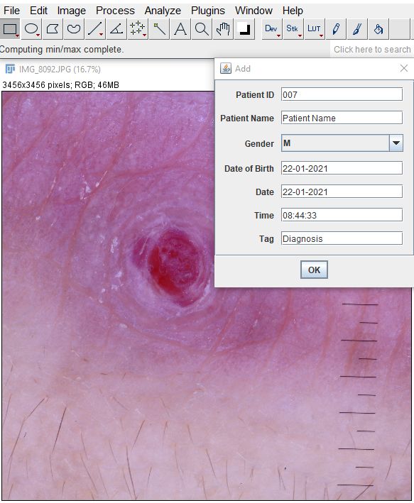
by these devices (this includes images captured on smartphones tions as submenus in the ImageJ. The ‘add tags’ function re-
too). EXIF captures a variety of details ranging from date and ceives the tags --- patient id, patient name, gender, date time
time information to camera settings such as aperture and shutter and diagnosis — from the user and converts them to a JSON
speed, and GPS coordinates for the location of capture. EXIF string and writes the string to the ‘User Comment’ EXIF tag
is part of the TIFF specification and can be found in image file of an image. The ‘StudyDescription’ tag is used to capture the
types such as JPG and PNG in addition to TIFF. The GIF format diagnosis ( Figure 1). The ImageJ provides the interface for
does not support EXIF. Some tags such as the EXIF version are inputting these tags ( Figure 2). DIT4IJ can display these tags
mandatory while most tags are optional such as the user com- for any image and provides an interface to search for these tags
ment tag. EXIF is a consumer specification and does not support in a folder structure. For example, it can open all images of a
any of the clinical tags in the DICOM header. However, some particular diagnosis such as lichen planus by searching in any
of the EXIF tags overlap with headers in the DICOM IODs. We specified file folder in the computer, including all subfolders
adopt a design approach that leverages the EXIF for clinical in the search. The consumer file formats such as JPEG can be
tags. converted into DICOM and saved anywhere in the system. This
converted DICOM (dcm) file can be used with any DICOM
aware application. See the attached video file to see the usage
5.2 ImageJ demonstration.
ImageJ is an image analysis program developed by the National
Institute of Health (NIH), widely used for biomedical image
analysis [11]. ImageJ is an open-source JAVA-based software 5.4 Advantages
with an extensible plug-in architecture. The first version which
We address the common limitation in the existing consumer
was released 25 years back was rewritten as ImageJ2 with addi-
image formats — the lack of support for patient metadata. This
tional functionalities. ImageJ2 and Fiji (ImageJ bundled with
need is addressed without affecting the images by the use of
a range of plugins that facilitate scientific image analysis) are
EXIF. The clinicians can still continue to use their imaging tools
widely used for biomedical image management [12].
for capture, processing and visualization of images. Some of the
DICOM SC IOD is for images that are converted from a non- visualization tools support viewing the EXIF metatags including
DICOM format such as JPEG and PNG. It is a modality indepen- UserComment, though the JSON formatted string is not meant
dent DICOM format with no constraints on the pixel data format. for direct visualization.
Though the initial specification was confined to single-frame
We introduce ImageJ, a popular biomedical imaging software
images, it has been expanded to include multi-frame images.
to the dermatology community. ImageJ is currently not a popu-
As SC IOD is modality independent PACS will not assign any
lar image viewer for clinical dermatology though it has use in
modality [13].
dermatopathology. Some of the image manipulation algorithms
We mapped common demographic and study-related tags from for clinical and cosmetic dermatology can be easily built us-
the DICOM SC IOD to a JSON structure as shown in Figure 1. ing the modular and extensible ImageJ framework. Some such
The DICODerma Java library (hereafter DICODerma) facilitates commercial products are available [15]. We believe that the
writing the JSON, represented as a string, to the ‘UserComment’ functions introduced by DIT4IJ will make ImageJ, a useful tool
section of EXIF. DICODerma can read and parse the JSON in dermatologists’ armamentarium and democratize imaging
string from EXIF. This enables mapping useful DICOM tags to workflows.Preprint – DICODerma: A practical approach for metadata management of images in dermatology 4
Figure 1: Mapping of DICOM tags to JSON for inclusion in the UserComment EXIF tag
Figure 2: The DIT4IJ interface for adding tags to an imagePreprint – DICODerma: A practical approach for metadata management of images in dermatology 5
The adoption of the DICOM standard in dermatology depends a increasingly accurate and useful for dermatologists and residents.
lot on the vendor support and the incorporation into commercial DICODerma method can improve the accuracy further because
software products. The open-source dicoderma library could of the availability of standard metadata.
facilitate the adoption of these standards by the software vendors.
Teledermatology is becoming increasingly important because of
Dermatologists increasingly use smartphones as a handy image
the scarcity of dermatologists, especially in resource-poor areas.
capture device. DICODerma can be used in smartphone apps to
The exchange of good quaity clinical images between patients
provide image tagging capability.
and dermatologists is vital in teledermatology [20]. The discus-
The inclusion of patient metadata in consumer file formats may sions related to skin findings in pandemics such as COVID-19
violate patient privacy if these images are inadvertently shared. is crucial for screening. DICODerma may improve the efficient
The metadata can be anonymized using the same techniques use of images for these purposes [21].
used for anonymizing DICOM resources. With wider adoption
Smartphone based image acquisition is the new normal in derma-
of this standard, patient privacy may paradoxically improve
tology, with dermoscopic addons becoming available for hand-
as EXIF can be easily checked for the presence of dicoderma
held devices [22]. Standardizing image capture from handheld
tags. The sharing platform can reject or block these images if
devices along with relevant metadata, is the need of the hour.
these tags are present. For example, social media platforms can
Vendors can incorporate simple solutions using DICODerma in
automatically reject any uploaded image if that image has the
apps that dermatologists routinely use [23].
dicoderma tags in them.
We propose a simple method and tool for managing imaging
The dicoderma tags will facilitate machine learning. One of the
metadata in dermatological images. Our method is suitable for
challenges with machine learning in dermatology is the lack of
an encounter-based workflow, commonly seen in dermatology.
availability of labelled images in a privacy-preserving manner.
In an encounter-based workflow, the imaging forms part of
Currently, labels associated with images should be supplied
other clinical documentation, unlike in an order-based workflow
as a separate file with unique identifiers. This is not ideal for
where the image-acquisition may be the primary purpose of
collaboration and sharing of resources between teams. Images
the visit [24]. The possibility of integrating with the enterprise
with dicoderma tags can be processed and tags extracted without
imaging systems with minimal change to the traditional and
the need for maintaining an associated metadata file.
straightforward imaging methods that dermatologists are used
to might lead to the development of more elaborate standards.
5.5 Limitations
DICODerma can only handle JPEG images with the traditional
EXIF structure. Sources that generate other file types such as References
PNG and GIF cannot be used with DICODerma. DICODerma
[1] Bell R. Eapen. Artificial Intelligence in Dermatology: A
uses the dcm4che library [14] to convert JPEG images to com-
Practical Introduction to a Paradigm Shift. Indian Derma-
pressed DICOM files. All DICOM readers do not yet support
tology Online Journal, 11(6):881–889, November 2020.
compressed DICOM files. The chance of inadvertently shar-
ISSN 2229-5178. doi: 10.4103/idoj.IDOJ_388_20.
ing sensitive patient information is a challenge in this method
though encryption of EXIF is a solution, again at the cost of [2] Bell Raj Eapen, Norm Archer, and Kamran Sartipi. Lesion-
increasing the the complexity [16]. DICODerm needs further Map: A Method and Tool for the Semantic Annotation of
development to support other modalities such as dermoscopy Dermatological Lesions for Documentation and Machine
and optical coherence tomography. Learning. JMIR Dermatology, 3(1):e18149, April 2020.
doi: 10.2196/18149.
The SC IOD is a general-purpose IOD for use with any digital
image. As the SC IOD is not associated with any modality, [3] Liam J. Caffery, David Clunie, Clara Curiel-Lewandrowski,
some PACS systems may not handle them well. SC IOD lacks Josep Malvehy, H. Peter Soyer, and Allan C. Halpern.
the meta-data model to cater to dermatologists’ unique needs Transforming Dermatologic Imaging for the Digital Era:
such as patient positioning and lighting. However, unlike other Metadata and Standards. Journal of Digital Imaging, 31
specialties that need specialty-specific metadata model, the der- (4):568–577, August 2018. ISSN 0897-1889. doi: 10.
matological community’s needs may be minimal. The machine- 1007/s10278-017-0045-8.
learning algorithms may be less tolerant of variability in colour [4] Taige Cao and Hong Liang Tey. High-definition optical
and lighting than human observers, and these requirements may coherence tomography – an aid to clinical practice and re-
change in the future [17]. We believe that our approach will search in dermatology. JDDG: Journal der Deutschen Der-
introduce dermatologists to the many advantages of standardiza- matologischen Gesellschaft, 13(9):886–890, 2015. ISSN
tion and ignite interest in developing a specialty-specific IOD in 1610-0387. doi: 10.1111/ddg.12768.
the future.
[5] H. Ohshima, S. Kinoshita, M. Futagawa, H. Takiwaki,
K. Washizaki, A. Ishiko, and H. Kanto. Quantification of
6 Discussion allergic and irritant patch test reactions using ImageJ. Skin
The standardization requirements for dermatological images are Research and Technology, 20(2):177–181, 2014. ISSN
beyond the handling of patient metadata. The proposed method 1600-0846. doi: 10.1111/srt.12103.
of using EXIF and interconversion with DICOM header fields [6] W. D. Bidgood and S. C. Horii. Modular extension of the
are easily extensible to capture other relevant metadata. Main- ACR-NEMA DICOM standard to support new diagnos-
stream [18] and specialized search engines [19] are becoming tic imaging modalities and services. Journal of DigitalPreprint – DICODerma: A practical approach for metadata management of images in dermatology 6
Imaging, 9(2):67–77, May 1996. ISSN 0897-1889. doi: Christye Sisson, Stein Skrøvseth, Darren Treanor, Paul
10.1007/BF03168859. Boynton, David Clunie, Michael J. Flynn, Tatsuo Heki,
[7] Hevner, March, Park, and Ram. Design Science in Infor- Stephen Hewitt, Hiroyuki Homma, Andy Masia, Takashi
mation Systems Research. MIS Quarterly, 28(1):75, 2004. Matsui, Balázs Nagy, Masahiro Nishibori, John Penczek,
ISSN 0276-7783. doi: 10.2307/25148625. Thomas Schopf, Yukako Yagi, Hideto Yokoi, and Summit
on Color in Medical Imaging. Consistency and standard-
[8] Haruhiko Kaiya. Meta-Requirements for Information ization of color in medical imaging: A consensus report.
System Requirements: Lesson Learned from Software Journal of Digital Imaging, 28(1):41–52, February 2015.
Ecosystem Researches. Procedia Computer Science, ISSN 1618-727X. doi: 10.1007/s10278-014-9721-0.
126:1243–1252, January 2018. ISSN 1877-0509. doi:
10.1016/j.procs.2018.08.066. [18] Montassar Amri and Kaliyadan Feroz. Google searches
help with diagnosis in dermatology. Informatics in Primary
[9] Afsheen Sharifzadeh, Shinjita Das, and Gideon P. Smith. Care, 21(2):70–72, 2014. ISSN 1475-9985. doi: 10.14236/
Google reverse image search using dermatology eConsult jhi.v21i2.52.
cases. Dermatologic Therapy, 33(6):e14372, 2020. ISSN
1529-8019. doi: 10.1111/dth.14372. [19] P. Tschandl, G. Argenziano, M. Razmara, and J. Yap. Di-
agnostic accuracy of content-based dermatoscopic image
[10] Bell Raj Eapen. DICOM for Dermatol-
retrieval with deep classification features. British Journal
ogy: GitHub:dermatologist/dicom-dermatology.
of Dermatology, 181(1):155–165, 2019. ISSN 1365-2133.
https://github.com/dermatologist/dicom-dermatology,
doi: 10.1111/bjd.17189.
2021.
[20] Nadiya Chuchvara, Rachel Patel, Radhika Srivastava,
[11] Caroline A. Schneider, Wayne S. Rasband, and Kevin W.
Catherine Reilly, and Babar K. Rao. The growth of
Eliceiri. NIH Image to ImageJ: 25 years of image analysis.
teledermatology: Expanding to reach the underserved.
Nature Methods, 9(7):671–675, July 2012. ISSN 1548-
Journal of the American Academy of Dermatology, 82
7105. doi: 10.1038/nmeth.2089.
(4):1025–1033, April 2020. ISSN 0190-9622. doi:
[12] Johannes Schindelin, Ignacio Arganda-Carreras, Erwin 10.1016/j.jaad.2019.11.055.
Frise, Verena Kaynig, Mark Longair, Tobias Pietzsch,
Stephan Preibisch, Curtis Rueden, Stephan Saalfeld, Ben- [21] Pavane L. Gorrepati and Gideon P. Smith. Analysis of
jamin Schmid, Jean-Yves Tinevez, Daniel James White, availability, types, and implementation of teledermatol-
Volker Hartenstein, Kevin Eliceiri, Pavel Tomancak, and ogy services during COVID-19. Journal of the American
Albert Cardona. Fiji: An open-source platform for Academy of Dermatology, 83(3):958–959, September 2020.
biological-image analysis. Nature Methods, 9(7):676–682, ISSN 0190-9622. doi: 10.1016/j.jaad.2020.06.022.
July 2012. ISSN 1548-7105. doi: 10.1038/nmeth.2019. [22] Linda Tognetti, Diletta Fiorani, Giulia Tonini, Lorenzo
[13] Oleg S. Pianykh. DICOM SOPs: Beyond Basic. In Zuliani, Gennaro Cataldo, Alberto Balistreri, Gabriele
Oleg S. Pianykh, editor, Digital Imaging and Communi- Cevenini, Elisa Cinotti, and Pietro Rubegni. Dermoscopy:
cations in Medicine (DICOM): A Practical Introduction Fundamentals and Technology Advances. In Michele Fimi-
and Survival Guide, pages 169–175. Springer, Berlin, Hei- ani, Pietro Rubegni, and Elisa Cinotti, editors, Technology
delberg, 2012. ISBN 978-3-642-10850-1. doi: 10.1007/ in Practical Dermatology: Non-Invasive Imaging, Lasers
978-3-642-10850-1_8. and Ulcer Management, pages 3–24. Springer Interna-
tional Publishing, Cham, 2020. ISBN 978-3-030-45351-0.
[14] Max J. Warnock, Christopher Toland, Damien Evans, Bill doi: 10.1007/978-3-030-45351-0_1.
Wallace, and Paul Nagy. Benefits of using the DCM4CHE
DICOM archive. Journal of Digital Imaging, 20 Suppl [23] Christine Jacob, Antonio Sanchez-Vazquez, and Chris
1:125–129, November 2007. ISSN 0897-1889. doi: 10. Ivory. Factors Impacting Clinicians’ Adoption of a Clinical
1007/s10278-007-9064-1. Photo Documentation App and its Implications for Clinical
Workflows and Quality of Care: Qualitative Case Study.
[15] De-Tian Xu, Jian-Na Yan, Yong Cui, and Wei Liu. Quan- JMIR mHealth and uHealth, 8(9):e20203, September 2020.
tifying facial skin erythema more precisely by analyz- doi: 10.2196/20203.
ing color channels of The VISIA Red images. Jour-
nal of Cosmetic and Laser Therapy: Official Publica- [24] Dawn Cram, Christopher J. Roth, and Alexander J. Towbin.
tion of the European Society for Laser Dermatology, Orders- Versus Encounters-Based Image Capture: Impli-
18(5):296–300, October 2016. ISSN 1476-4180. doi: cations Pre- and Post-Procedure Workflow, Technical and
10.3109/14764172.2016.1157360. Build Capabilities, Resulting, Analytics and Revenue Cap-
ture: HIMSS-SIIM Collaborative White Paper. Journal
[16] Hidear Talirongan, Ariel M. Sison, and Ruji P. Medina. of Digital Imaging, 29(5):559–566, October 2016. ISSN
A New Advanced Encryption Standard-Butterfly Effect 1618-727X. doi: 10.1007/s10278-016-9888-7.
in Protecting Image of Copyright Piracy. In Proceedings
of the 6th International Conference on Information Tech-
nology: IoT and Smart City, ICIT 2018, pages 214–218,
New York, NY, USA, December 2018. Association for
Computing Machinery. ISBN 978-1-4503-6629-8. doi:
10.1145/3301551.3301603.
[17] Aldo Badano, Craig Revie, Andrew Casertano, Wei-Chung
Cheng, Phil Green, Tom Kimpe, Elizabeth Krupinski,You can also read