High-resolution handheld rigid endomicroscope based on full-field optical coherence tomography
←
→
Page content transcription
If your browser does not render page correctly, please read the page content below
High-resolution handheld rigid
                 endomicroscope based on full-field
                 optical coherence tomography

                 Emilie Benoit a la Guillaume
                 Franck Martins
                 Claude Boccara
                 Fabrice Harms

Downloaded From: https://www.spiedigitallibrary.org/journals/Journal-of-Biomedical-Optics on 10 Aug 2021
Terms of Use: https://www.spiedigitallibrary.org/terms-of-use
Journal of Biomedical Optics 21(2), 026005 (February 2016)

                High-resolution handheld rigid endomicroscope
                based on full-field optical coherence tomography
                Emilie Benoit a la Guillaume,a,* Franck Martins,a Claude Boccara,a,b and Fabrice Harmsa
                a
                 LLTech, Pépinière Paris Santé Cochin, 29 rue du Faubourg Saint-Jacques, 75014 Paris, France
                b
                 ESPCI ParisTech, PSL Research University, UMR 7587 CNRS, U979 INSERM, Institut Langevin, 1 rue Jussieu, 75005 Paris, France

                              Abstract. Full-field optical coherence tomography (FF-OCT) is a powerful tool for nondestructive assessment of
                              biological tissue, i.e., for the structural examination of tissue in depth at a cellular resolution. Mostly known as a
                              microscopy device for ex vivo analysis, FF-OCT has also been adapted to endoscopy setups since it shows
                              good potential for in situ cancer diagnosis and biopsy guidance. Nevertheless, all the attempts to perform endo-
                              scopic FF-OCT imaging did not go beyond lab setups. We describe here, to the best of our knowledge, the first
                              handheld FF-OCT endoscope based on a tandem interferometry assembly using incoherent illumination.
                              A common-path passive imaging interferometer at the tip of an optical probe makes it robust and insensitive
                              to environmental perturbations, and a low finesse Fabry–Perot processing interferometer guarantees a compact
                              system. A good resolution (2.7 μm transverse and 6 μm axial) is maintained through the long distance, small
                              diameter relay optics of the probe, and a good signal-to-noise ratio is achieved in a limited 100 ms acquisition
                              time. High-resolution images and a movie of a rat brain slice have been recorded by moving the contact endo-
                              scope over the surface of the sample, allowing for tissue microscopic exploration at 20 μm under the surface.
                              These promising ex vivo results open new perspectives for in vivo imaging of biological tissue, in particular, in the
                              field of cancer and surgical margin assessment. © 2016 Society of Photo-Optical Instrumentation Engineers (SPIE) [DOI: 10.1117/1.
                              JBO.21.2.026005]

                              Keywords: endoscopy; endomicroscopy; full-field optical coherence tomography; tandem interferometry; medical and biomedical im-
                              aging; optical biopsy.
                              Paper 150630RR received Oct. 8, 2015; accepted for publication Jan. 13, 2016; published online Feb. 8, 2016.

                1     Introduction                                                                 in order to apply two-photon fluorescence (TPE) microscopy
                Optical endomicroscopy is an expanding field of research that                      and second-harmonic generation (SHG) microscopy in vivo.
                consists of the integration of a high-resolution optical imaging                   Although its development has been slowed down, mainly by
                                                                                                   the high cost and bulky size of the light source, a high-sensitivity
                technique into a small diameter probe in order to perform optical
                                                                                                   compact system was recently built and gave TPE and SHG
                biopsies at the cellular level in situ. The development of these
                                                                                                   images of liver, skin, and retina without using any contrast
                new technologies targets early cancer detection through a
                                                                                                   agent.7 The other main endomicroscopy technique is based on
                fast and minimally invasive process. Flexible endomicroscopes
                                                                                                   Fourier domain optical coherence tomography (FD-OCT) and is
                based on confocal microscopy and marketed by Mauna Kea
                                                                                                   referred to as volumetric laser endomicroscopy. The imaging
                Technologies are already used in gastroenterology, pulmonol-
                                                                                                   part of the OCT system is encapsulated either in an optome-
                ogy, and urology medical departments in a few hundred hospi-
                                                                                                   chanically engineered pill that is swallowed by the patient8 or
                tals in the world since they have demonstrated good accuracy in
                                                                                                   in a catheter that is introduced into the body through the
                several clinical trials.1–4 Developed more recently, spectrally
                                                                                                   classical endoscopy instrument channel and positioned against
                encoded confocal microscopy (SECM)5 has the potential to
                                                                                                   the imaged lumen by endoscopic guidance9 or by inflation for
                overcome two significant limitations of conventional confocal
                                                                                                   balloon-based probes.10,11 In its most common use in gastroin-
                microscopy, which are the need for a contrast agent (fluorescein)
                                                                                                   testinal examination, the imaging spot is helically scanned into
                and the small field of view (less than 500 × 500 μm2 ). Based on                   the digestive tract so that thousands of A-scans are quickly
                endogenous reflectance, SECM acquires multiple points along a                      recorded during one rotation, leading to possible three-dimen-
                transverse line simultaneously, using spatial and spectral sepa-                   sional (3-D) visualization of the volumetric data set. Preliminary
                ration of light through the use of a grating. Thus, performing                     clinical studies have already been conducted on human volun-
                parallel acquisition releases the need for mechanical scanning in                  teers in vivo and demonstrated good adequacy between the OCT
                one direction and allows acquiring images at least three times                     images and the gold standard histology.8–10 A commercial sys-
                faster than conventional confocal endomicroscopy with a sys-                       tem (NvisionVLE Imaging System, Bedford) has also been
                tem that fits into small diameter probes. For instance, Kang                       developed by NinePoint Medical.11
                et al. demonstrated the imaging of 33 cm2 of swine esophagus                           Despite the above-mentioned promising results, endomicro-
                in vivo in 2 min using a 7-mm diameter SECM endoscopic                             scopy still requires improvements to achieve widespread clinical
                probe.6 Nonlinear endomicroscopy has also been developed                           adoption, which explains the thriving variety of newly created
                                                                                                   devices and prototypes. The main efforts are focused on enhancing

                *Address all correspondence to: Emilie Benoit a la Guillaume, E-mail: ebenoit@
                 lltech.fr                                                                         1083-3668/2016/$25.00 © 2016 SPIE

                Journal of Biomedical Optics                                                026005-1                                       February 2016   •   Vol. 21(2)

Downloaded From: https://www.spiedigitallibrary.org/journals/Journal-of-Biomedical-Optics on 10 Aug 2021
Terms of Use: https://www.spiedigitallibrary.org/terms-of-use
Benoit a la Guillaume, et al.: High-resolution handheld rigid endomicroscope. . .
the image quality in terms of resolution and contrast, and found in time domain OCT to extract the useful signal
decreasing the operation time. Moreover, the current challenge from the background;14
is to upgrade the systems working without exogenous contrast ii. a common-path “passive” interferometer, called the im-
agents in order to minimize the regulatory issues linked to per- aging interferometer, located at the distal end of the opti-
mitted dose limits, risk of allergy, etc., and to allow for easier cal probe. It is a simple Fizeau interferometer defined by
access to the medical market. Among noninvasive optical biopsy the partially reflective contact window and the imag-
techniques, full-field OCT (FF-OCT) is known to offer one of ing plane.
the best resolutions since it is able to catch details at micron
resolution within the tissues in three dimensions. As a conse- The idea of cascading two interferometers was introduced in
quence, it is an interesting candidate for endomicroscopy, which fiber-coupled low-coherence interferometry to prevent signal
is evidenced by the work that has already been done to transfer loss induced by polarization mismatch between the reference
the FF-OCT technique from a microscope to an endoscope.12,13 and signal beams.15,16 In the present endoscopy configuration,
Latrive and Boccara13 presented a new FF-OCT design based on the reference beam is the light back-reflected by the window
the coupling of two interferometers and validated it by imaging at the tip of the probe and the signal beam is the light back-
biological tissues ex vivo and skin in vivo. reflected by the sample in contact with this window within a
However a long integration time (1 s∕image acquisition) was slice of thickness LC , where LC is the light source coherence
needed to ensure a good signal-to-noise ration (SNR) and the length. The probe is thus insensitive to ambient perturbations
system was still too bulky for handheld use, so fast in situ im- since the two beams follow a common path through it. In the
aging was not achievable. In this paper, we report significant absence of a second interferometer, the maximum signal recorded
performance improvements on the system described in would come from the slice of the sample in direct contact with the
Ref. 13 and a new instrumental design allowing for handheld probe, corresponding to the zero path length difference. With the
use of a fast FF-OCT rigid endomicroscope. A movie obtained processing interferometer, a controlled path length difference is
with this endoscope on a rat brain coronal slice is presented and introduced, which needs to be compensated in the second inter-
compared to the images recorded with a standard FF-OCT ferometer so that the imaging plane is shifted further inside the
microscope. sample.
2 Materials and Methods
2.2 Full-Field Optical Coherence Tomography
2.1 Tandem Interferometry Endoscope
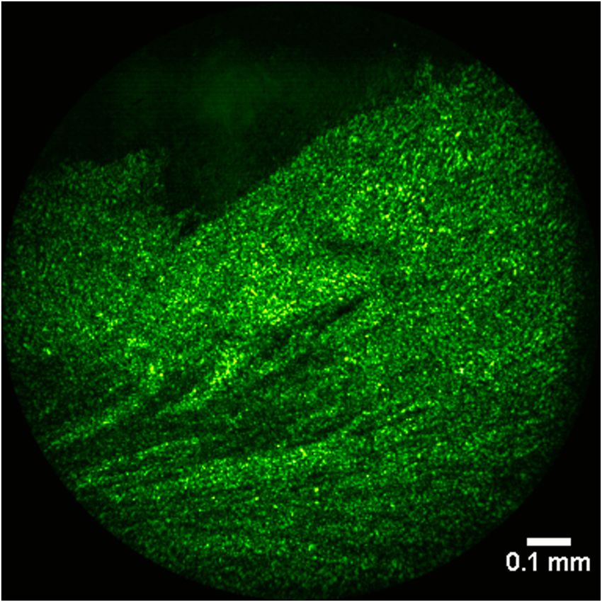
The conventional FF-OCT optical setup for microscopy is based Figure 1(a) gives an overview of the endoscope prototype opti-
on a Linnik interferometer.14 When considering its implementa- cal design. As depicted in Fig. 1(b), the system presents a gun
tion as an endoscope, constraints of miniaturization, stability shape, where the grip carries the light source and associated
and resistance to environmental perturbations impose to divert optics, the barrel is the optical probe, and the back that supports
from the classical configuration toward a design where the most the camera is the imaging arm. With a diameter of 5 cm and a
exposed part—the probe—is as passive as possible. To do so, length of 15 cm, the grip allows easy handling of the 1-kg total
a tandem interferometry system is used, composed of: weight device. The 20 cm ðlengthÞ × 5 mm ðdiameterÞ probe is
long and thin enough to reach many of the endoscopic exami-
i. an “active” interferometer, called the processing inter- nation areas.
ferometer, since it defines the imaging depth within A high-power LED (M660L3, Thorlabs) is used as a low
the tissue and provides the path modulation usually temporal and spatial light source (λ0 ¼ 660 nm, Δλ ¼ 25 nm),
Fig. 1 (a) FF-OCT endoscope prototype setup and (b) pictures. L1 − L3 , lenses; BS, beam splitter; FPI,
Fabry–Perot interferometer; PZT, piezoelectric actuator; PR, partially reflective.
Journal of Biomedical Optics 026005-2 February 2016 • Vol. 21(2)
Downloaded From: https://www.spiedigitallibrary.org/journals/Journal-of-Biomedical-Optics on 10 Aug 2021
Terms of Use: https://www.spiedigitallibrary.org/terms-of-useBenoit a la Guillaume, et al.: High-resolution handheld rigid endomicroscope. . .
with a pair of lenses to conjugate the LED chip with the entrance a section of 35 mm × 45 mm transversally. The thickness of
of the optical probe (L1: 6-mm diameter, 21-mm focal length; the air space between the two plates is precisely defined and
and L2: 9-mm diameter, 22-mm focal length). The processing controlled through the measurement of the fringes period with
interferometer is positioned between the two conjugation lenses, a spectrometer before integration into the endoscope. The thick-
where the light is collimated. After passing the processing inter- ness e determines the depth of the slice imaged with the
ferometer, the light is reflected by a beam splitter plate before FF-OCT device, which is equal to e∕n, where n is the optical
being injected into the optical probe so that 50% of the light index of the sample (average 1.38 for biological tissue).
power is lost and another 50% of the signal power is lost on By changing the piezoelectric actuator offset voltage, the
its way back to the camera. Contrary to conventional medical thickness of the Fabry–Perot interferometer can be modified,
endoscopes with peripheral illumination and central signal col- thus making it possible to scan the imaging plane through
lection, the beam splitter plate is crossed both by the injected the sample depth and retrieve 3-D information. However, the
light emitted by the LED source and by the light coming from depth of imaging is limited to the depth of focus of the optical
the sample, which explains such a significant power loss and the probe13 defined by the objective lens and the wavelength used,
need for a high-power light source. The optical power delivered here set at 50 μm. For this first prototype endoscope, we did not
to the sample is 1mW. The probe is composed of a 20-cm include an axial scanning option, and the position of the imaging
long pitch 2 relay gradient index (GRIN) lens (GRINTECH, plane is fixed at 20 μm behind the surface of the tissue thanks to
Germany) showing a diameter of 2 mm and a relatively small the Fabry–Perot interferometer thickness being tuned to 28 μm
numerical aperture of 0.11 for image transfer, and of an objec- (fringes period of 7.5 nm). The piezoelectric actuator thus only
tive lens (49271, Edmund Optics) with a focal length of 6 mm provides the low amplitude modulation (3.5 VPP ) needed to
and a higher numerical aperture of 0.17 in order to ensure good introduce a π phase shift during one image acquisition in two.
lateral resolution. The probe ends with a planar partially reflec- The displayed OCT signal is the difference between two con-
tive window (thickness 0.3 mm, diameter 2 mm) in order to cre- secutive images.
ate a surface where a plane contact is established with the tissue
and where the reference wave originates. When in contact with 2.4 Performance
biological tissue, the distal window reflects 8% of the incident
light. This reference beam, as well as the signal beam coming The main characteristics and performance of the endoscope are
from the imaging plane, which is the focal plane of the objective summarized in Table 1. The transverse resolution and the field
lens, travel back through the probe and are transmitted through of view are measured experimentally by imaging a high-resolu-
the beam splitter plate, then conjugated with the camera tion 1951 USAF target, and the axial resolution is also exper-
(1440 × 1440 pixels, 12 μm pixel side, 700 Hz maximum imentally evaluated by translating a mirror through the depth of
frame rate, 2 million electrons full well capacity, Adimec, the focus and measuring the full width at half maximum of the
Netherlands) thanks to an achromatic doublet lens of focal fringe envelope. Compared to the system presented in Ref. 13,
length 75 mm and diameter 15 mm. The camera integrated in the transverse resolution evaluated at 2.7 μm is better with this
the FF-OCT endoscope is also a prototype, which has been endoscope thanks to the improvement of the numerical aperture
developed jointly by CMOSIS (Belgium) and Adimec in the of the distal optics, but the axial resolution of 6 μm is poorer
framework of the CAReIOCA European FP7 research project to because of the spectrum of the light source. It was decided for
meet the specific technical requirements of FF-OCT in terms of the light source to prioritize optical power over spectral width so
full well capacity and speed. Internal mechanical mounting that the imaging speed is higher (to minimize the influence of
elements are custom-made in order to minimize space loss. An movements), but the axial resolution is altered. Moreover, this
external protective housing is made from light and thin plastics sectioning ability of 6 μm stays close to the typical thickness of
to reduce weight. Custom-made Labview software is used to histology slides, typically 4 μm.
control the instrument and the acquisition. In particular, the soft- The detection sensitivity is affected by the tandem interferom-
ware synchronizes the camera triggering with the movement of etry configuration, as explained in Ref. 16. As a consequence, a
the piezoelectric actuator located on the processing interferom-
eter in order to perform phase shifting OCT detection. Table 1 FF-OCT endoscope prototype technical data.
2.3 Processing Interferometer
Dimensions 80 mm × 165 mm × 350 mm
In previous realizations of FF-OCT endoscopic setups using
tandem interferometry, the processing interferometer is a Weight 1 kg
Michelson-type one.12,13 Because it is bulky, it has to be sepa-
Probe length 195 mm
rated from the imaging probe in order to optimize the access
possibilities of the device. This leads to the use of a multimode Probe diameter 5 mm
fiber to prolong the optical path between the two interferome-
ters, which increases alignment issues and enhances the critical Axial resolution 6 μm
aspect of good parallelism tuning of the first interferometer.
Transverse resolution 2.7 μm
In our system, a low finesse, tunable Fabry–Perot interferometer
is used (labeled FPI in Fig. 1) and easily inserted after the Field of view Circular, diameter 1.1 mm
source and its collimating lens. Composed of two 50∶50
plate beam splitters of diameter 12.5 mm, one of them being Imaging depth 20 μm
attached to a piezoelectric actuator (PA 8/14, Piezosystem
Frame rate 1 to 10 Hz depending on the sample
Jena, Germany), the Fabry–Perot interferometer extends on and the required averaging
25 mm only in the direction of light propagation and presents
Journal of Biomedical Optics 026005-3 February 2016 • Vol. 21(2)
Downloaded From: https://www.spiedigitallibrary.org/journals/Journal-of-Biomedical-Optics on 10 Aug 2021
Terms of Use: https://www.spiedigitallibrary.org/terms-of-useBenoit a la Guillaume, et al.: High-resolution handheld rigid endomicroscope. . .
6-dB (four-fold) decrease in the SNR is theoretically expected in the difference of axial resolution, and the existence of larger
comparison with the traditional single Linnik interferometer.16 speckle grains on the endoscopic images, which reduces the
In order to evaluate the SNR loss in practice, a rat brain slice possibility of finding uniform areas where the SNR can be com-
fixed in formalin is imaged ex vivo in several areas consecutively pared with the Light-CT scanner images, are the possible rea-
with the endoscope and with the LLTech commercial FF-OCT sons the image quality loss is lower than theoretically expected.
microscope (Light-CT scanner). All the experiments reported in The sensitivity of −88 dB usually expected from the Light-CT
this paper followed European Union and institutional guidelines scanner under 30 accumulations is thus reduced to −83 dB with
for the care and use of laboratory animals. For the purposes of the endoscope prototype. In spite of the SNR alteration, ana-
this comparative study as well as to evaluate the performance tomical features such as axons and myelin fibers or bundles
enhancement brought by the new camera, the endoscope is are easily recognized on the endoscopic images, which is an
tested either with the Adimec camera or with the so-called con- important pillar of the diagnostic.
ventional camera (1024 × 1024 pixels, 10.6 μm pixel side, When the endoscope is equipped with the Adimec camera
140 Hz maximum frame rate, 0.2 million electrons full well instead of the conventional camera, the SNR measured on
capacity, Photon Focus, Switzerland), which is the camera clas- the images increases by a factor of 2.7. This enhancement is
sically used in FF-OCT setups. The result of the comparison on the consequence of a higher full well capacity, provided that
two different areas of the rat brain slice is given in Fig. 2. On the the light power is enough to saturate the sensor pixel wells.
left, the images recorded with a Light-CT scanner are shown. In As the theoretical gain factor in full well capacity between
the middle and on the right, the images taken with the prototype the two cameras is 10, the expected gain factor in SNR is 3.2,
endoscope equipped with the Adimec camera and with the since the SNR is proportional to the square root of the full well
conventional camera, respectively, are shown. The images have capacity (shot noise limited acquisition). As the Adimec camera
is a prototype, the gain in full well capacity may be reduced,
been resized and cut in order to limit the comparison to a
which explains the difference between the measurement and
common zone at a common scale. The conditions of illumina-
the theory. Another way to increase the SNR of the OCT images
tion (camera saturated at 80%) and averaging (30 accumula-
is to average more. Following this strategy, seven times more
tions) during the acquisition are also the same for each device.
accumulations are needed to reach the same image quality
A tolerance of about 10 μm should be considered regarding the
with the conventional camera. Consequently, the second advan-
adequacy of the imaging planes with the Light-CT scanner and
tage of the Adimec camera is that at a given SNR, the imaging
the endoscope given that the axial resolution and the compres-
speed is multiplied by 4.
sion of the sample are not the same in the two experiments.
The experiment shows a three-fold (5 dB) decrease in the
SNR between the microscope and the endoscope equipped 3 Results
with the same camera. The possible error of adequacy between The same rat brain slice sample is scanned with the FF-OCT
the two imaging planes, the difference of slice thickness due to endoscope prototype equipped with the Adimec camera
Fig. 2 Comparative study between (middle) FF-OCT images taken with the endoscope equipped with
the Adimec camera or (right) with the conventional camera and (left) with the commercial Light-CT scan-
ner. The sample is a rat brain coronal slice, imaged at 20 μm in depth with 30 averaging. The two rows
correspond to two different areas of the sample.
Journal of Biomedical Optics 026005-4 February 2016 • Vol. 21(2)
Downloaded From: https://www.spiedigitallibrary.org/journals/Journal-of-Biomedical-Optics on 10 Aug 2021
Terms of Use: https://www.spiedigitallibrary.org/terms-of-useBenoit a la Guillaume, et al.: High-resolution handheld rigid endomicroscope. . .
in areas 4 and 6. These smaller details are more easily caught
when watching the images in motion than when looking at
a static image taken with the endoscope.
4 Conclusion
In this paper, we presented, to the best of our knowledge, the
first handheld FF-OCT endoscope. A tandem inteferometry
design including a modulated Fabry–Perot etalon as the process-
ing interferometer results in a compact and robust device. The
high-resolution characteristics of FF-OCT are well preserved
transversally thanks to the combination of a GRIN lens and
an objective lens in the optical probe, slightly altered axially
because of the use of a light source of smaller bandwidth. The
movie recorded with this endoscope ex vivo on a rat brain slice
shows a good level of structural detail at 20 μm within the sam-
ple after the surface of contact. Our current results suggest good
expectactions for future in vivo experiments.
The comparison with the commercial FF-OCT microscope
reveals some limitations of the endoscope that need to be pushed
to reach the same image quality. Indeed, some further efforts can
be made to get rid of the parasitic light coming from the several
Fig. 3 Frame of Video 1 (QuickTime, 25 MB) showing rat brain slice
interface air/optical components crossed by the light injected
imaging at 5 fps with the FF-OCT endoscope. [URL: http://dx.doi.org/
10.1117/1.JBO.21.2.026005.1]. into the sample, via the use of antireflective coatings and dia-
phragms, in order to increase SNR. Current developments also
plan to allow new functionalities such as axial scanning of the
(Fig. 3). A “green hot” look-up table is chosen for the visuali- sample. The diversity and the constant renewal of LED light
zation of the series of images, since it improves visual percep- sources commercially available also give the opportunity to
tion of the contrast between the different structures. With 30 improve the axial resolution of the system by increasing the
averaging/frame, the acquisition is done at 5 Hz. A wide-field spectral bandwidth and to obtain a higher speed gain by increas-
image of the sample achieved with a Light-CT scanner is shown ing the available light power. Finally, visualization and interpre-
in Fig. 4 for comparison, where the path followed by the endo- tation of FF-OCT images can also be improved through image
scope is notified by the dashed line. Several areas can be iden- processing, but this work should considerably benefit from the
tified on the endoscopic movie depending on their structural first users’ feedback.
architecture. The numbering of these areas is defined in Fig. 4. In the framework of the CAReIOCA project, the prototype
The axon fibers are easily recognized on the endoscopic movie endoscope was transferred to Gustave Roussy Institute (IGR) in
with different orientations depending on their location inside Villejuif (France) in early June in order to be tested in a preclini-
the brain slice. In the areas labeled 1 and 5, the axon fibers cal study on biopsy samples taken in the oral cavity (head,
are seen in a longitudinal view, whereas their cross-section is tongue, throat) from patients suspected for or diagnosed with
visible in area 3. Much thinner fibers of myelin also appear head and neck cancer. About 50 samples are planned to be
Fig. 4 Wide-field image (5 × 8.3 mm2 ) of the coronal rat brain slice studied in Video 1 taken with a Light-
CT scanner for comparison. The field of view and path of the endoscope are represented by the white
dashed circles and arrow, respectively.
Journal of Biomedical Optics 026005-5 February 2016 • Vol. 21(2)
Downloaded From: https://www.spiedigitallibrary.org/journals/Journal-of-Biomedical-Optics on 10 Aug 2021
Terms of Use: https://www.spiedigitallibrary.org/terms-of-useBenoit a la Guillaume, et al.: High-resolution handheld rigid endomicroscope. . .
imaged. A preliminary work was done before to evaluate the 6. D. Kang et al., “Comprehensive confocal endomicroscopy of the
potential of FF-OCT in the diagnosis of head and neck cancer. esophagus in vivo,” Endosc. Int. Open 2, E135–E140 (2014).
7. J. Liu et al., “Low cost and compact nonlinear (SHG/TPE) laser scan-
It consisted of the observation and analysis of biopsy samples
ning endoscope for bio-medical application,” Proc. SPIE 9304, 93041K
with a Light-CT scanner by two pathologists after training, in (2015).
comparison with the gold standard histology.17 The diagnosis 8. M. J. Gora et al., “Tethered capsule endomicroscopy enables less inva-
accuracy of the presence or not of a cancer reached 87%. sive imaging of the gastrointestinal tract microstructure,” Nat. Med.
The same pathologists are involved in the endoscopic study 19(2), 238–241 (2013).
so that they are already familiar with FF-OCT images. The endo- 9. T.-H. Tsai et al., “Ultrahigh speed endoscopic optical coherence tomog-
scopic movie review at IGR is still in progress and will result in raphy for gastroenterology,” Biomed. Opt. Express 5(12), 4387–4404
(2014).
the evaluation of the diagnostic capability of the FF-OCT endo- 10. M. J. Suter et al., “Esophageal-guided biopsy with volumetric laser
scope prototype by the end of 2015. In vivo tests on an animal endomicroscopy and laser cautery marking: a pilot clinical study,”
model will also give the opportunity to estimate the impact of Gastrointest. Endosc. 79(6), 886–896 (2014).
the current speed limitations of the system and to appreciate 11. H. C. Wolfsen et al., “Safety and feasibility of volumetric laser endo-
the difficulty of maintaining the contact between the endoscope microscopy in patients with Barrett’s esophagus (with videos),”
and the tissues despite the natural constraining movements and Gastrointest. Endosc. 82(4), 631–640 (2015).
12. W. Y. Oh et al., “Spectrally-modulated full-field optical coherence
shapes of a living body. We believe that with further work on microscopy for ultrahigh-resolution endoscopic imaging,” Opt. Express
miniaturization and after the regulatory procedure is complete, 14(19), 8675–8684 (2006).
the device will be ready for clinical evaluation in head and neck 13. A. Latrive and A. C. Boccara, “In vivo and in situ cellular imaging
cancer diagnosis for biopsy guidance and margin assesment. full-field optical coherence tomography with a rigid endoscopic probe,”
Other diseases routinely diagnosed or monitored with rigid Biomed. Opt. Express 2(10), 2897–2904 (2011).
endoscopy are direct potential applications of FF-OCT endos- 14. A. Dubois et al., “High-resolution full-field optical coherence tomog-
raphy with a Linnik microscope,” Appl. Opt. 41(4), 805–812 (2002).
copy (e.g., arthroscopy), in particular where removal of tissue
15. K. Bamford et al., “Optical radar detection of precancerous bronchial
is of particular caution at a microscopic scale (neurosurgery). tissue,” Lasers Med. Sci. 15(3), 188–194 (2000).
The introduction of FF-OCT endoscopy in standard medical 16. H. D. Ford et al., “Comparative signal-to-noise analysis of fibre-optic
procedures could give access to this microscopic view with min- based optical coherence tomography systems,” J. Mod. Opt. 52(14),
imal invasion and without any contrast agent. 1965–1979 (2005).
17. F. De Leeuw et al., “Full-field OCT for fast diagnostic of head and neck
cancer,” Proc. SPIE 9303, 93031Z (2015).
Acknowledgments
We would like to thank LLTech’s partners in the CAReIOCA Emilie Benoit a la Guillaume received her PhD degree in physics in
2013 from Pierre et Marie Curie University for the work she did on
project. This research was part of the CAReIOCA project and acousto-optic imaging. She then joined LLTech as a research and
was supported by the European Union Seventh Framework development project manager to work on the latest evolutions of full-
Program FP7-ICT-2011-8 under Grant Agreement No. 318729 field optical coherence tomography technique, such as endoscopy.
(www.careioca.eu).
Franck Martins is the manufacturing manager of LLTech. He has
17 years of experience in the development of medical devices with
a cutting-edge expertise in opto-mechanical design and fabrication
References processes, as well as in CE marking and regulatory affairs.
1. M. Giovannini et al., “Results of a phase I–II study on intraductal con-
focal microscopy in patients with common bile duct stenosis,” Surg. Claude Boccara is a former dean of research at ESPCI and has more
Endosc. 25(7), 2247–2253 (2011). than 300 scientific publications, 11 awards and H index 48. He was
2. M. W. Shahid et al., “Diagnostic accuracy of probe-based confocal laser involved in light-matter interactions, introduced new instruments and
methods mostly limited in their performances by physical laws such as
endomicroscopy in detecting residual colorectal neoplasia after EMR:
new kind of microscopies to increase depth and lateral resolution.
a prospective study,” Gastrointest. Endosc. 75(3), 525–533 (2012). Recently, optical approaches to ultimate measurements have
3. S. P. Chen and J. C. Liao, “Confocal laser endomicroscopy of bladder found new fields of application from optical detection of gravitational
and upper tract urothelial carcinoma: a new era of optical diagnosis?” waves to imaging though scattering media.
Curr. Urol. Rep. 15, 437 (2014).
4. T. Hassan et al., “A novel method for in vivo imaging of solitary lung Fabrice Harms is the former CTO of LLTech (until September 2015).
nodules using navigational bronshoscopy and confocal laser microen- He has 14 years of experience in biophotonics and biomedical fields,
doscopy,” Lung 193, 773–778 (2015). managing research and product development programs as well as
5. G. J. Tearney, R. H. Webb, and B. E. Bouma, “Spectrally encoded building and leading teams. He holds a PhD in physics from Pierre
confocal microscopy,” Opt. Lett. 23(15), 1152–1154 (1998). et Marie Curie University and is among the inventors of 6 patents.
Journal of Biomedical Optics 026005-6 February 2016 • Vol. 21(2)
Downloaded From: https://www.spiedigitallibrary.org/journals/Journal-of-Biomedical-Optics on 10 Aug 2021
Terms of Use: https://www.spiedigitallibrary.org/terms-of-useYou can also read