Do Proteins Adhere to Gold ITC Cells? - TA Instruments - Technical Note
←
→
Page content transcription
If your browser does not render page correctly, please read the page content below
TA Instruments – Technical Note
  __________________________________________________________________________________
                         Do Proteins Adhere to Gold ITC Cells?
                                         Colette Quinn, PhD.
      Microcalorimetry, TA Instruments – Waters LLC, 890 West 410 North, Lindon, UT 84042, USA

Introduction

           The Nano ITC ultrasensitive isothermal titration instrument contains a sample cell and
reference cell made entirely of 99.999% Au(0). Gold was selected for this instrument because of its
desirable thermodynamic properties as an excellent conductor and its inert nature. When the thermal
signal from inside an ITC cell can be rapidly and quantitatively transferred to the sensors attached to the
outside of the cell, the result is a rapid response time and the highest sensitivity that can be achieved.

           There is a mistaken belief that thiol containing or highly charged proteins will nonspecifically
adsorb to or chelate the gold of an ITC cell, thus rendering the titration data generated while the
nonspecific reaction is taking place useless. Published reports do show that many proteins with a
reactive cysteine residues will in some instances bind to Au(I) particles or surfaces. Even more
publications exist that demonstrate that specialized gold surfaces can be functionalized in such a way
that proteins are easily coupled to this chemically altered surface.

The gold cells in a Nano ITC are Au(0) not Au(I) and are not functionalized in any way.

         ITC is not the only system that takes advantage of gold’s unique properties. Surface Plasmon
Resonance (SPR), also commonly known by one of the most frequently used instrument names,
BiaCore®, is a technique where one partner of an interacting pair is immobilized on a functionalized,
gold-coated slide. In the binding assay, the other partner is flowed across the gold surface (1). For SPR,
as well as ITC, it is assumed that nonspecific proteins do not passively or chemically adsorb to the gold
surfaces during the binding assay, otherwise both techniques would give invalid results.

         One effective method to show the non-reactivity of the ITC Au(0) cells in the Nano ITC cells is to
design an actual titration experiment where any nonspecific binding of the reagents to the cell surfaces
would be evident in the raw data. In this report, a cysteine containing protein, that is also highly
charged and in theory might have a tendency to nonspecifically bind to the ITC gold cells, was used in a
typical titration. The results generated with such a model protein can be extrapolated to many proteins
with similar structures and chemical characteristics. This report shows raw titration data from one
representative model that demonstrates the non-reactivity of the Nano ITC gold cells. It also provides
guidelines for ITC users that may be considering ITC experiments with other samples that may have
nonspecific binding reactions.

 ____________________________________________________________________________________
TA Instruments – Waters LLC            Page 1 of 6                      MCTN-2011-03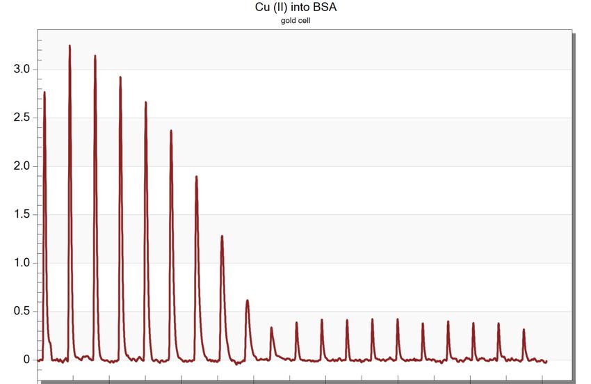
TA Instruments – Technical Note
__________________________________________________________________________________
Results and Discussion
Will cysteine (-SH) containing proteins will adsorb onto the gold surface of an ITC instrument?
In order to address this question, the protein bovine serum albumin (BSA) was used as a test
protein. Charged proteins sticking to the surface of glass and polystyrene can be problematic; this
adhesion has even been quantified for BSA, which is considered a notoriously “sticky” protein (2).
Bovine serum albumin is highly charged (-18 overall) at neutral pH and it contains 35 cysteine residues,
where only one residue is not involved in an intermolecular disulfide bond at neutral pH (3). The free
cysteine, Cys-34, has been known to bind gold salts for several decades and this interaction is of interest
to those studying gold-containing therapeutics (4). BSA with the reactivity of the Cys-34, as well as its
overall charge, has lent itself to being a useful protein in the studying adhesion, especially of late, with
the increasing utility of bare or functionalized gold nanoparticles (5). Concern with adhesion of BSA to a
Biacore X system, has also been tested for nonspecific binding. It was found that after exposing the
surfaces to BSA and then rinsing the protein, all BSA had been easily removed from the gold surface (6).
It is true that BSA may bind to some gold surfaces; however, this does not mean that BSA will bind to the
gold cell of an ITC instrument.
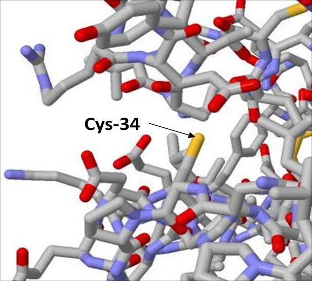
Figure 1. The high-resolution x-ray crystallographic structure of bovine serum albumin (BSA). Cysteine 34 (Cys-34) located
near the surface of BSA.
To test the hypothesis that most proteins do not adhere to the gold of an ITC cell, BSA was an
excellent model system to show the non-reactivity of the Nano ITC cells. Since TA Instruments
manufactures the Nano ITC Standard Volume with either Hastelloy or gold cells, titration experiments
were performed simultaneously on both models of the Nano ITC. Slight changes in the stirring speed and
injection spacing were made so that both instruments were operating under optimal conditions. The
____________________________________________________________________________________
TA Instruments – Waters LLC Page 2 of 6 MCTN-2011-03TA Instruments – Technical Note __________________________________________________________________________________ titrand BSA solutions of either 30 or 35 µM were prepared from the same stock solution and the final concentration of the protein was determined by measuring the absorbance at 280 nM, using the molar extinction coefficient of 43,000M-1cm-1 (7). The titrant was 660 µM of Cu(II) and both titrant and titrand were prepared in 100 mM Tris (µ= 0.1 M) at pH 7.4. Tris buffer was used because the heat of interaction of BSA and Cu(II) is quite large. However, because the of the competition for the Cu(II) with Tris (Cu(Tris)42+formation log β = 14.1 (8)), the binding constant would be in a measurable range for the Nano ITC SV. The titration of Cu(II) into BSA has been previously studied by ITC under different conditions (9). This is a buffer and pH dependent system and under different conditions the results would vary from those reported here (Figure 2, 3 and Table 1). Figure 2. Representative raw data for the titration of Cu(II) into BSA in a Nano ITC SV with a gold cell. Exotherm events are plotted upward. ____________________________________________________________________________________ TA Instruments – Waters LLC Page 3 of 6 MCTN-2011-03
TA Instruments – Technical Note
  __________________________________________________________________________________

Figure 3. Representative titration of the integrated (blue diamond), and fit (red line) data for Cu(II) into BSA in a Nano ITC SV
with a gold cell.

                                             Ka                     ΔH(kJ/mol)        Stoichiometry (n)
                    SV-ITC gold              4.7 ± 0.7 x 10⁷        -31 ± 1           0.93 ± 0.05
                    SV-ITC Hastelloy         4.0 ± 0.8 x 10⁷        -35 ± 1           0.88 ± 0.08

Table 1. Average values and standard error for six titration of Cu(II) into BSA in 100 mM Tris, pH 7.4.

            If BSA did nonspecifically adhere to the gold cell of the instrument during the BSA – Cu(II)
titration, the resulting stoichiometry would be lower and the other thermodynamic parameters may
not agree with each other. In the experiments performed for this report, the Ka fit of the data in Table 1
are similar in magnitude and both the stoichiometry values of 0.93 and 0.88 can be interpreted to be
the same at a value of 1.
 ____________________________________________________________________________________
TA Instruments – Waters LLC                      Page 4 of 6                                MCTN-2011-03
TA Instruments – Technical Note
__________________________________________________________________________________
A second, easy method to address any concern of protein adhesion to the cell surface is to
perform a fast colorimetric or UV-Vis validation test. Using the molar extinction coefficient of the
protein of interest, the final concentration can be determined accurately before and after an
experiment.
This method was utilized with another protein, insulin. A concentrated solution of insulin was
prepared in 2% acetic acid. At this pH the zinc would be displaced by H+, leaving a potential gold surface
binding site available. The initial absorbance of a 2% dilution of the concentrated protein at 280 nm was
0.236. The concentrated solution of protein was then loaded (300 µL) into a Nano ITC Low Volume (LV)
and allowed to incubate for 4 hours while stirring at 350 rpm. The same solution (300 µL) was also
pipetted into a polystyrene tube that was placed into a cooling plate during the same time period at the
same temperature (25 ˚C). After the incubation period, the absorbance at 280 nm was measured. The
insulin incubation experiment was performed in duplicate and it was found that the protein
concentration of the sample removed from the ITC after the incubation decreased by 4% (Abs = 0.227)
in the ITC and by 3 % (Abs avg. = 0.228) in the polystyrene tube. The change in the concentration is
considered insignificant; since errors associated with pipetting, the UV-Vis instrument, and protein
stability could easily contribute to the small discrepancy in the protein concentration over the 4 hour
time.
Summary
The data generated in this report has shown that the proteins BSA and insulin do not adhere
to the Nano ITC gold cells under the conditions of this study. Two separate methods were utilized to
demonstrate the non-reactivity of the Nano ITC gold cells. The typical ITC titration performed with BSA
in either a Nano ITC with Hastelloy or a Nano ITC with gold cells resulted in data that when analyzed
demonstrated identical stoichiometry at 1:1. The second technique, using standard OD 280
concentration determinations to measure the concentration of a concentrated insulin solution before
and after a period of incubation in a Nano ITC instrument with gold cells, demonstrated no significant
difference in concentration pre or post incubation. The results in this report are clear indications that
the two model cysteine rich proteins do not nonspecifically adhere to the gold surfaces of the Nano ITC
cells.
Although BSA and insulin do not represent all possible samples that would be considered for use in a
Nano ITC gold cell instrument, they do demonstrate the non-reactive nature of the gold cells and
certainly indicate the utility of the Nano ITC gold cell instrument when analyzing typical biological
samples. As with all binding assay surfaces, any unexpected values in stoichiometry or binding constant
should be investigated for all nonspecific reactions that may be influencing the raw data.
____________________________________________________________________________________
TA Instruments – Waters LLC Page 5 of 6 MCTN-2011-03TA Instruments – Technical Note
__________________________________________________________________________________
References
1. Homola, J. (2003) Anal. Bioanal. Chem. 377:528-539.
2. Sagvolden, G. (1999) Biophys. J. 77: 526-532.
3. Peters, T., Jr. (1985) Adv. Protein Chem. 37: 161-245.
4. Shaw, P. (1979) Inorg. Perspect. Biol Med. 2: 287-355.
5. Brewer, et al. (2005) Langmuir 21: 9303–9307.
6. Baker, F.R.; Laiwalla, A.N.; Yoon, J-Y; Canavate, J.; Garrell, R.L. (2001) Polm. Mater. Sci. Eng. 85:
115-116.
7. Pace, CN; Vajdos, F.; Fee, L. Grimsely, G.; and Gray, T (1995) Protein Sci 4:2411-2423.
8. Martell, A.E.; Smith, R.M.; Simeon VI (1989) Critical stability constants, vol 5, Plenum, New York.
9. Zhang, Y.; Wilcox, D.E. (2002) J. Biol. Inorg. Chem. 7:327-337.
____________________________________________________________________________________
TA Instruments – Waters LLC Page 6 of 6 MCTN-2011-03You can also read