Does Dominant Prolapse in Other Compartments Affect the Assessment of Bladder Prolapse on Translabial Ultrasound? - Authorea

Page created by Linda Brooks
 
CONTINUE READING
Does Dominant Prolapse in Other Compartments Affect the Assessment of Bladder Prolapse on Translabial Ultrasound? - Authorea
Posted on Authorea 2 Apr 2021 — The copyright holder is the author/funder. All rights reserved. No reuse without permission. — https://doi.org/10.22541/au.161735657.71843341/v1 — This a preprint and has not been peer reviewed. Data may be preliminary.

                                                                                                                                                                                                                                                              Does Dominant Prolapse in Other Compartments Affect the
                                                                                                                                                                                                                                                              Assessment of Bladder Prolapse on Translabial Ultrasound?
                                                                                                                                                                                                                                                              Manli Wu1 , Xudong Wang2 , Zhijuan Zheng1 , Junyan Cao1 , Jing Xu1 , Shuangyu Wu1 , Ying
                                                                                                                                                                                                                                                              Chen1 , Jiawei Tian2 , and Xinling Zhang3
                                                                                                                                                                                                                                                              1
                                                                                                                                                                                                                                                                Third Affiliated Hospital of Sun Yat-Sen University
                                                                                                                                                                                                                                                              2
                                                                                                                                                                                                                                                                Second Affiliated Hospital of Harbin Medical University
                                                                                                                                                                                                                                                              3
                                                                                                                                                                                                                                                                Affiliation not available

                                                                                                                                                                                                                                                              April 2, 2021

                                                                                                                                                                                                                                                              Abstract
                                                                                                                                                                                                                                                              Objective: To explore the impact of dominant prolapse in other compartments in assessing bladder prolapse, and to establish
                                                                                                                                                                                                                                                              cutoffs for staging bladder prolapse among Chinese women using translabial ultrasound. Design: Prospective multicentre
                                                                                                                                                                                                                                                              observational study. Setting: Tertiary referral urogynaecology unit. Population: A total of 741 women with symptoms of
                                                                                                                                                                                                                                                              lower urinary tract dysfunction and/or pelvic floor dysfunction were included. Methods: Women underwent interview, pelvic
                                                                                                                                                                                                                                                              organ prolapse quantification (POP-Q) examinations and 4D translabial ultrasounds. Main outcome measures: The ROC
                                                                                                                                                                                                                                                              statistic was used to assess accuracy and define the optimal cutoffs. Results: The mean patient age was 42.7 years (range,
                                                                                                                                                                                                                                                              18-82). There were 456 women without dominant prolapse in the apical/posterior compartments and 285 women with dominant
                                                                                                                                                                                                                                                              prolapse in the apical/posterior compartments. Among patients without and with dominant prolapse, similar cutoffs (-10.9 mm
                                                                                                                                                                                                                                                              vs. -9.1 mm) were determined for predicting POP-Q stage [?] 2 in the anterior compartment, with AUCs of 0.87 and 0.79,
                                                                                                                                                                                                                                                              respectively. In contrast, significantly different cutoffs (-5.7 mm vs. +3.5 mm) were determined for predicting POP-Q stage
                                                                                                                                                                                                                                                              [?] 1 among patients with and without dominant prolapse, with AUCs of 0.85 and 0.77, respectively. Conclusion: Dominant
                                                                                                                                                                                                                                                              prolapse in the apical/posterior compartments affected the accuracy and cutoffs of translabial ultrasound for staging bladder
                                                                                                                                                                                                                                                              prolapse. Thus, competition of various organs in women with multi-compartment prolapse should be considered as a potential
                                                                                                                                                                                                                                                              complicating factor in assessing pelvic organ prolapse. Funding: The study is supported by grants from the National Natural
                                                                                                                                                                                                                                                              Science Foundation of China (No. 91859115). Key words: Bladder prolapse; translabial ultrasound.

                                                                                                                                                                                                                                                              Title: Does Dominant Prolapse in Other Compartments Affect the Assessment of Bladder Prolapse on
                                                                                                                                                                                                                                                              Translabial Ultrasound?
                                                                                                                                                                                                                                                              Short title: Assessment of Bladder Prolapse on Ultrasound
                                                                                                                                                                                                                                                              Manli Wu1 + , Xudong Wang2 + , Zhijuan Zheng1 , Junyan Cao1 , Jing Xu1 , Shuangyu Wu1 , Ying Chen1 ,
                                                                                                                                                                                                                                                              Jiawei Tian2 *, Xinling Zhang1 *
                                                                                                                                                                                                                                                              +These authors contributed equally to this work and should be considered co-first authors.
                                                                                                                                                                                                                                                              *Contributed equally as co-correspondence authors.
                                                                                                                                                                                                                                                              1
                                                                                                                                                                                                                                                               Department of Ultrasound, the Third Affiliated Hospital of Sun Yat-sen University, Guangzhou, Guangdong
                                                                                                                                                                                                                                                              Province, China; 2 Department of Ultrasound, the Second Affiliated Hospital of Harbin Medical University,
                                                                                                                                                                                                                                                              Harbin, Heilongjiang Province, China.
                                                                                                                                                                                                                                                              Correspondence authors:

                                                                                                                                                                                                                                                                                                                           1
Does Dominant Prolapse in Other Compartments Affect the Assessment of Bladder Prolapse on Translabial Ultrasound? - Authorea
Jiawei Tian, Department of Ultrasound, the Second Affiliated Hospital of Harbin Medical University, No.246,
Posted on Authorea 2 Apr 2021 — The copyright holder is the author/funder. All rights reserved. No reuse without permission. — https://doi.org/10.22541/au.161735657.71843341/v1 — This a preprint and has not been peer reviewed. Data may be preliminary.

                                                                                                                                                                                                                                                              Xuefu Road, Nangang District, Harbin, Heilongjiang Province, PR China. Zip code:150001; Tel: 0451-
                                                                                                                                                                                                                                                              86662961; Fax: 0451-86662961; Email: jwtian2004@163.com.
                                                                                                                                                                                                                                                              Xinling Zhang , Department of Ultrasound, the Third Affiliated Hospital of Sun Yat-Sen University, 600
                                                                                                                                                                                                                                                              Tianhe Road, Guangzhou, Guangdong Province, PR China. ZIP code: 510630; Tel: 020-85253030; Fax:
                                                                                                                                                                                                                                                              020-85252416; Email: zhxinl@mail.sysu.edu.cn.
                                                                                                                                                                                                                                                              Authors’ emails:
                                                                                                                                                                                                                                                              Manli Wu’s e-mail: wumanli@mail2.sysu.edu.cn; Xudong Wang’s e-mail:
                                                                                                                                                                                                                                                              xudongwang1984@163.com; Zhijuan Zheng’s email: zhzhij27@mail.sysu.edu.cn;
                                                                                                                                                                                                                                                              Junyan Cao’s email: caojyan@mail.sysu.edu.cn;Jing Xu’s email: xujing38@mail.sysu.edu.cn;
                                                                                                                                                                                                                                                              Shuangyu Wu’s email: wushy67@mail2.sysu.edu.cn;Ying Chen’s email: cheny393@mail2.sysu.edu.cn;Jiawei
                                                                                                                                                                                                                                                              Tian’s email: jwtian2004@163.com; Xinlin Zhang ’s email: zhxinl@mail.sysu.edu.cn.
                                                                                                                                                                                                                                                              Abbreviations: FPOP, Female pelvic organ prolapse; ICS POP-Q, International Continence Society pelvic
                                                                                                                                                                                                                                                              organ prolapse quantification; AUC, the area under the receiver operating characteristic curve.
                                                                                                                                                                                                                                                              Word count:
                                                                                                                                                                                                                                                              Main text: 2681 words.
                                                                                                                                                                                                                                                              Structured Abstract
                                                                                                                                                                                                                                                              Objective: To explore the impact of dominant prolapse in other compartments in assessing bladder prolapse,
                                                                                                                                                                                                                                                              and to establish cutoffs for staging bladder prolapse among Chinese women using translabial ultrasound.
                                                                                                                                                                                                                                                              Design: Prospective multicentre observational study.
                                                                                                                                                                                                                                                              Setting: Tertiary referral urogynaecology unit.
                                                                                                                                                                                                                                                              Population: A total of 741 women with symptoms of lower urinary tract dysfunction and/or pelvic floor
                                                                                                                                                                                                                                                              dysfunction were included.
                                                                                                                                                                                                                                                              Methods: Women underwent interview, pelvic organ prolapse quantification (POP-Q) examinations and
                                                                                                                                                                                                                                                              4D translabial ultrasounds.
                                                                                                                                                                                                                                                              Main outcome measures: The ROC statistic was used to assess accuracy and define the optimal cutoffs.
                                                                                                                                                                                                                                                              Results: The mean patient age was 42.7 years (range, 18-82). There were 456 women without dominant
                                                                                                                                                                                                                                                              prolapse in the apical/posterior compartments and 285 women with dominant prolapse in the apical/posterior
                                                                                                                                                                                                                                                              compartments. Among patients without and with dominant prolapse, similar cutoffs (-10.9 mm vs. -9.1
                                                                                                                                                                                                                                                              mm) were determined for predicting POP-Q stage [?] 2 in the anterior compartment, with AUCs of 0.87
                                                                                                                                                                                                                                                              and 0.79, respectively. In contrast, significantly different cutoffs (-5.7 mm vs. +3.5 mm) were determined
                                                                                                                                                                                                                                                              for predicting POP-Q stage [?] 1 among patients with and without dominant prolapse, with AUCs of 0.85
                                                                                                                                                                                                                                                              and 0.77, respectively.
                                                                                                                                                                                                                                                              Conclusion: Dominant prolapse in the apical/posterior compartments affected the accuracy and cutoffs
                                                                                                                                                                                                                                                              of translabial ultrasound for staging bladder prolapse. Thus, competition of various organs in women with
                                                                                                                                                                                                                                                              multi-compartment prolapse should be considered as a potential complicating factor in assessing pelvic organ
                                                                                                                                                                                                                                                              prolapse.
                                                                                                                                                                                                                                                              Funding: The study is supported by grants from the National Natural Science Foundation of China (No.
                                                                                                                                                                                                                                                              91859115).
                                                                                                                                                                                                                                                              Key words: bladder prolapse; translabial ultrasound.
                                                                                                                                                                                                                                                              Tweetable abstract

                                                                                                                                                                                                                                                                                                                   2
Dominant prolapse in other compartments affects the accuracy and cutoffs of translabial ultrasound for
Posted on Authorea 2 Apr 2021 — The copyright holder is the author/funder. All rights reserved. No reuse without permission. — https://doi.org/10.22541/au.161735657.71843341/v1 — This a preprint and has not been peer reviewed. Data may be preliminary.

                                                                                                                                                                                                                                                              staging bladder prolapse.
                                                                                                                                                                                                                                                              Main body of text
                                                                                                                                                                                                                                                              Introduction
                                                                                                                                                                                                                                                              Female pelvic organ prolapse (FPOP), defined as the descent of one or more of the female pelvic organs, is
                                                                                                                                                                                                                                                              a common gynecological condition1 . Symptomatic FPOP significantly impacts quality of life among women,
                                                                                                                                                                                                                                                              with an estimated risk as high as 20% for the need of gynecological surgical intervention2 . FPOP has been
                                                                                                                                                                                                                                                              shown to be significantly associated with symptoms of issues such as stress incontinence, voiding dysfunction,
                                                                                                                                                                                                                                                              fecal incontinence, and others3-5 .
                                                                                                                                                                                                                                                              If FPOP is considered as a possible form of hernia6 , the levator hiatus could be regarded as the largest
                                                                                                                                                                                                                                                              potential hernia portal in the human body, allowing for the descent of female pelvic organs. Unsurprisingly,
                                                                                                                                                                                                                                                              there may exist competition between different compartments during pelvic organ descent, so the evaluation
                                                                                                                                                                                                                                                              of pelvic organ prolapse within one compartment could potentially be affected by prolapse occurring in
                                                                                                                                                                                                                                                              another compartment.
                                                                                                                                                                                                                                                              Imaging techniques have played an important role in the investigation of FPOP7 . Of the available techniques,
                                                                                                                                                                                                                                                              translabial ultrasound is currently regarded as one of the most promising modalities in the evaluation of
                                                                                                                                                                                                                                                              prolapses6 . The development of translabial ultrasound has contributed greatly to the understanding of
                                                                                                                                                                                                                                                              FPOP, and it has also helped settle some arguments in the field. However, research appears to be limited
                                                                                                                                                                                                                                                              on the subject of translabial ultrasound and potential competition between different compartments in multi-
                                                                                                                                                                                                                                                              compartment prolapse. In one study8 , it was suggested that the optimal cutoffs for uterine prolapse on
                                                                                                                                                                                                                                                              translabial ultrasound were not affected by dominant prolapse in other compartments. However, to date there
                                                                                                                                                                                                                                                              are no known studies regarding the impact of dominant prolapse in other compartments on the assessment of
                                                                                                                                                                                                                                                              bladder prolapse. Additionally, the best cutoffs for defining bladder prolapse among the Chinese population
                                                                                                                                                                                                                                                              have not been well established on translabial ultrasound.
                                                                                                                                                                                                                                                              Therefore, this study aimed to explore the impact of dominant prolapse in other compartments on the
                                                                                                                                                                                                                                                              assessment of bladder prolapse on translabial ultrasound. This study also sought to establish the best cutoff
                                                                                                                                                                                                                                                              values for bladder prolapse among Chinese women on translabial ultrasound.
                                                                                                                                                                                                                                                              Patients and Methods
                                                                                                                                                                                                                                                              Study population
                                                                                                                                                                                                                                                              This prospective, multicenter study involved ten clinical centers located in Mainland China (Chinese Clinical
                                                                                                                                                                                                                                                              Trial Registration No. ChiCTR ROC 16009855). The study protocol was approved by the local Institutional
                                                                                                                                                                                                                                                              Human Research Ethics Committee. Informed consent was obtained from each study participant. For quality
                                                                                                                                                                                                                                                              control, the data from the entire study was controlled and analyzed by the principal investigator.
                                                                                                                                                                                                                                                              For this study, women were recruited who were seen at tertiary gynecological centers for lower urinary
                                                                                                                                                                                                                                                              tract symptoms and/or symptoms of pelvic organ prolapse between February 2017 and September 2018.
                                                                                                                                                                                                                                                              All participants went through a local standardized interview, a clinical examination using the ICS POP-
                                                                                                                                                                                                                                                              Q assessment, and a 4D translabial ultrasound examination. For the purposes of the research, prolapse
                                                                                                                                                                                                                                                              symptoms were defined as “the sensation of a lump in the vagina and/or a dragging sensation in the vagina”
                                                                                                                                                                                                                                                              9
                                                                                                                                                                                                                                                                . Women with either a history of hysterectomy or pelvic floor surgery for prolapse or incontinence was
                                                                                                                                                                                                                                                              excluded from the data.
                                                                                                                                                                                                                                                              The ICS POP-Q assessment
                                                                                                                                                                                                                                                              The clinical examinations at each center were assessed using the International Continence Society (ICS)
                                                                                                                                                                                                                                                              POP-Q method10,11 by experienced gynecologists (each with [?] 2 years of experience with ICS POP-Q
                                                                                                                                                                                                                                                              examinations). The gynecologists who performed the assessments were blinded to the patients’ clinical data,
                                                                                                                                                                                                                                                              as well as the results of the 4D translabial ultrasound examinations.

                                                                                                                                                                                                                                                                                                                    3
The ICS POP-Q examination assessed anterior, central, and posterior compartment descent by measuring
Posted on Authorea 2 Apr 2021 — The copyright holder is the author/funder. All rights reserved. No reuse without permission. — https://doi.org/10.22541/au.161735657.71843341/v1 — This a preprint and has not been peer reviewed. Data may be preliminary.

                                                                                                                                                                                                                                                              the maximum downward displacement of the anterior vaginal wall, the cervix, and the posterior vaginal wall
                                                                                                                                                                                                                                                              relative to the hymen with the patient in a maximal Valsalva maneuver. The quantitative descriptions of the
                                                                                                                                                                                                                                                              ICS POP-Q assessments from this study were recorded using the five stages of pelvic organ support according
                                                                                                                                                                                                                                                              to the ICS POP-Q system (Table S1). Dominant prolapse in the apical and/or posterior compartment was
                                                                                                                                                                                                                                                              defined as follows: (a) apical compartment prolapses had the same or higher POP-Q stage than the anterior
                                                                                                                                                                                                                                                              compartment prolapse; and/or (b) the posterior compartment prolapse had the higher POP-Q stage than
                                                                                                                                                                                                                                                              the anterior compartment. This is because based on previous findings12 , stage 1 uterine prolapse is just as
                                                                                                                                                                                                                                                              significant in predicting prolapse symptoms as stage 2 anterior or posterior compartment prolapse on ICS
                                                                                                                                                                                                                                                              POP-Q assessments.
                                                                                                                                                                                                                                                              4D translabial ultrasound examination
                                                                                                                                                                                                                                                              The 4D translabial pelvic floor ultrasound examinations were performed using a GE Voluson S6 or S8
                                                                                                                                                                                                                                                              ultrasound device with a 4-8MHz curved array volume transducer and with an acquisition angle of 85° (GE,
                                                                                                                                                                                                                                                              Kretztechnik, GmbH, Zipf, Austria). All ultrasounds were conducted by senior sonographers (each trained for
                                                                                                                                                                                                                                                              at least 50 consecutive examinations). The sonographers had no access to the patients’ other clinical data.
                                                                                                                                                                                                                                                              The patients were examined in the supine position after bladder emptying and defecation, as previously
                                                                                                                                                                                                                                                              described. Volume acquisitions were obtained at rest, on contraction, and on Valsalva, and at least three
                                                                                                                                                                                                                                                              volumes were acquired on Valsalva. Great care had to be taken to have the Valsalva maneuver performed
                                                                                                                                                                                                                                                              for a minimum duration of 6 seconds without levator coactivation13,14 .
                                                                                                                                                                                                                                                              Archived 4D translabial ultrasound datasets were analyzed offline at a later date using 4D View software
                                                                                                                                                                                                                                                              (version 10.0, GE Medical Systems) by two investigators, who each had at least two years of experience with
                                                                                                                                                                                                                                                              4D translabial pelvic floor ultrasound examination. These investigators were both blinded to all other data.
                                                                                                                                                                                                                                                              Using the offline system, they evaluated anterior, central, and posterior compartment descent by measuring
                                                                                                                                                                                                                                                              the leading edge of the bladder, cervix, and rectum relative to the inferior margin of the symphysis pubis.
                                                                                                                                                                                                                                                              Positions above the symphysis pubis were given as positive values, while positions below were given as
                                                                                                                                                                                                                                                              negative values.
                                                                                                                                                                                                                                                              Statistical methods
                                                                                                                                                                                                                                                              Statistical analysis was performed using IBM SPSS Statistics 22 software for Windows (SPSS Inc., Chicago,
                                                                                                                                                                                                                                                              IL, USA) and Medcalc (version 11.2; 2011 MedCalc Software Bvba, Mariakerke, Belgium).
                                                                                                                                                                                                                                                              The quantitative variables (age and body mass index) were found to be normally distributed using a one-
                                                                                                                                                                                                                                                              sample Kolmogorov-Smirnov test and therefore were expressed as mean values and standard deviations. The
                                                                                                                                                                                                                                                              qualitative variables were expressed as counts and percentages. Comparisons of the quantitative variables
                                                                                                                                                                                                                                                              between the cohorts were assessed using an unpaired t-test. A chi-squared test was performed for comparisons
                                                                                                                                                                                                                                                              of the qualitative variables between the cohorts. The performance of measuring bladder descent on translabial
                                                                                                                                                                                                                                                              ultrasound in order to predict a POP-Q stage [?] 1 and POP-Q stage [?] 2 for the anterior compartment in
                                                                                                                                                                                                                                                              clinical examination was evaluated using the area under the receiver operating characteristic curve (AUC) and
                                                                                                                                                                                                                                                              a 95% confidence interval (CI). Differences between the AUCs of separate cohorts were compared using the
                                                                                                                                                                                                                                                              Z-test15 , according to the following formula: Z=|AUC1-AUC2|/sqrt[SE(Standard Errors)1ˆ2+SEˆ2]. The
                                                                                                                                                                                                                                                              sensitivity, specificity, and 95% CIs were also presented and compared using a chi-squared test. The highest
                                                                                                                                                                                                                                                              Youden index was used to generate the optimal cutoffs of bladder descent on Valsalva for the prediction of
                                                                                                                                                                                                                                                              different POP-Q stages: Youden’s index = sensitivity + specificity – 116 . P < 0.05 was considered statistically
                                                                                                                                                                                                                                                              significant.
                                                                                                                                                                                                                                                              Results
                                                                                                                                                                                                                                                              Participant Characteristics
                                                                                                                                                                                                                                                              Between February 2017 and September 2018, a total of 967 women were initially recruited for the study.
                                                                                                                                                                                                                                                              Women less than one year postpartum were excluded (n = 159). Seventeen women were excluded for either
                                                                                                                                                                                                                                                              a previous history of hysterectomy or pelvic floor surgery for prolapse or incontinence. Two were excluded

                                                                                                                                                                                                                                                                                                                     4
for being under the age of 18, and five others declined to participate in the study. Forty-three data sets were
Posted on Authorea 2 Apr 2021 — The copyright holder is the author/funder. All rights reserved. No reuse without permission. — https://doi.org/10.22541/au.161735657.71843341/v1 — This a preprint and has not been peer reviewed. Data may be preliminary.

                                                                                                                                                                                                                                                              further excluded due to missing POP-Q information, missing US data, or technically suboptimal ultrasound
                                                                                                                                                                                                                                                              volume data. In total, 741 women were included in the final analysis (76.6%, 741/967).
                                                                                                                                                                                                                                                              Table S2 presents the demographic data and symptom assessments of the total cohort. The mean age
                                                                                                                                                                                                                                                              of the 741 study participants was 42.7 years (range, 18– 82), and the mean body mass index was 29.4
                                                                                                                                                                                                                                                              kg/m2 (range, 15.4–35.5). There were 204 post-menopausal women (27.5%, 204/741) who participated in
                                                                                                                                                                                                                                                              the study. A total of 293 women, or 39.5% of the cohort, reported prolapse symptoms (a lump or drag felt
                                                                                                                                                                                                                                                              in the vagina). Of the 293 symptomatic participants, 38 cases were classified as POP-Q stage 0 in clinical
                                                                                                                                                                                                                                                              examinations, leaving 255 who were actually experiencing symptomatic prolapse. One hundred and ninety-
                                                                                                                                                                                                                                                              seven women presented symptomatic multi-compartment prolapse, while 58 women showed symptomatic
                                                                                                                                                                                                                                                              single compartment prolapse. Numbers for isolated symptomatic cystoceles, uterine prolapses, and rectoceles
                                                                                                                                                                                                                                                              were 41, 15, and 2, respectively. Symptomatic isolated cystoceles descended on average to 8.2 mm below the
                                                                                                                                                                                                                                                              symphysis pubis (range, 20.6 mm above to 31.9 mm below). There were no cases of isolated enteroceles in
                                                                                                                                                                                                                                                              this study.
                                                                                                                                                                                                                                                              To better evaluate the impact of dominant prolapse in the apical/posterior compartments on the assessment of
                                                                                                                                                                                                                                                              bladder prolapse using translabial ultrasound, the study participants were divided into two groups: a cohort
                                                                                                                                                                                                                                                              excluding dominant prolapse(N = 456), and a cohort including dominant prolapse (N = 285). Comparisons
                                                                                                                                                                                                                                                              of the demographic data and symptom assessments from both cohorts are shown in Table 1. Participants
                                                                                                                                                                                                                                                              in the cohort including dominant prolapse tended to be older compared to those in the cohort excluding
                                                                                                                                                                                                                                                              dominant prolapse(mean age: 44.5 years old vs. 41.5 years old). The percentage of women who reported
                                                                                                                                                                                                                                                              prolapse symptoms in the cohort including dominant prolapse was higher than in the cohort excluding
                                                                                                                                                                                                                                                              dominant prolapse (49.5% vs. 33.3%). There were no statistical differences in other demographic data or
                                                                                                                                                                                                                                                              symptom assessments between the two cohorts.
                                                                                                                                                                                                                                                              Assessment of bladder descent on translabial ultrasound
                                                                                                                                                                                                                                                              In the total population, the receiver operating characteristic curve (ROC) statistics proved that measuring
                                                                                                                                                                                                                                                              bladder descent using translabial ultrasound showed solid performance for predicting both ICS POP-Q
                                                                                                                                                                                                                                                              stage [?]1 and ICS POP-Q stage [?] 2, with AUCs of 0.82 (95% CI: 0.80-0.85) and 0.84 (95%: 0.81-0.87),
                                                                                                                                                                                                                                                              respectively (Table S3). Optimal cutoff values of 5.7 mm below the symphysis pubis and 9.1 mm below the
                                                                                                                                                                                                                                                              symphysis pubis were determined on translabial ultrasound for predicting ICS POP-Q stage [?]1 and ICS
                                                                                                                                                                                                                                                              POP-Q stage [?] 2, respectively (Table S3).
                                                                                                                                                                                                                                                              Table 2 shows the comparisons of the optimal cutoff values and AUCs of bladder descent for predicting ICS
                                                                                                                                                                                                                                                              POP-Q stages in different cohorts. Figure 1 and 2 present the examples of translabial ultrasound images
                                                                                                                                                                                                                                                              (midsagittal plane) in woman with multi-compartment prolapse. The ROC statistics suggested an optimal
                                                                                                                                                                                                                                                              cutoff value of 5.7 mm below the symphysis pubis of bladder descent on the ultrasound for ICS POP-Q
                                                                                                                                                                                                                                                              stage [?]1 in the cohort excluding dominant prolapse (AUC: 0.85; 95% CI: 0.81-0.88), while they pointed
                                                                                                                                                                                                                                                              to a markedly different optimal cutoff value of 3.5 mm above the symphysis pubis of bladder descent on
                                                                                                                                                                                                                                                              the ultrasound for ICS POP-Q stage [?]1 in the cohort including dominant prolapse (AUC: 0.77; 95% CI:
                                                                                                                                                                                                                                                              0.71-0.81). In contrast, comparable cutoffs of bladder descent on ultrasound images for ICS POP-Q stage [?]
                                                                                                                                                                                                                                                              2 were obtained through ROC analysis in both cohorts, with an optimal cutoff value of 10.9 mm below the
                                                                                                                                                                                                                                                              symphysis pubis in the cohort excluding dominant prolapse, and a cutoff value of 9.1 mm below the symphysis
                                                                                                                                                                                                                                                              pubis in the cohort including dominant prolapse, respectively. The AUCs for staging bladder descent in the
                                                                                                                                                                                                                                                              cohort including dominant prolapse were significantly impaired compared to the cohort excluding dominant
                                                                                                                                                                                                                                                              prolapse (P values < 0.001).
                                                                                                                                                                                                                                                              Discussion
                                                                                                                                                                                                                                                              Main findings
                                                                                                                                                                                                                                                              This prospective multicenter study mainly focused on the impact of dominant prolapse in the apical and/or
                                                                                                                                                                                                                                                              posterior compartments on the assessment of bladder descent in clinical examinations. The data suggested

                                                                                                                                                                                                                                                                                                                    5
that dominant prolapse in other compartments significantly impaired the performance of ultrasound parame-
Posted on Authorea 2 Apr 2021 — The copyright holder is the author/funder. All rights reserved. No reuse without permission. — https://doi.org/10.22541/au.161735657.71843341/v1 — This a preprint and has not been peer reviewed. Data may be preliminary.

                                                                                                                                                                                                                                                              ters for staging bladder prolapse. Interestingly, dominant prolapse in other compartments seemed to have no
                                                                                                                                                                                                                                                              impact on the optimal cutoffs of bladder descent for predicting POP-Q stage [?] 2 on translabial ultrasound,
                                                                                                                                                                                                                                                              but it might affect the optimal cutoffs for POP-Q stage [?] 1 according to ROC statistics.
                                                                                                                                                                                                                                                              This study also aimed to determine the optimal cutoffs of bladder descent using translabial ultrasound
                                                                                                                                                                                                                                                              to assess signs of prolapse among Chinese women for the first time. Similar to a previous finding17 for
                                                                                                                                                                                                                                                              the definition of significant bladder descent on pelvic floor ultrasound (descent of the bladder to [?]10 mm
                                                                                                                                                                                                                                                              below the symphysis pubis on maximal Valsalva), this study proposed that significant bladder descent on an
                                                                                                                                                                                                                                                              ultrasound should be defined as a descent of the bladder to 10.9 mm or more below the symphysis pubis on
                                                                                                                                                                                                                                                              Valsalva, excluding patients with dominant prolapse in the apical or posterior compartments.
                                                                                                                                                                                                                                                              Interpretation
                                                                                                                                                                                                                                                              Multi-compartment prolapse is commonly seen in real-world clinical practice. Indeed, female pelvic organ
                                                                                                                                                                                                                                                              prolapse is like a form of hernia, and the levator hiatus is a space that allows for the downward displacement of
                                                                                                                                                                                                                                                              organs from different compartments18 . Dominant prolapse in other compartments is a particularly interesting
                                                                                                                                                                                                                                                              confounder for the diagnosis of bladder descent on a translabial ultrasound. This study confirmed that
                                                                                                                                                                                                                                                              dominant prolapse in other compartments might reduce the accuracy of the assessment of pelvic organ
                                                                                                                                                                                                                                                              prolapse using translabial ultrasound. Concerning the impact on the optimal cutoffs of bladder descent,
                                                                                                                                                                                                                                                              the data suggested that dominant prolapse in other compartment(s) did not change the cutoffs of bladder
                                                                                                                                                                                                                                                              descent for predicting significant POP, which is consistent with the results of previous research8 . However,
                                                                                                                                                                                                                                                              the descent of the bladder was less pronounced on Valsalva for POP-Q stage [?] 1 among patients with
                                                                                                                                                                                                                                                              dominant prolapse in other compartments, according to ROC statistics. Possible reasons for this difference
                                                                                                                                                                                                                                                              are complex. One thing to consider, for example, is that participants with dominant prolapse in the apical
                                                                                                                                                                                                                                                              and/or posterior compartments in this study presented higher POP-Q stages compared to those without
                                                                                                                                                                                                                                                              dominant prolapse, which suggested that these patients might present larger areas of levator hiatus for the
                                                                                                                                                                                                                                                              downward displacement of various organs. Slight bladder descent might be masked with a smaller area of
                                                                                                                                                                                                                                                              levator hiatus. Future studies are needed to better explore the reason for this discrepancy.
                                                                                                                                                                                                                                                              Different from a previous study mainly conducted on Caucasian women17 , the percentage of participants
                                                                                                                                                                                                                                                              with multi-compartment prolapse in this study was higher than the percentage with single compartment
                                                                                                                                                                                                                                                              prolapse among those with prolapse symptoms. Additionally, in the data from the current study, women who
                                                                                                                                                                                                                                                              presented uterine prolapse or rectocele were also usually accompanied by bladder prolapse. Such differences
                                                                                                                                                                                                                                                              could possibly be explained by the ethnic differences in the female pelvic floor19-22 . It has been demonstrated
                                                                                                                                                                                                                                                              that Asian women tend to present apical compartment prolapse, while Caucasian women are more likely to
                                                                                                                                                                                                                                                              have posterior compartment prolapse21 .
                                                                                                                                                                                                                                                              Also worth mentioning is that women with dominant prolapse in the apical and/or posterior compartments in
                                                                                                                                                                                                                                                              this study were found to be older and among a higher percentage of patients who reported prolapse symptoms.
                                                                                                                                                                                                                                                              This phenomenon might suggest that older Asian women are more likely to present a larger levator hiatus
                                                                                                                                                                                                                                                              area and therefore, possibly have more severe apical compartment prolapse. The larger the size of the hernia,
                                                                                                                                                                                                                                                              the higher the risk the women might have severe apical compartment prolapse23,24 . Unsurprisingly, among
                                                                                                                                                                                                                                                              such women, prolapse symptoms were usually more serious.
                                                                                                                                                                                                                                                              Strengths and limitations
                                                                                                                                                                                                                                                              To the best of our knowledge, this prospective, multicenter study was performed with the largest sample
                                                                                                                                                                                                                                                              size conducted among Chinses women. Unlike other studies which only sought to establish the optimal
                                                                                                                                                                                                                                                              cutoffs on translabial ultrasound, the current study also aimed to explore the confounders for assessing
                                                                                                                                                                                                                                                              prolapses. However, this study had several limitations. First, there exists a possibility of selection bias
                                                                                                                                                                                                                                                              in the study population, as there might yet be other potential confounders in the assessment of bladder
                                                                                                                                                                                                                                                              prolapse on translabial ultrasound. Second, this study was limited in scope to examining those of Chinese
                                                                                                                                                                                                                                                              nationality. However, the optimal cutoffs determined in this study for defining bladder prolapse on translabial
                                                                                                                                                                                                                                                              ultrasound among Chinese women are similar to the definitions established among an Australian population

                                                                                                                                                                                                                                                                                                                     6
by a previous study17 , which might suggest that anterior prolapse assessment does not vary much among
Posted on Authorea 2 Apr 2021 — The copyright holder is the author/funder. All rights reserved. No reuse without permission. — https://doi.org/10.22541/au.161735657.71843341/v1 — This a preprint and has not been peer reviewed. Data may be preliminary.

                                                                                                                                                                                                                                                              different ethnic groups.
                                                                                                                                                                                                                                                              Conclusion
                                                                                                                                                                                                                                                              In summary, this study proposed that dominant prolapse in other compartments might significantly impair
                                                                                                                                                                                                                                                              the diagnostic accuracy for bladder descent on ultrasound, and therefore should be considered as a potential
                                                                                                                                                                                                                                                              confounder. This study also suggested that the descent of the bladder to 10.9 mm or more below the sym-
                                                                                                                                                                                                                                                              physis pubis on Valsalva should be proposed as an optimal cutoff for the ultrasonic prediction of significant
                                                                                                                                                                                                                                                              anterior compartment prolapse among the Chinese population. Great caution should be taken when exam-
                                                                                                                                                                                                                                                              ining women with multi-compartment prolapse in order to more accurately assess their prolapse. Future
                                                                                                                                                                                                                                                              studies will be needed to explore other potential confounders in the assessment of pelvic organ prolapse
                                                                                                                                                                                                                                                              during the translabial pelvic floor ultrasound procedure.
                                                                                                                                                                                                                                                              Disclosure statement :
                                                                                                                                                                                                                                                              The authors report no conflict of interest.
                                                                                                                                                                                                                                                              Contribution to authorship:
                                                                                                                                                                                                                                                              Manli Wu and Xudong Wang: Concept and design of data; Formal analysis, Writing - Original Draft;
                                                                                                                                                                                                                                                              Approval of the version of the manuscript submitted.
                                                                                                                                                                                                                                                              Zhijuan Zheng, Junyan Cao and Jing Xu: Concept and design of data; Project administration; Approval of
                                                                                                                                                                                                                                                              the version of the manuscript submitted.
                                                                                                                                                                                                                                                              Shuangyu Wu and Ying Chen: Analysis and interpretation of data; Project administration; Approval of the
                                                                                                                                                                                                                                                              version of the manuscript submitted.
                                                                                                                                                                                                                                                              Jiawei Tian: Concept and design of data; Supervision; Project administration; Approval of the version of
                                                                                                                                                                                                                                                              the manuscript submitted.
                                                                                                                                                                                                                                                              Xinling Zhang: Concept and design of data; Supervision; Project administration; Funding; Writing - Review
                                                                                                                                                                                                                                                              & Editing; Approval of the version of the manuscript submitted.
                                                                                                                                                                                                                                                              Funding
                                                                                                                                                                                                                                                              This study was supported by grants from the National Natural Science Foundation of China (No. 91859115).
                                                                                                                                                                                                                                                              Acknowledgments
                                                                                                                                                                                                                                                              We thanked Yuanchun Fu (Hunan Provincial Maternal and Child Health Care Hospital, China), Haiyan Chen
                                                                                                                                                                                                                                                              (Zhongshan People’s Hospital, China), Xiangping Guan (Shaanxi Provincial People’s Hospital, China), Yu
                                                                                                                                                                                                                                                              Chen (the Fourth Hospital of Shijiazhuang, China), Li Zhang (Tangdu Hospital, Fourth Military Medical
                                                                                                                                                                                                                                                              University, China), Chunli Jing (Dalian Maternal and Child Health Care Hospital, China), Jianhui Wei
                                                                                                                                                                                                                                                              (Jiujiang City Maternal and Child Health Care Hospital, China), Weijun Huang (the First People’s Hospital
                                                                                                                                                                                                                                                              of Foshan, China) for their work involved in data collection in this prospective multicenter study.
                                                                                                                                                                                                                                                              Details of ethics approval:
                                                                                                                                                                                                                                                              Ethics approval was obtained from Human Research Ethics Committee of the principle clinical centre (the
                                                                                                                                                                                                                                                              Second Affiliated Hospital of Harbin Medical University, KY2016-203, approved on 7 November 2016).
                                                                                                                                                                                                                                                              References
                                                                                                                                                                                                                                                              1. Brincat CA. Pelvic Organ Prolapse: Reconsidering Treatment, Innovation, and Failure. Jama 2019; 322:
                                                                                                                                                                                                                                                              1047-8.
                                                                                                                                                                                                                                                              2. Smith FJ, Holman CD, Moorin RE, Tsokos N. Lifetime risk of undergoing surgery for pelvic organ
                                                                                                                                                                                                                                                              prolapse. Obstet Gynecol 2010; 116: 1096-100.

                                                                                                                                                                                                                                                                                                                   7
3. Lukacz ES, Santiago-Lastra Y, Albo ME, Brubaker L. Urinary Incontinence in Women: A Review. Jama
Posted on Authorea 2 Apr 2021 — The copyright holder is the author/funder. All rights reserved. No reuse without permission. — https://doi.org/10.22541/au.161735657.71843341/v1 — This a preprint and has not been peer reviewed. Data may be preliminary.

                                                                                                                                                                                                                                                              2017; 318: 1592-604.
                                                                                                                                                                                                                                                              4. Slieker-Ten Hove MC, Pool-Goudzwaard AL, Eijkemans MJ, Steegers-Theunissen RP, Burger CW, Vier-
                                                                                                                                                                                                                                                              hout ME. The prevalence of pelvic organ prolapse symptoms and signs and their relation with bladder and
                                                                                                                                                                                                                                                              bowel disorders in a general female population. Int Urogynecol J Pelvic Floor Dysfunct 2009; 20: 1037-45.
                                                                                                                                                                                                                                                              5. Verbeek M, Hayward L. Pelvic Floor Dysfunction And Its Effect On Quality Of Sexual Life. Sex Med
                                                                                                                                                                                                                                                              Rev 2019; 7: 559-64.
                                                                                                                                                                                                                                                              6. Shek KL, Dietz HP. Assessment of pelvic organ prolapse: a review.Ultrasound Obstet Gynecol 2016; 48:
                                                                                                                                                                                                                                                              681-92.
                                                                                                                                                                                                                                                              7. Dietz HP. Pelvic floor ultrasound: a review. Am J Obstet Gynecol 2010; 202: 321-34.
                                                                                                                                                                                                                                                              8. Shek KL, Dietz HP. What is abnormal uterine descent on translabial ultrasound? Int Urogynecol J 2015;
                                                                                                                                                                                                                                                              26: 1783-7.
                                                                                                                                                                                                                                                              9. Dietz HP, Kamisan Atan I, Salita A. Association between ICS POP-Q coordinates and translabial
                                                                                                                                                                                                                                                              ultrasound findings: implications for definition of ’normal pelvic organ support’. Ultrasound Obstet Gynecol
                                                                                                                                                                                                                                                              2016; 47: 363-8.
                                                                                                                                                                                                                                                              10. Madhu C, Swift S, Moloney-Geany S, Drake MJ. How to use the Pelvic Organ Prolapse Quantification
                                                                                                                                                                                                                                                              (POP-Q) system? Neurourol Urodyn2018; 37: S39-s43.
                                                                                                                                                                                                                                                              11. Wang YT, Jiang JY, Han JS. A review of the pelvic organ prolapse quantification system in China. Int
                                                                                                                                                                                                                                                              Urogynecol J 2016; 27: 287-90.
                                                                                                                                                                                                                                                              12. Dietz HP, Mann KP. What is clinically relevant prolapse? An attempt at defining cutoffs for the clinical
                                                                                                                                                                                                                                                              assessment of pelvic organ descent.Int Urogynecol J 2014; 25: 451-5.
                                                                                                                                                                                                                                                              13. Dietz HP. Ultrasound imaging of the pelvic floor. Part I: two-dimensional aspects. Ultrasound Obstet
                                                                                                                                                                                                                                                              Gynecol 2004; 23: 80-92.
                                                                                                                                                                                                                                                              14. Dietz HP. Ultrasound imaging of the pelvic floor. Part II: three-dimensional or volume imaging. Ultra-
                                                                                                                                                                                                                                                              sound Obstet Gynecol 2004; 23: 615-25.
                                                                                                                                                                                                                                                              15. Hanley JA, Mcneil BJ. The meaning and use of the area under a receiver operating characteristic (ROC)
                                                                                                                                                                                                                                                              curve. Radiology 1982; 143: 29-36.
                                                                                                                                                                                                                                                              16. Fluss R, Faraggi D, Reiser B. Estimation of the Youden Index and its associated cutoff point. Biom J
                                                                                                                                                                                                                                                              2005; 47: 458-72.
                                                                                                                                                                                                                                                              17. Dietz HP, Lekskulchai O. Ultrasound assessment of pelvic organ prolapse: the relationship between
                                                                                                                                                                                                                                                              prolapse severity and symptoms.Ultrasound Obstet Gynecol 2007; 29: 688-91.
                                                                                                                                                                                                                                                              18. Ashton-Miller JA, Delancey JO. Functional anatomy of the female pelvic floor. Ann N Y Acad Sci 2007;
                                                                                                                                                                                                                                                              1101: 266-96.
                                                                                                                                                                                                                                                              19. Shek KL, Krause HG, Wong V, Goh J, Dietz HP. Is pelvic organ support different between young
                                                                                                                                                                                                                                                              nulliparous African and Caucasian women?Ultrasound Obstet Gynecol 2016; 47: 774-8.
                                                                                                                                                                                                                                                              20. Cheung RY, Shek KL, Chan SS, Chung TK, Dietz HP. Pelvic floor muscle biometry and pelvic organ
                                                                                                                                                                                                                                                              mobility in East Asian and Caucasian nulliparae. Ultrasound Obstet Gynecol 2015; 45: 599-604.
                                                                                                                                                                                                                                                              21. Cheung RYK, Chan SSC, Shek KL, Chung TKH, Dietz HP. Pelvic organ prolapse in Caucasian and
                                                                                                                                                                                                                                                              East Asian women: a comparative study.Ultrasound Obstet Gynecol 2019; 53: 541-5.
                                                                                                                                                                                                                                                              22. Abdool Z, Dietz HP, Lindeque BG. Ethnic differences in the levator hiatus and pelvic organ descent: a
                                                                                                                                                                                                                                                              prospective observational study.Ultrasound Obstet Gynecol 2017; 50: 242-6.

                                                                                                                                                                                                                                                                                                                   8
23. Thiagamoorthy G, Cardozo L, Srikrishna S, Toozs-Hobson P, Robinson D. Management of prolapse in
Posted on Authorea 2 Apr 2021 — The copyright holder is the author/funder. All rights reserved. No reuse without permission. — https://doi.org/10.22541/au.161735657.71843341/v1 — This a preprint and has not been peer reviewed. Data may be preliminary.

                                                                                                                                                                                                                                                              older women. Post Reprod Health2014; 20: 30-5.
                                                                                                                                                                                                                                                              24. Handa VL, Roem J, Blomquist JL, Dietz HP, Munoz A. Pelvic organ prolapse as a function of levator
                                                                                                                                                                                                                                                              ani avulsion, hiatus size, and strength. Am J Obstet Gynecol 2019; 221: 41.e1-.e7.
                                                                                                                                                                                                                                                              Table Legends
                                                                                                                                                                                                                                                              Table 1 Comparison of demographic data and symptoms assessments between cohorts with and without
                                                                                                                                                                                                                                                              dominant prolapse in apical/posterior compartments
                                                                                                                                                                                                                                                              Table 2 Influence of dominant prolapse in apical/posterior compartments on the assessment of bladder
                                                                                                                                                                                                                                                              descent on Valsalva using translabial ultrasound for predicting POP-Q stage [?] 1 and POP-Q stage [?] 2
                                                                                                                                                                                                                                                              Figure Legends
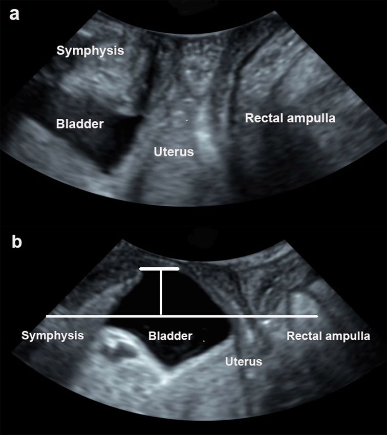
                                                                                                                                                                                                                                                              Figure 1 Translabial ultrasound images (midsagittal plane) in a woman with dominant prolapse in the
                                                                                                                                                                                                                                                              anterior compartment (POP-Q stage 3 for anterior compartment and POP-Q stage 1 for apical compartment)
                                                                                                                                                                                                                                                              at rest (a) and on maximal Valsalva maneuver (b). The leading edge of the bladder relative to the inferior
                                                                                                                                                                                                                                                              margin of the symphysis pubis was -19 mm. POP-Q, pelvic organ prolapse quantification.
                                                                                                                                                                                                                                                              Figure 2 Translabial ultrasound images (midsagittal plane) in a woman with a severe symptomatic, multi-
                                                                                                                                                                                                                                                              compartment compartment prolapse (POP-Q stage 3 for anterior compartment, POP-Q stage 4 for apical
                                                                                                                                                                                                                                                              compartment and POP-Q stage 3 for posterior compartment) at rest (a) and on maximal Valsalva maneuver
                                                                                                                                                                                                                                                              (b). The leading edge of the bladder, cervix, and rectum relative to the inferior margin of the symphysis
                                                                                                                                                                                                                                                              pubis were -21 mm, -26 mm and -19 mm, respectively. POP-Q, pelvic organ prolapse quantification.
                                                                                                                                                                                                                                                              Supplementary Table Legends
                                                                                                                                                                                                                                                              Table S1 The five stages of pelvic organ support using ICS POP-Q system
                                                                                                                                                                                                                                                              Table S2 Demographic data and symptoms assessments of the total cohort
                                                                                                                                                                                                                                                              Table S3 Assessment of bladder descent on Valsalva using translabial ultrasound for predicting POP-Q
                                                                                                                                                                                                                                                              stage [?] 1 and POP-Q stage [?] 2 in the total population
                                                                                                                                                                                                                                                              Hosted file
                                                                                                                                                                                                                                                              Tables.pdf available at https://authorea.com/users/405429/articles/516405-does-dominant-
                                                                                                                                                                                                                                                              prolapse-in-other-compartments-affect-the-assessment-of-bladder-prolapse-on-translabial-
                                                                                                                                                                                                                                                              ultrasound

                                                                                                                                                                                                                                                                                                                  9
Posted on Authorea 2 Apr 2021 — The copyright holder is the author/funder. All rights reserved. No reuse without permission. — https://doi.org/10.22541/au.161735657.71843341/v1 — This a preprint and has not been peer reviewed. Data may be preliminary.

10
Posted on Authorea 2 Apr 2021 — The copyright holder is the author/funder. All rights reserved. No reuse without permission. — https://doi.org/10.22541/au.161735657.71843341/v1 — This a preprint and has not been peer reviewed. Data may be preliminary.

                                                                                                                                                                                         Hosted file

                                                                                              on-translabial-ultrasound

11
                                                                                              does-dominant-prolapse-in-other-compartments-affect-the-assessment-of-bladder-prolapse-
                                                                                              Supplementary tables.pdf available at https://authorea.com/users/405429/articles/516405-
You can also read