Effects of airway management and tidal volume feedback ventilation during pediatric resuscitation in piglets with asphyxial cardiac arrest
←
→
Page content transcription
If your browser does not render page correctly, please read the page content below
www.nature.com/scientificreports
OPEN Effects of airway management
and tidal volume feedback
ventilation during pediatric
resuscitation in piglets
with asphyxial cardiac arrest
Gema Manrique1,2,3, Gema Pérez1,2,3, Laura Butragueño‑Laiseca1,2,3, Miriam García1,2,3,
María Slöcker1,2,3, Rafael González1,2,3, Laura Herrera1,2, Santiago Mencía1,2,3,4,
Jimena del Castillo1,2,3, María José Solana1,2,3, Débora Sanz1,2, Raquel Cieza1,2,
Sarah N. Fernández1,2,3, Jorge López1,2,3, Javier Urbano1,2,3,4* & Jesús López‑Herce1,2,3,4*
To compare the effect on the recovery of spontaneous circulation (ROSC) of early endotracheal
intubation (ETI) versus bag-mask ventilation (BMV), and expiratory real-time tidal volume (VTe)
feedback (TVF) ventilation versus without feedback or standard ventilation (SV) in a pediatric
animal model of asphyxial cardiac arrest. Piglets were randomized into five groups: 1: ETI and
TVF ventilation (10 ml/kg); 2: ETI and TVF (7 ml/kg); 3: ETI and SV; 4: BMV and TVF (10 ml/kg) and
5: BMV and SV. Thirty breaths-per-minute guided by metronome were given. ROSC, pCO2, pO2,
EtCO2 and VTe were compared among groups. Seventy-nine piglets (11.3 ± 1.2 kg) were included.
Twenty-six (32.9%) achieved ROSC. Survival was non-significantly higher in ETI (40.4%) than BMV
groups (21.9%), p = 0.08. No differences in ROSC were found between TVF and SV groups (30.0%
versus 34.7%, p = 0.67). ETI groups presented lower pCO2, and higher pO2, EtCO2 and VTe than BMV
groups (p < 0.05). VTe was lower in TVF than in SV groups and in BMV than in ETI groups (p < 0.05).
Groups 1 and 3 showed higher pO2 and lower pCO2 over time, although with hyperventilation values
(pCO2 < 35 mmHg). ETI groups had non significantly higher survival rate than BMV groups. Compared
to BMV groups, ETI groups achieved better oxygenation and ventilation parameters. VTe was lower
in both TVF and BMV groups. Hyperventilation was observed in intubated animals with SV and with
10 ml/kg VTF.
Ventilation is an important maneuver of cardiopulmonary resuscitation (CPR) that provide oxygenation to tis-
sues. It plays a greater role in pediatric cardiac arrest (CA) than in adult CA, because in children, CA is manly
caused by hypoxia1. Endotracheal intubation (ETI) provides better ventilation and oxygenation and allows
continuous chest compressions (CC). However, it requires specific training, and in non-expert rescuers may be
detrimental, due to prolonged interruptions of CC and delay of other maneuvers. Several observational studies2,3
have found that intubation is not superior to bag-mask ventilation (BMV)3. There is controversy regarding the
optimal time for intubation and whether it is always necessary during pediatric CPR.
In children suffering from CA, visual observation of the chest provides qualitative information about tidal
volume of ventilations, although hyperventilation, either by rate or tidal volume, is r eported4–8. There is still
lack of evidence of the optimal tidal volume and respiratory rate9 during pediatric CPR. Expiratory tidal volume
(VTe) is a surrogate marker of ventilation effectiveness, so it could be used to guide ventilation during resuscita-
tion. As far as we know, there are no studies that have analyzed tidal volume feedback ventilation and survival
in pediatric cardiac arrest.
1
Pediatric Intensive Care Department, Hospital General Universitario Gregorio Marañón, Dr Castelo
47, 28009 Madrid, Spain. 2Health Research Institute of the Gregorio Marañón Hospital, Madrid,
Spain. 3Research Network on Maternal and Child Health and Development (RedSAMID), Madrid,
Spain. 4Maternal and Child Public Health Department. School of Medicine, Complutense University of Madrid,
Madrid, Spain. *email: javierurbanovillaescusa@gmail.com; pielvi@hotmail.com
Scientific Reports | (2021) 11:16138 | https://doi.org/10.1038/s41598-021-95296-w 1
Vol.:(0123456789)
www.nature.com/scientificreports/
Figure 1. Brief summary of the experimental protocol. CA cardiac arrest, CPR cardiopulmonary resuscitation,
ROSC recovery of spontaneous circulation.
The main objective of the study was to compare the effect on the return of spontaneous circulation (ROSC)
of ETI versus BMV and real-time VTe feedback ventilation (TVF) versus standard ventilation (SV) without
guidance. The secondary outcomes were to analyze the influence of intubation and ventilation guidance in
hemodynamic and respiratory parameters.
Materials and methods
We designed a randomized controlled experimental clinical trial developed in the Department of Experimental
Medicine and Surgery of a tertiary hospital in Madrid, Spain. The experimental protocol was approved by Ethics
Committee in Animal Research of the Gregorio Marañón Hospital and it was authorized by the Autonomous
Community of Madrid, Spain. All methods were carried out in accordance with guidelines and regulations. The
study was developed in compliance with the ARRIVE guidelines.
Animal preparation and monitoring. Seventy-nine holoxenic, 3-month-old miniature piglets were
included in the study. The model could be equivalent in weight to a child between one and two years. CA was
induced as described in previously published articles from our research g roup10–12.
Arterial, peripheral and central venous lines were placed to drug administration, blood draw and hemody-
namic monitoring. Central venous and arterial catheters were connected to a PiCCO system for hemodynamic
monitoring (heart rate, arterial blood pressure cardiac index and temperature). ECG and pulse oximetry were
also monitored continuously. Cerebral (ScO2) and splanchnic (SsO2) oxygen saturations were monitored by
near-infrared spectroscopy (NIRS) (INVOS Cerebral Oximeter monitor, Somanetics, Troy, Michigan, USA). An
arterial blood flow sensor was surgically placed in the left carotid artery and connected to a flow monitor (Tran-
sonic Systems Inc, Ithaca, New York, USA) to assess carotid arterial blood flow (CaBF). Respiratory parameters
were monitored continuously using a sensor placed at the Y piece and connected to a Respironics NM3 monitor
(Philips Healthcare, Markham, ON, Canada).
Maintenance fluids containing glucose and saline were infused. Animal temperature was kept between 37
and 39ºC with a heating blanket.
Pediatric CPR electrodes were applied and connected to a Zoll monitor/defibrillator Z series (ZOLL Medical
Corporation, Chelmsford, MA, USA) to guide and record the CC quality.
Experimental protocol. Baseline data were collected after a stabilization period, when ventilation was
checked to have a normal PCO2 (35–45 mmHg). To cause the asphyxial CA, animals were extubated after
administering a bolus of atracurium. CA was defined as a mean arterial pressure (MAP) under 25 mmHg. CPR
was started 2 min after CA was diagnosed and was carried out by qualified staff. CPR was started with five rescue
breaths. Manual chest compressions were performed with a depth target of 4–5 cm (cm) and a rate between
100 and 120 compressions per minute (cpm). Resuscitation was continued until ROSC or up to a maximum of
24 min. Protocol overview is shown Fig. 1.
The animals were randomized into five groups according to two variables. Firstly, depending on the airway
management: ETI or BMV. Secondly, based on delivery of ventilation: with real-time tidal volume feedback
(target VTe of 7 or 10 ml/kg) or without feedback, depending on chest expansion (standard ventilation). Resus-
citation groups were: Group 1: ETI and TVF ventilation with a VTe target of 10 ml/kg; group 2: ETI and TVF
ventilation with a VTe target of 7 ml/kg; group 3: ETI and SV; group 4: BMV and TVF ventilation of 10 ml/kg
and group 5: BMV and SV.
In groups 1, 2 and 3 intubation procedure started after 5 rescue breaths. Intubation was performed by
advanced pediatric airway high trained staff and with adequate skills for this piglet animal model. Chest com-
pressions were not interrupted for laryngoscopy but were interrupted for tube insertion. In all animals, venti-
lations were delivered at a metronome-tailored rate of 30 bpm, according to the results of a previous study10.
Respiratory parameters were continuously registered by volumetric capnography, but the rescuer only received
monitor visual information to perform feedback of VTe values in groups 1, 2 and 4.
Scientific Reports | (2021) 11:16138 | https://doi.org/10.1038/s41598-021-95296-w 2
Vol:.(1234567890)
www.nature.com/scientificreports/
Group 1 Group 2 Group 3 Group 4 Group 5
Parameters ETI + TVF 10 ml/kg ETI + TVF 7 ml/kg ETI + SV BMV + TVF 10 ml/kg BMV + SV p
N 17 15 15 17 15
Weight (kg) 11.5 (10.8–12) 11.8 (10.7–12.2) 11.05 (10.1–12.2) 10.35 (10–11.5) 11 (10.15–12) 0.22
Length (cm) 69 (68–72) 68 (67–72) 70 (67–72) 67 (66–70) 69 (67–73) 0.25
Heart rate (bpm) 96 (86–117) 89 (81–102) 88 (80–110) 111 (84–127) 100 (88–116) 0.28
SAP (mmHg) 95 (85–106) 101 (86–108) 94 (87–105) 104 (88–110) 100 (92–109) 0.78
DAP (mmHg) 50 (43–64) 52 (44–59) 52 (43–55) 49 (47–62) 50 (45–60) 0.95
MAP (mmHg) 71 (60–82) 71 (62–77) 69 (58–74) 69 (65–80) 68 (64–80) 0.89
CVP (mmHg) 6 (5–7) 6 (4–8) 7 (5–9) 6 (4–8) 7 (6–9) 0.61
SatO2 (%) 100 (99–100) 100 (97–100) 99 (97–100) 100 (99–100) 99 (99–100) 0.61
ScO2 (%) 51 (42–68) 46 (34–49) 55 (48–62) 48 (45–55) 54 (41–60) 0.12
SsO2 (%) 58 (51–61) 54 (50–54) 60 (50–67) 56 (53–64) 50 (41–56) 0.04
CaBF (ml/min) 45 (34–57) 42 (38–59) 49 (42–59) 46 (34–49) 41 (37–50) 0.77
CI (L/min/m2) 2.8 (2.4–3.1) 2.8 (2.5–2.9) 2.8 (2.2–3.1) 2.9 (2.5–3.4) 2.8 (2.4–3.3) 0.96
Temperature (ºC) 37.8 (37.4–38.6) 37.9 (37.3–38.6) 37.5 (37.5–38.5) 37.9 (37.5–38.9) 38.3 (37.6–38.7) 0.82
pCO2 (mmHg) 37 (35–40) 38 (37–41) 39 (36–41) 37 (36–40) 40 (37–42) 0.53
pO2 (mmHg) 132 (116–139) 126 (116–138) 127 (107–146) 133 (123–139) 120 (107–128) 0.63
Table 1. Comparison in baseline data between groups. Values were expressed as median and interquartile
range and significance calculated with Kruskal–Wallis Test. ETI endotracheal intubation, BMV bag-mask
ventilation, SAP systolic blood pressure, DAP diastolic blood pressure, MAP mean blood pressure, CVP central
venous pressure, NIRS near-infrared spectroscopy, CI cardiac index, CaBF carotid artery blood flow.
During CPR, epinephrine 0.02 mg/kg/dose every 3 min and sodium bicarbonate (1 mEq/kg/dose) at 9 and
18 min of CPR were administered. If a shockable rhythm was detected, animals were defibrillated (4 J/kg) and
epinephrine 0.02 mg/kg/dose and amiodarone 5 mg/kg/dose was administered after the third, fifth and seventh
shock if the shockable rhythm persisted (maximum of two doses)10–12.
Study variables. The following parameters were collected at baseline, 5 min after extubation, before start-
ing CPR and every 3 min during resuscitation: Heart rate and rhythm, systolic arterial pressure (SAP), diastolic
arterial pressure (DAP), mean arterial pressure (MAP), SpO2, ScO2, SsO2, CaBF and temperature. Time from
extubation to CA, time necessary for delivery intubation and number of intubation attempts were also registered.
Arterial blood gases were drawn at baseline and after 3, 6, 9, 12, 18, 21 and 24 min of CPR.
Depth, rate and release velocity of CC and time with CC and without CC were registered. Respiratory param-
eters were also recorded: inspiratory tidal volume (Vti), expiratory tidal volume (VTe), inspiratory peak-flow,
expiratory peak-flow, breath rate, peak inspiratory pressure, mean airway pressure, positive end expiratory
pressure, end tidal CO2 (EtCO2) and alveolar tidal volume (VTalv).
Statistical analysis. The SPSS statistical package, version 25.0 (SPSS Inc, Chicago, USA) was used for sta-
tistical analysis. Normal distribution of variables was tested with the Kolmogorov–Smirnov test. Continuous
variables are expressed as means with standard deviation and categorical variables as percentages. Chi-squared
(χ2) test was used to compare categorical variables and Kruskal Wallis and U-Mann Whitney tests for continu-
ous variables. A linear mixed model was used to analyze the behaviour of hemodynamic and respiratory param-
eters between groups over time and at different resuscitation time points. The parameters analyzed include
pCO2, pO2, VTe, MAP, DAP and EtCO2. Logistic regression was used to control possible confounding factors.
P values less than 0.05 were considered significant.
Results
Seventy-nine piglets weighting 11.3 ± 1.2 kg were included in the study: 17 in group 1 (21.5%), 15 in group 2
(19%), 15 in group 3 (19%), 17 in group 4 (21.5%) and 15 in group 5 (19%). No differences were found in baseline
parameters among groups, except in somatic NIRS (see Table 1). Mean time to CA was 6.9 ± 0.8 min, p = 0.69.
CPR was started in all cases after 2 min of CA.
Return of spontaneous circulation. ROSC was achieved in 26 animals (32.9%): 7 in group 1 (41.2%),
6 in group 2 (40%), 6 in group 3 (40.0%), 4 in group 4 (23.5%) and 3 in group 5 (20%), p = 0.55. ROSC rate was
40.4% in ETI groups and 21.9% in BMV groups, p = 0.08. No differences in survival rate were found between SV
and TVF ventilation (34.7% vs 30%, p = 0.67).
Rhythm of cardiac arrest. The most frequent CA electrocardiographic rhythm was pulseless electrical
activity (77.2%), followed by ventricular fibrillation (VF) (12.7%), sinus bradycardia (7.6%) and asystole (2.5%).
During CPR, 42 (53.2%) animals presented a shockable rhythm and 41 (51.9%) were defibrillated. One piglet
presented VF prior to the beginning of CPR, but it shifted to asystole before the attempted of defibrillation. No
Scientific Reports | (2021) 11:16138 | https://doi.org/10.1038/s41598-021-95296-w 3
Vol.:(0123456789)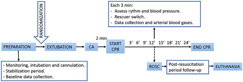
www.nature.com/scientificreports/

                                            Figure 2.  Linear mixed models comparing the following parameters over time among the five groups: pCO2,
                                            pO2, VTe, MAP and EtCO2. The differences between groups over the study period for each parameter were
                                            as follows: pCO2 < 0.01, pO2 p < 0.01, VTe p < 0.01, MAP p = 0.55 and EtCO2 p < 0.01. Significant differences
                                            (p < 0.05) among groups at specific time points during CPR are marked as *.

                                            differences were found regarding the presence or absence of shockable rhythms among resuscitation groups
                                            (p = 0.92). Animals with VF were less likely to achieve ROSC (14.3%) than those with non-shockable rhythm
                                            (54.1%; p < 0.01).

                                            Airway management.              Mean intubation attempts were 1.5 ± 0.7. Sixty-three percent of animals were intu-
                                            bated on a first attempt, 28.3% with 2 attempts, and 8.7% required 3 or more attempts. The mean time from the
                                            start of CPR to intubation was 1.7 ± 2.0 min, with equal distribution among groups. Esophageal intubation, tube
                                            misplacement or dislodgment was not initially detected in 5 animals (10.6%), being noticed after 4.2 to 12.5 min
                                            of CPR. Two of these animals survived. The time required to intubate significantly decreased to 1.1 ± 0.7 min
                                            (p < 0.01) if these 5 piglets were not considered.

                                            Comparison of hemodynamic and respiratory parameters among groups. There were differ-
                                            ences among groups in the evolution over time of the following variables: pCO2, pO2, VCe, and EtCO2 p < 0.01
                                            (Fig. 2). Analyzing MAP and DAP, statistical differences at some resuscitation time points were found among
                                            groups (Fig. 2 and Supplementary Fig. S1). Groups 1 and 3 showed higher pO2 and lower pCO2 over time,
                                            although these groups reached hyperventilation values (pCO2 < 35 mmHg) after 3 min of resuscitation. VTe was
                                            higher in group 3 compared to the other groups from minute 6 to 21 (p < 0.05). MAP was higher in groups 3
                                            and 4 at minute 3 of CPR and in group 2 and 3 at minute 6 of CPR (p < 0.05). EtCO2 was higher in group 1 and
                                            2 throughout resuscitation (p < 0.05).

                                            Hemodynamic and respiratory parameters and their relationship with airway manage‑
                                            ment. ETI groups compared to BMV groups presented significant lower pCO2, and higher EtCO2, pO2 and

          Scientific Reports |   (2021) 11:16138 |               https://doi.org/10.1038/s41598-021-95296-w                                                 4

Vol:.(1234567890)
www.nature.com/scientificreports/
Figure 3. Linear mixed models comparing the following parameters over time between the two airway
strategies (BMV and ETI groups): pCO2, pO2, EtCO2 and VTe. The differences between groups over the
study period for each parameter were as follows: pCO2 p < 0.01, pO2 p < 0.01, VTe p = 0.04 and EtCO2 < 0.01.
Significant differences (p < 0.05) between groups at specific time points during CPR are marked as *.
VTe throughout resuscitation (Fig. 3). No differences were found regarding MAP or DAP (see Supplementary
Fig S2 and S3). Comparison of hemodynamic ventilatory parameters at 3 min of resuscitation between both
groups were shown in Table 2. Although there were no differences concerning VTe between ETI groups and
BMV groups, VTi was significantly higher in BMV groups.
Hemodynamic and respiratory parameters in relation to ventilation strategies (tidal volume
feedback). Compared with those receiving SV, piglets receiving TVF ventilation exhibited higher EtCO2
and lower VTe, p < 0.02. Nevertheless, there were no significant differences in the evolution of pCO2, pO2, MAP
and DAP throughout CPR (Supplementary Fig. S4–S9). If only intubated animals were considered, SV achieved
significant higher pO2 and VTe and lower pCO2 during CPR than TVF ventilation, p < 0.01 (see Supplementary
Fig. S10 and S11).
Hemodynamic and respiratory parameters related to ROSC. ROSC animals presented during
resuscitation higher values of pO2, EtCO2, MAP and DAP than non-ROSC (p < 0.01) (Fig. 4 and Supplementary
Fig. S12–S14), with no differences in other parameters. Hemodynamic and respiratory parameters at 3 min of
resuscitation in ROSC and non-ROSC piglets were shown in Table 3. Survivors showed significantly higher SAP,
MAP, DAP, carotid blood flow and ETCO2 and lower VTi than non-survivors.
Chest compressions quality. Regarding the quality of CC, 151,442 CC were analyzed. No differences
were found in depth or rate of CC between survivors and non-survivors. Mean release velocity was higher in
survival than non-survival animals (323.2 ± 52.8 vs 301.7 ± 24.0 mm/s, p = 0.024). The percentage of time of CPR
with CC (compression fraction) was lower in piglets which achieve ROSC (93.0 ± 6.9 vs 96.2 ± 1.2, p < 0.01).
Scientific Reports | (2021) 11:16138 | https://doi.org/10.1038/s41598-021-95296-w 5
Vol.:(0123456789)www.nature.com/scientificreports/
Parameters ETI BMV p
N 47 32
Heart rate (bpm) 117 (106–126) 115 (106–122) 0.68
SAP (mmHg) 61 (51–88) 61 (49–99) 0.79
DAP (mmHg) 17 (14–27) 24.5 (16–29) 0.12
MAP (mmHg) 26 (20–36) 26 (14–37.5) 0.82
ScO2 (%) 26 (16–58) 15 (15–41) 0.07
SsO2 (%) 37 (30.5–41) 28 (18–36) < 0.01
CaBF (ml/min) 10 (4–16) 7 (3–13) 0.48
VTi (ml) 179 (133–219) 387 (272–432) < 0.01
VTe (ml) 92 (71–129) 77 (49–112) 0.13
Inspiratory peak-flow (liter/min) 28 (22–33) 47 (42–59) < 0.01
Expiratory peak-flow (liter/min) 19 (15–22) 22 (16–27) 0.04
Breath rate (bpm) 25 (21–27) 26 (25–27) 0.014
Peak inspiratory pressure (cmH2O) 45 (35–58) 31 (25–39) < 0.01
Mean airway pressure (cmH2O) 10 (8–14) 9 (7–10) < 0.01
Positive end expiratory pressure (cmH2O) 2 (1.4–5.1) 0.7 (0.3–1.5) < 0.01
Inspiratory airway resistance (cmH2O/liter/sec) 48 (35–61) 33 (28–46) < 0.01
Expiratory airway resistance (cmH2O/liter/sec) 54 (41–72) 48 (37–64) 0.33
EtCO2 (mmHg) 19 (12–28) 13 (8–16) < 0.01
VCO2 (ml/min) 32.3 (9.1–38.9) 7.6 (0.6–19) < 0.01
VTalv (ml) 64 (47–91) 44 (34–56) 0.04
Table 2. Comparison of hemodynamic and ventilatory parameters at 3 min of resuscitation between BMV
groups and ETI groups. Values were expressed as median and interquartile range, comparisons were performed
with U-Mann Whitney test. p values less than 0.05 were marked in bold. ETI endotracheal intubation, BMV
bag-mask ventilation, VTi inspiratory tidal volume, VTe expiratory tidal volume, EtCO2 end tidal CO2, VCO2
carbon dioxide output, VTalv alveolar tidal volume.
These differences persisted (p = 0.02) after removing intubation and defibrillation as possible confounding fac-
tors as both procedures interrupt CC. In piglets that were defibrillated, time from interruption of CC to defibril-
lation was shorter in animals that achieved ROSC (2.4 ± 3.1 s) than in those that did not (7.7 ± 9.2 s; p = 0.01).
These differences did not persist when intubation was considered as confounding factor (p = 0.057).
No differences were found in depth and rate of CC among all groups, between TVF and SV groups and
between ETI and BMV groups. In ETI groups the compression fraction was lower than in BMV groups
(94.1 ± 5.2% vs 96.6 ± 1.6%, p < 0.01).
In 16 animals (20.3%) airway bleeding appeared during resuscitation. Of the 16 animals, 3 (18.8%) were in
group 1, 2 (2.5%) in group 2, 4 (25%) in group 3, 2 (12.5%) in group 4 and 5 (31.3%) in group 5 (p = 0.53). No
differences were found in the appearance of this event between ETI and BMV groups and between TVF and
SV groups. In all of these animals the bleeding was observed after the first 3 min of resuscitation. None of the
animals with this complication survived.
Discussion
In this animal model of asphyxial CA, animals that were intubated reached ROSC twice than those BMV was
delivered, although the differences did not reach statistical significance. ROSC animals showed higher pO2,
EtCO2 and MAP than non-ROSC. VTe was higher in groups in which SV is delivered, although VTF ventilation
did not modify survival.
International recommendations emphasize the importance of the quality of CPR to improve outcomes. Dif-
ferent studies showed that, the use of devices that provide CC feedback improve CPR quality13–16 and ROSC12,17.
However, especially in children good quality CPR is not only related to the improvement of CC18, but also to
optimizing ventilation9. Despite this, ventilation metrics are not analyzed in most of the studies and it is still
unknown the optimal tidal volume or respiratory rate and if ventilation feedback is associated with survival.
On the other hand, some observational studies found better or equal survival in BMV than in ETI
patients3,19,20. In a prospective randomized study of pediatric out-of-hospital-CA no difference in survival
between both techniques was found21. However, in-hospital and out-of-hospital settings and pediatric patients
and adults are not c omparable22. Moreover, observational studies have several limitations: ETI patients may be
more severely ill and these studies did not analyze quality of CC or post-resuscitation care23. In our study, the
survival rate was practically twice in ETI groups than in the BMV groups. Nevertheless, our study is an experi-
mental randomized study (without selection bias), in which other parameters that may influence on ROSC are
controlled, such as the quality of CC. A posteriori sample size calculation with the ROSC proportion observed
in this study showed that 97 animals in each group would have been required to reach statistical significance.
Scientific Reports | (2021) 11:16138 | https://doi.org/10.1038/s41598-021-95296-w 6
Vol:.(1234567890)www.nature.com/scientificreports/
Figure 4. Linear mixed models comparing the following parameters over time between the animals which
achieved ROSC and did not (non-ROSC): pO2, MAP and EtCO2. The differences between groups over the
study period for each parameter were as follows: pO2 p < 0.01, MAP p < 0.01 and EtCO2 < 0.01. Significant
differences (p < 0.05) between groups at specific time points during CPR are marked as *.
EtCO2 was higher in groups with ETI, it is consistent with the fact that this group of animals reached a non-
significant high proportion of ROSC than the BMV groups.
Despite the staff were trained for invasive airway management, intubation during CPR carries potential
complications. The most relevant problem in our study were esophageal intubation or tube misplacement which
occurred in 10.6% of the animals. This fact could cause deficient oxygenation and ventilation, although two of
these 5 animals survived. Previous studies found that only 40–60% of intubations are achieved on a first attempt
and there are complications or mistakes in a third of the patients. Factors related to intubation problems or failure
are: age under 18 months24–26, non-interruption of CC27, and the presence of a difficult a irway26.
International recommendations highlight to avoid hyperventilation, which could increase intrathoracic pres-
sure, limiting venous return and decreasing cardiac output. Most authors paid attention on respiratory rate rather
than tidal volume4,5. In our study, respiratory rate was the same in all groups, but higher VTe was delivered in
piglets receiving SV and intubation.
We found no differences in oxygenation and ventilation between ETI groups and BMV groups in the first
3 min of CPR, but significant differences appeared later, with higher pO2 and lower pCO2 in intubated animals.
Moreover, in intubated animals with SV or with VTF of 10 ml/kg, hyperventilation values were found after
3 min of resuscitation. These data are consistent with previous studies showing that hyperventilation is more
common in intubated p atients28. However, most of the studies considered hyperventilation as high ventilatory
parameters but not as a low arterial pCO2. In our study, no differences were found in the evolution of pCO2
between TVF and SV groups. Nevertheless, within intubated animals, SV resulted in lower pCO2 during CPR
than TVF ventilation. This suggests that greater tidal volumes are associated to higher pO2, but also to lower
Scientific Reports | (2021) 11:16138 | https://doi.org/10.1038/s41598-021-95296-w 7
Vol.:(0123456789)www.nature.com/scientificreports/
Parameters ROSC Non-ROSC p
N 25 53
Heart rate (bpm) 122 (115–173) 107 (103–117) < 0.01
SAP (mmHg) 90 (57–144) 55 (50–72) < 0.01
DAP (mmHg) 33 (20–58) 16 (12–23) < 0.01
MAP (mmHg) 39 (29–87) 21.5 (14–28) < 0.01
Arterial lactic (mmol/L) 7.2 (6.5–8.1) 7.3 (6.9–7.7) 0.71
ScO2 (%) 28 (16–58) 22 (15–41) 0.30
SsO2 (%) 36 (30–41) 33.5 (23–39) 0.23
CaBF (ml/min) 16 (12–30) 6 (2–10) < 0.01
VTi (ml) 179 (125–268) 258 (172–415) 0.045
VTe (ml) 86 (73–114) 86 (55–128) 0.72
Inspiratory peak-flow (l/min) 32 (22–43) 33 (28–45) 0.18
Expiratory peak-flow (l/min) 20 (15.0–25.0) 20 (16–22) 0.60
Breath rate (bpm) 26 (22–28) 25 (23–27) 0.50
Peak inspiratory pressure (cmH2O) 42 (32–58) 39 (30–50) 0.28
Mean airway pressure (cmH2O) 10 (8–12) 9 (7–11) 0.35
Positive end expiratory pressure (cmH2O) 2.0 (1.2–5.1) 1.5 (0.5–2.0) 0.09
Inspiratory airway resistance (cmH2O/l/s) 40 (29–54) 44 (31–53) 0.84
Expiratory airway resistance (cmH2O/l/s) 48 (37–66) 55 (44–65) 0.32
EtCO2 (mmHg) 23 (16–32) 14 (9–19) < 0.01
VCO2 (ml/min) 26 (9–42) 13 (1–33) 0.048
VTalv (ml) 53 (33–64) 59 (42–90) 0.48
Table 3. Comparison of haemodynamic and ventilatory parameters at 3 min of resuscitation between
animals which achieved ROSC and those did not. Values were expressed as median and interquartile range,
comparisons were performed using U-Mann Whitney test. p values less than 0.05 were marked in bold. SAP
systolic blood pressure, DAP diastolic blood pressure, MAP mean blood pressure, CaBF carotid artery blood
flow, VTi inspiratory tidal volume, VTe expiratory tidal volume, EtCO2 end tidal CO2, VCO2 carbon dioxide
output, VTalv alveolar tidal volume.
pCO2 and hyperventilation (pCO2 < 35 mmHg). Inadvertent excessive tidal volume is probably more frequent
than expected as it is not usually measured.
Some devices, based on impedance, can accurately measure the respiratory rate but are less precise to estimate
the tidal volume29. In recent years, simulation CA studies that analyze real-time tidal volume feedback have been
developed8,30–34, showing that the feedback increases the proportion of target VT ventilations8,30–34, and reduces
the variability of tidal v olume30,34, and the peak p
ressure34. Without feedback, VT is usually higher than with feed-
back, although You et al.31 observed a large proportion of hypoventilation. In our knowledge, there are no clinical
studies that analyze the influence of TVF on survival or in the pediatric population. It is still unknown which
is the optimal VTe or respiratory rate during pediatric resuscitation, so the implementation of its measurement
could contribute to determine its optimal values, its influence on survival, and if it could avoid hyperventilation.
Our study has some limitations. Although we have used a validated pediatric animal model for this purpose,
the results from experimental studies must be interpreted with caution and could not be directly extrapolated
to children. Intubation in pigs is slightly different than in children. Piglets have a long mouth, a large epiglottis
and mobile larynx, although the morphological structure and distribution of the porcine airways is similar to
the human and has been used previously in studies of CA35. There are also differences between humans and pigs
in BMV, differing the mask and the opening of the airway. Advanced airway management was performed by
experienced staff, so the results obtained could not be applicable to rescuers without advanced pediatric training.
Furthermore, the animals were not autopsied. Nevertheless, in previous studies published by our group, lung
injury was found in most of the animals that had airway b leeding11. Another limitation is that coronary perfu-
sion pressure was not measured, which could provide relevant data on the hemodynamic effects of ventilation
strategies.
Conclusions
In this animal model of asphyxial CA, ETI groups had higher survival rate than BMV groups without statistical
significance. No differences were found between SV and TVF ventilation. Piglets that achieved ROSC had higher
pO2, EtCO2, MAP, DAP and compression fraction than non-survivors.
ETI groups had higher pO2, EtCO2 and lower pCO2 than BMV groups. VTe was higher both in ETI groups
and SV groups. After 3 min of resuscitation, in intubated piglets, SV and ventilation with VTe of 10 ml/kg could
be excessive, but TVF with 7 ml/kg could prevent hyperventilation.
Scientific Reports | (2021) 11:16138 | https://doi.org/10.1038/s41598-021-95296-w 8
Vol:.(1234567890)www.nature.com/scientificreports/
Received: 25 March 2021; Accepted: 20 July 2021
References
1. Girotra, S. et al. Survival trends in pediatric in-hospital cardiac arrests an analysis from get with the guidelines-resuscitation. Circ.
Cardiovasc. Qual. Outcomes 6, 42–49 (2013).
2. Gupta, P. et al. Association of presence and timing of invasive airway placement with outcomes after pediatric in-hospital cardiac
arrest. Resuscitation 92, 53–58 (2015).
3. Andersen, L. W. et al. Association between tracheal intubation during pediatric in-hospital cardiac arrest and survival. JAMA 316,
1786 (2016).
4. Niebauer, J. M., White, M. L., Zinkan, J. L., Youngblood, A. Q. & Tofil, N. M. Hyperventilation in pediatric resuscitation: perfor-
mance in simulated pediatric medical emergencies. Pediatrics 128, e1195–e1200 (2011).
5. McInnes, A. D. et al. The first quantitative report of ventilation rate during in-hospital resuscitation of older children and adoles-
cents. Resuscitation 82, 1025–1029 (2011).
6. Park, S. O. et al. A clinical observational study analysing the factors associated with hyperventilation during actual cardiopulmonary
resuscitation in the emergency department. Resuscitation 84, 298–303 (2013).
7. Losert, H. et al. Quality of cardiopulmonary resuscitation among highly trained staff in an emergency department setting. Arch.
Intern. Med. 166, 2375–2380 (2006).
8. Gould, J. R., Campana, L., Rabickow, D., Raymond, R. & Partridge, R. Manual ventilation quality is improved with a real-time
visual feedback system during simulated resuscitation. Int. J. Emerg. Med. 13, 1–5 (2020).
9. Sutton, R. M. et al. Ventilation rates and pediatric in-hospital cardiac arrest survival outcomes. Crit. Care Med. 47, 1627–1636
(2019).
10. López, J. et al. Different respiratory rates during resuscitation in a pediatric animal model of asphyxial cardiac arrest. PLoS ONE
11, 1–14 (2016).
11. López, J. et al. Comparison between manual and mechanical chest compressions during resuscitation in a pediatric animal model
of asphyxial cardiac arrest. PLoS ONE 12, 1–14 (2017).
12. Manrique, G. et al. Comparison between synchronized and non-synchronized ventilation and between guided and non-guided
chest compressions during resuscitation in a pediatric animal model after asphyxial cardiac arrest. PLoS ONE 14, e0219660 (2019).
13. Vahedian-Azimi, A. et al. Effect of the Cardio First A ngelTM device on CPR indices: a randomized controlled clinical trial. Crit.
Care 20, 1–8 (2016).
14. Buléon, C. et al. Improvement in chest compression quality using a feedback device (CPRmeter): a simulation randomized crossover
study. Am. J. Emerg. Med. 31, 1457–1461 (2013).
15. Lin, C. Y. et al. Effect of audiovisual cardiopulmonary resuscitation feedback device on improving chest compression quality. Sci.
Rep. 10, 1–7 (2020).
16. Yeung, J. et al. The use of CPR feedback/prompt devices during training and CPR performance: a systematic review. Resuscitation
80, 743–751 (2009).
17. Goharani, R. et al. Real-time compression feedback for patients with in-hospital cardiac arrest: a multi-center randomized con-
trolled clinical trial. J. Intensive Care 7, 1–11 (2019).
18. Talikowska, M., Tohira, H. & Finn, J. Cardiopulmonary resuscitation quality and patient survival outcome in cardiac arrest: a
systematic review and meta-analysis. Resuscitation 96, 66–77 (2015).
19. McMullan, J. et al. Airway management and out-of-hospital cardiac arrest outcome in the CARES registry. Resuscitation 85,
617–622 (2014).
20. Hasegawa, K., Hiraide, A., Chang, Y. & Brown, D. F. M. Association of prehospital advanced airway management with neurologic
outcome and survival in patients with out-of-hospital cardiac arrest. JAMA J. Am. Med. Assoc. 309, 257–266 (2013).
21. Gausche, M. et al. Effect of out-of-hospital pediatric endotracheal intubation on survival and neurological outcome: a controlled
clinical trial. JAMA 283, 783–790 (2000).
22. Moler, F. W. et al. In-hospital versus out-of-hospital pediatric cardiac arrest: a multicenter cohort study. Crit. Care Med. 37,
2259–2267 (2009).
23. Decaen, A. R., Guerra, G. G. & MacOnochie, I. Intubation during pediatric CPR early, late, or not at all?. JAMA J. Am. Med. Assoc.
316, 1772–1774 (2016).
24. Losek, J. D. et al. Prehospital pediatric endotracheal intubation performance review. Pediatr. Emerg. Care 5, 1–4 (1989).
25. Goto, T. et al. Factors associated with First-Pass success in pediatric intubation in the emergency department. West. J. Emerg. Med.
17, 129–134 (2016).
26. Graciano, A. L. et al. Incidence and associated factors of difficult tracheal intubations in pediatric ICUs: a report from National
Emergency Airway Registry for Children: NEAR4KIDS. Intensive Care Med. 40, 1659–1669 (2014).
27. Donoghue, A., Hsieh, T. C., Nishisaki, A. & Myers, S. Tracheal intubation during pediatric cardiopulmonary resuscitation: a
videography-based assessment in an emergency department resuscitation room. Resuscitation 99, 38–43 (2016).
28. Aufderheide, T. P. & Lurie, K. G. Death by hyperventilation: a common and life-threatening problem during cardiopulmonary
resuscitation. Crit. Care Med. 32, S345–S351 (2004).
29. Terndrup, T. E. & Rhee, J. Available ventilation monitoring methods during pre-hospital cardiopulmonary resuscitation. Resuscita-
tion 71, 10–18 (2006).
30. Scott, J. B., Schneider, J. M., Schneider, K. & Li, J. An evaluation of manual tidal volume and respiratory rate delivery during
simulated resuscitation. Am. J. Emerg. Med. https://doi.org/10.1016/j.ajem.2020.09.091 (2020).
31. You, K. M. et al. Real-time tidal volume feedback guides optimal ventilation during simulated CPR. Am. J. Emerg. Med. 35, 292–298
(2017).
32. Heo, S. et al. Effectiveness of a real-time ventilation feedback device for guiding adequate minute ventilation: a manikin simulation
study. Medicine 56, 1–11 (2020).
33. Kim, J. W., Park, S. O., Lee, K. R., Hong, D. Y. & Baek, K. J. Efficacy of Amflow®, a real-time-portable feedback device for delivering
appropriate ventilation in critically ill patients: a randomised, controlled, cross-over simulation study. Emerg. Med. Int. 2020, 1–7
(2020).
34. Khoury, A., De Luca, A., Sall, F. S., Pazart, L. & Capellier, G. Ventilation feedback device for manual ventilation in simulated
respiratory arrest: a crossover manikin study. Scand. J. Trauma. Resusc. Emerg. Med. 27, 1–8 (2019).
35. Vognsen, M. et al. Contemporary animal models of cardiac arrest: a systematic review. Resuscitation 113, 115–123 (2017).
Acknowledgements
The authors gratefully acknowledge the work of all the personnel from the Department of Experimental Medicine
and Surgery of Hospital.
Scientific Reports | (2021) 11:16138 | https://doi.org/10.1038/s41598-021-95296-w 9
Vol.:(0123456789)www.nature.com/scientificreports/
Author contributions
G.M., J.L.H. and J.U. conceived and designed the study and obtained research funding. G.P., L.B.L., M.G., M.S.,
R.G., L.H., S.M., J.C., M.J.S., D.S., J.L., R.C. and S.N.F. developed the experimental procedures and data collec-
tion. G.M. and G.P. managed the data. G.M. and R.G. provided statistical advice on study design and analyzed
the data. G.M. and L.B.L. drafted the manuscript, and all authors contributed substantially to its revision. G.M.,
J.L.H. and J.U. takes responsibility for the paper as a whole.
Funding
This work was supported by the PI 18/01632 of Carlos III Health Institute, responsible for Spain’s Health Research
and Development Strategy, co-financed by the European Union ERDF funds (European Regional Development
Fund) and RETICS “Maternal and Child Health and Development Network” (SAMID Network), funded by the
PN I+D+i 2013-2016 (Spain), ISCIII-Sub-Directorate General for Research Assessment and Promotion and the
European Regional Development Fund (ERDF), ref. RD16/0022. Funders did not participate in the study design,
dataset collection and analysis and writing the manuscript.
Competing interests
The authors declare no competing interests.
Additional information
Supplementary Information The online version contains supplementary material available at https://doi.org/
10.1038/s41598-021-95296-w.
Correspondence and requests for materials should be addressed to J.U. or J.L.-H.
Reprints and permissions information is available at www.nature.com/reprints.
Publisher’s note Springer Nature remains neutral with regard to jurisdictional claims in published maps and
institutional affiliations.
Open Access This article is licensed under a Creative Commons Attribution 4.0 International
License, which permits use, sharing, adaptation, distribution and reproduction in any medium or
format, as long as you give appropriate credit to the original author(s) and the source, provide a link to the
Creative Commons licence, and indicate if changes were made. The images or other third party material in this
article are included in the article’s Creative Commons licence, unless indicated otherwise in a credit line to the
material. If material is not included in the article’s Creative Commons licence and your intended use is not
permitted by statutory regulation or exceeds the permitted use, you will need to obtain permission directly from
the copyright holder. To view a copy of this licence, visit http://creativecommons.org/licenses/by/4.0/.
© The Author(s) 2021
Scientific Reports | (2021) 11:16138 | https://doi.org/10.1038/s41598-021-95296-w 10
Vol:.(1234567890)You can also read