Effects of Intermittent Treatment with Topical Corticosteroids and Calcineurin Inhibitors on Epidermal and Dermal Thickness Using Optical ...
←
→
Page content transcription
If your browser does not render page correctly, please read the page content below
Research Article

                                                          Skin Pharmacol Physiol 2022;35:41–50                                                         Received: December 30, 2020
                                                                                                                                                       Accepted: June 3, 2021
                                                          DOI: 10.1159/000518214                                                                       Published online: July 8, 2021

Effects of Intermittent Treatment with Topical
Corticosteroids and Calcineurin Inhibitors on
Epidermal and Dermal Thickness Using Optical
Coherence Tomography and Ultrasound
Roland Aschoff a Awena Lang a Edmund Koch b
aDepartment
            of Dermatology, University Hospital Carl Gustav Carus, Technische Universität Dresden,
Dresden, Germany; bDepartment of Clinical Sensoring and Monitoring, Technische Universität Dresden,
Dresden, Germany

Keywords                                                                                     BMV led to significant epidermal thinning on both sites. Four
Atopic dermatitis · Corticosteroid · Optical coherence                                       weeks after the end of treatment, epidermal thickness re-
tomography · Pimecrolimus · Skin atrophy                                                     turned to baseline. No dermal thinning, atrophy, or telangi-
                                                                                             ectasia was observed. Conclusions: MPA, HC, and PIM may
                                                                                             be more suitable for repeated and prolonged treatment, es-
Abstract                                                                                     pecially in chronic diseases.        © 2021 The Author(s)
Introduction: Proactive therapy with topical corticosteroids                                                                                     Published by S. Karger AG, Basel

(TCSs) is the standard treatment for chronic inflammatory
diseases such as atopic dermatitis; however, skin atrophy as
TCS side effect remains a concern. Methods: This 16-week,                                        Introduction
evaluator-blinded, within-patient placebo-controlled, ran-
domized study enrolled volunteers with healthy skin condi-                                       Atopic dermatitis (AD) is an inflammatory, highly pru-
tions. For 12 weeks, their volar forearm and the back of their                               ritic, chronic, and chronically relapsing skin disease that
hand were applied with hydrocortisone acetate 1% cream                                       negatively affects one’s quality of life [1, 2]. Application of
(HC), methylprednisolone aceponate 0.1% cream (MPA), be-                                     topical corticosteroids (TCSs) remains to be the mainstay
tamethasone valerate 0.1% cream (BMV), or an active agent-                                   of treatment for AD; however, long-term use of TCSs
free base cream (Dermatop® Basiscreme) once daily twice                                      causes various side effects, such as skin atrophy, striae dis-
weekly, and pimecrolimus 1% cream (PIM) twice daily twice                                    tensae, telangiectasia, and skin barrier function impair-
weekly. Epidermal and dermal thickness was measured by                                       ment [3, 4]. Additional effects include increased water
optical coherence tomography and high-frequency ultra-                                       loss, decreased hydration level, and increased skin trans-
sound, respectively. Furthermore, skin atrophy and telangi-                                  parency. These side effects depend on the strength, dura-
ectasia were determined by contact dermatoscopic photog-                                     tion, and dose of the treatment and on the morphological
raphy (Dermaphot®). Results: After 8 and 12 weeks, only                                      properties of the skin in various anatomical areas [3–5].

karger@karger.com     © 2021 The Author(s)                                                   Correspondence to:
www.karger.com/spp    Published by S. Karger AG, Basel                                       Roland Aschoff, roland.aschoff @ uniklinikum-dresden.de
                      This article is licensed under the Creative Commons Attribution 4.0
                      International License (CC BY) (http://www.karger.com/Services/
                      OpenAccessLicense). Usage, derivative works and distribution are
                      permitted provided that proper credit is given to the author and the
                      original publisher.
Fortunately, an intermittent therapy regimen has been                applied twice daily twice weekly. Meanwhile, 5 volunteers did not
established to reduce or prevent the frequencies of relaps-             apply any cream (placebo group). Because of the different frequen-
                                                                        cy of application or nonapplication, complete blinding of the sub-
es and to decrease the intensity of exacerbations in the                jects was not given.
long-term management of AD [6–8]. This proactive ap-                        Sequence generation was performed using a computer-based
proach starts with an intensive topical anti-inflammatory               random-number generator (STATA 10 for Windows; StatCorp,
therapy that lasts until the visible lesions are completely             College Station, TX, USA). To exclude possible bias by the treated
cleared, followed by the intermittent application of low-               sides, we set the same number of volunteers that applied creams
                                                                        with the same ingredients on the right and left sides. As each of the
dose anti-inflammatory drugs on the previously affected                 30 subjects had one site on one volar forearm and one site on the
skin areas [7]. The rationale of this approach is based on              back of the hand treated with an investigational product and one
the fact that the skin of patients with AD, though appears              site on the contralateral side treated with a different investigation-
normal, may have an impaired barrier function and a                     al product, a total of 10 subjects were investigated in each group.
subclinical inflammatory infiltrate [9, 10]. Unlike TCSs,               They were instructed to use a different finger for each preparation
                                                                        to avoid intermixing the drug components. In addition, the origi-
topical calcineurin inhibitors (TCIs) do not induce skin                nal tubes were labeled with “volar forearm and back of the hand
atrophy [2, 11, 12]. Proactive application of tacrolimus                left” and “volar forearm and back of the hand right,” and individ-
ointment in adults and children with AD also remarkably                 ual stencils were made for each volunteer to reproduce the applica-
reduces the frequencies of disease exacerbations and in-                tion sides easily. Prior to follow-up observation, the skin stayed
creases the time to first disease exacerbation [13, 14].                untreated for 4 weeks. Emollients could be additionally used but
                                                                        only for twice a week; however, those containing active substances
   Although TCSs and TCIs do not exhibit skin atrophy                   were prohibited during the study period. Furthermore, treatment
when applied twice weekly, prolonged application of                     application was not allowed 12 h before the measurement sessions.
TCSs to a nonlesional skin might lead to skin thinning                      Using OCT, Dermaphot®, ultrasound, transepidermal water
[6]. Therefore, this study mainly aimed to investigate the              loss (TEWL), and corneometry, a blinded study nurse measured
effects of TCS and TCI treatment for 12 weeks on epider-                all 4 treatment areas on days 0 (start of treatment), 7, 28, 56, 84
                                                                        (end of treatment period), and 112 (end of follow-up period). The
mal and dermal thickness by using optical coherence to-                 outcome assessor and the statistician were also blinded to the treat-
mography (OCT), ultrasound, and Dermaphot®.                             ment. During each visit, the adverse events (AEs) and serious AEs,
                                                                        their severity, and their relationship to the study drugs were docu-
                                                                        mented.
     Materials and Methods                                                  This study was approved by the institutional review board of
                                                                        the Technische Universität Dresden (Approval No: EK 145052012)
    Study Design and Volunteers                                         and the German Federal Institute for Pharmaceuticals and Medical
    This 16-week, evaluator-blinded, within-patient placebo-con-        Products (EudaCT-No. 2011-004953-17, Bundesinstitut für Arz-
trolled, randomized, and single-center study enrolled 30 adult vol-     neimittel und Medizinprodukte). In addition, this study con-
unteers with healthy skin conditions. The inclusion criteria were       formed to the ethical principles of the Declaration of Helsinki and
as follows: age between 18 and 55 years, no skin disease, and skin      was posted in clinicaltrial.gov. All volunteers gave their written
type I–III according to the Fitzpatrick scale [15]. Conversely, the     informed consent to participate in the study.
exclusion criteria were the following: participation in a clinical
study within the last 30 days prior to screening; systemic or topical       Skin Measurements
treatment within the last 6 months with drugs (e.g., retinoids, glu-        After an acclimatization period of 15 min in an air-conditioned
cocorticosteroids, calcineurin inhibitors, and tar) that are suspect-   room, the skin was measured using the OCT device, followed by
ed or known to influence skin thickness or telangiectasia forma-        TEWL and corneometry. Thereafter, the skin was moistened with
tion; systemic therapy with immunosuppressants; severe systemic         water and then examined using Dermaphot® and ultrasound con-
disease; genetic effects of the epidermal barrier (e.g., Netherton      secutively.
syndrome); intensive natural or artificial UV-light exposure with-          Epidermal thickness was measured using the OCT system Der-
in the last 4 weeks or during the study period; known intolerance       maSR (DermaRadar 830; Technische Universität, Dresden, Ger-
to drug components used in the study; and drug or alcohol abuse.        many), which uses an 830.3-nm superluminescence diode (spec-
    The volunteers randomly applied 2 of the following 5 different      tral width: 46.4 nm; optical power: 1 mW), as previously described
study drugs: pimecrolimus 1% cream (PIM) (Elidel®, Novartis             [16]. To determine epidermal thickness, we calculated the mean
Pharma GmbH, Germany), hydrocortisone acetate 1% cream                  difference between the signals of the air-skin transition and the
(HC) (Hydrogalen® Creme, GALENpharma GmbH, Germany),                    dermal-epidermal junction of the 600 A-scans of the B-scan image
methylprednisolone aceponate 0.1% cream (MPA) (Advantan®                [17]. The arithmetic mean was calculated from 3 individual mea-
Creme, Intendis GmbH, Germany), betamethasone valerate 0.1%             surements.
cream (BMV) (Betagalen® Crème, GALENpharma GmbH, Ger-                       For measuring dermal thickness, we used a 22-MHz ultrasound
many), and an active agent-free base cream (DBC) (Dermatop®             device (DUB20, tpm – taberna pro medicum GmbH, Germany, CE
Basiscreme; Sanofi-Aventis Deutschland GmbH, Germany).                  0482) with a maximum physical resolution of 72 μm and a signal
These drugs were topically applied on the right or left volar fore-     penetration of up to 8 mm. Epidermal thinning and telangiectasia
arm and back of the hand once daily twice weekly. Only PIM was          formation were detected by contact dermatoscopic photography

42                      Skin Pharmacol Physiol 2022;35:41–50                                    Aschoff/Lang/Koch
                        DOI: 10.1159/000518214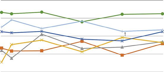
Table 1. Dermaphot® score for the evaluation of skin atrophy and telangiectasia
Score Definition
Atrophy
0 No change
1 Slight transparency increase
2 Moderate thinning of the epidermis with moderate increase in transparency
3 Severe thinning and increase in transparency
4 Very severe thinning of the epidermis, the vasculature appearing to be directly under the surface
Telangiectasia
0 Normal vascular pattern
1 Capillary hyperemia with slight elongation and dilatation of blood vessels not visible to the naked eye
2 Moderate telangiectasia, just visible with naked eye
3 Severe telangiectasia
4 Very severe telangiectasia with large blunt vessels
72
Mean epidermal thickness, µm
70
68
66
BMV
64 MPA *
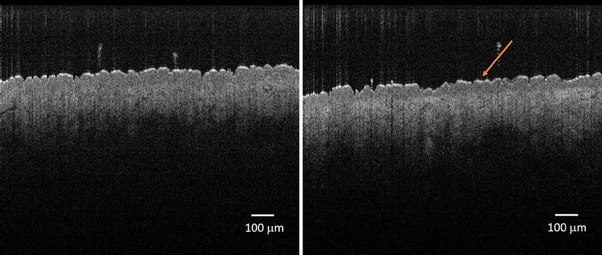
Fig. 1. Mean change of epidermal thickness
measured by OCT on the volar forearm
HC *
PIM
(*p < 0.05: significant difference). OCT, 62
DBC
optical coherence tomography; BMV, be- Placebo
tamethasone valerate; MPA, methylpred- 60
nisolone aceponate; HC, hydrocortisone 0 7 14 21 28 35 42 49 56 63 70 77 84 91 98 105 112
acetate; PIM, pimecrolimus; DBC, active Days
agent-free base cream.
with Dermaphot® (Heine Dermaphot Optotechnik GmbH, measured by OCT. Data were analyzed by paired t-test. A 20% de-
Herrsching, Germany) and Nikon® Digital Camera D70 modified crease in epidermal thickness was assumed to be relevant. Sample
for contact dermatoscopic use (Nikon Corporation, Tokyo, Ja- size calculation indicated that 8 volunteers per group were re-
pan). The images were evaluated using a modified Frosh Score quired for a study with 5% significance level and 90% power. The
(Table 1) [18]. secondary end points were the determination of dermal thickness,
Furthermore, TEWL was measured using the open-chamber skin atrophy and telangiectasia formation, skin hydration changes,
evaporimeter Tewameter® TM 300 (Courage + Khazaka Electron- and water loss.
ic GmbH, Köln, Germany), and skin hydration was determined
using Corneometer® CM 825 (Courage + Khazaka Electronic
GmbH, Köln, Germany). Capacitance was performed 5 times in
each test area. The highest and lowest values were deleted, and the Results
mean value of the 3 remaining measurements was calculated; the
results are expressed in grams per square meters per hour or arbi-
trary units (au) accordingly. The measurement of TEWL and skin Patients and Safety
hydration was in accordance with the established guidelines [19, This study included 30 volunteers (22 females and 8
20]. males); all of them completed the trial. The mean age was
40 years (range: 23–54 years). In addition, 22 study par-
Statistical Methods ticipants reported 43 AEs in which 23 were mild, 19 were
The statistical analysis was performed on the intent-to-treat
population. In this study, the primary end point was the change of moderate, and 1 was severe (tooth root resection with ra-
epidermal thickness under treatment with PIM, HC, MPA, BMV, diculodental cyst treatment). These AEs included head-
and DBC and without treatment (placebo) between days 0 and 84 ache (n = 16), flu-like symptoms (n = 9), rhinitis (n = 4),
Intermittent Treatment of AD with Skin Pharmacol Physiol 2022;35:41–50 43
Corticosteroids DOI: 10.1159/000518214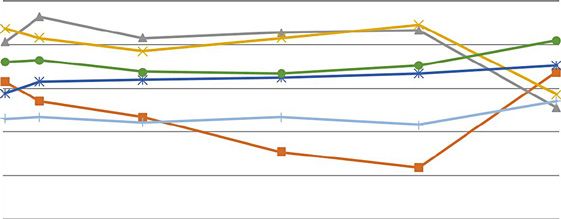
105

                                                                                 100

                                                  Mean epidermal thickness, µm
                                                                                  95

                                                                                  90

                                                                                  85
Fig. 2. Mean change of epidermal thickness
                                                                                  80
measured by OCT on the back of the hand                                                        BMV         PIM                                       *
(*p < 0.05, **p < 0.01: significant differ-                                       75           MPA         DBC                 **
ence). OCT, optical coherence tomogra-                                                         HC          Placebo
phy; BMV, betamethasone valerate; MPA,                                            70
                                                                                       0   7    14    21   28   35   42   49   56     63   70   77   84   91   98 105 112
methylprednisolone aceponate; HC, hy-
drocortisone acetate; PIM, pimecrolimus;                                                                                       Days
DBC, active agent-free base cream.

 a                                                                                                   b

                       Fig. 3. Example of OCT measurement at baseline (a) and epidermal thinning (arrow) after 12 weeks of treatment
                       with BMV on the volar forearm. OCT, optical coherence tomography; BMV, betamethasone valerate.

herpes labialis (n = 2), dental surgery (n = 2), shoulder                                            Fig. 1), reaching from 68.31 ± 8.09 μm to 64.35 ± 7.96 μm
surgery (n = 1), hallux rigidus operation (n = 1), gastro-                                           after 84 days. The thinning was −7.96% and statistically
intestinal disease (n = 1), cough (n = 1), menstrual pain                                            significant (p = 0.017). On the back of the hand, the epi-
(n = 1), folliculitis (n = 1), vitamin B12 deficiency (n = 1),                                       dermis thinned continuously up to visit 4 (day 56) and
perlèche (n = 1), hypertensive crisis (n = 1), and calca-                                            remained stable until visit 5 (day 84) (shown in Fig. 2).
neodynia (n = 1). All AEs were unrelated to the study                                                The epidermal thickness decreased from 85.05 ± 11.42
drugs. Furthermore, we found no AE on the treatment                                                  μm to 79.47 ± 11.99 μm (day 84, −6.56%), indicating a
sites, no severe AE, and no protocol violation.                                                      statistical significance (p = 0.025). An example of epider-
                                                                                                     mal thinning under BMV is shown in Figure 3. After
   OCT Assessment                                                                                    treatment completion, the epidermal thickness resumed
   The epidermal thickness decreased continuously on                                                 to baseline on both sites. Meanwhile, treatment with
the volar forearm during BMV treatment (shown in                                                     MPA, HC, PIM, and DBC and placebo did not result in

44                     Skin Pharmacol Physiol 2022;35:41–50                                                                    Aschoff/Lang/Koch
                       DOI: 10.1159/000518214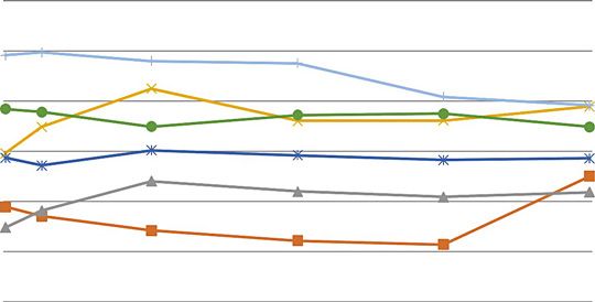
1,250

                                                                              1,200

                                                  Mean dermal thickness, µm
                                                                              1,150

                                                                              1,100

                                                                              1,050

                                                                              1,000
Fig. 4. Mean change of epidermal thickness                                                                                                        BMV          PIM
measured by ultrasound on the volar fore-                                       950                                                               MPA          DBC
arm (*p < 0.05: significant difference).                                                                                                          HC           Placebo
BMV, betamethasone valerate; MPA,                                               900
                                                                                      0   7   14   21   28   35   42   49    56    63   70   77   84    91   98 105 112
methylprednisolone aceponate; HC, hy-
drocortisone acetate; PIM, pimecrolimus;                                                                                      Days
DBC, active agent-free base cream.

                                                                              1,250

                                                                              1,200
                                             Mean dermal thickness, µm

                                                                              1,150                                                                *
                                                                                                                                                  *
                                                                              1,100

                                                                              1,050                                                               BMV          PIM
Fig. 5. Mean change of epidermal thickness
                                                                                                                                                  MPA          DBC
measured by ultrasound on the back of the                                                                                                         HC           Placebo
hand (*p < 0.05: significant difference).                                     1,000
BMV, betamethasone valerate; MPA,                                                     0   7   14   21   28   35   42   49    56    63   70   77   84    91   98 105 112
methylprednisolone aceponate; HC, hy-                                                                                       Days
drocortisone acetate; PIM, pimecrolimus;
DBC, active agent-free base cream.

significant epidermal thinning on the forearm and the                                          ± 132.41 μm to 1136.80 ± 126.88 μm (−25.7 μm, −2.2%)
back of the hand (shown in Fig. 1, 2).                                                         on the back of the hand (p = 0.001), whereby no change
                                                                                               could be seen up to day 28, and the baseline value was
    20-MHz Ultrasound Assessment                                                               reached after the therapy was completed. Throughout the
    In BMV treatment, dermal thickness decreased con-                                          observation period, the values on the forearm did not
tinuously from 1044.60 ± 120.91 μm to 1007.00 ± 117.20                                         change. After 84 days, a reduction of only 2.2 μm (−0.2%)
μm (−37.6 μm, −3.6%) on the volar forearm and from                                             was observed (shown in Fig. 4–6).
1116.40 ± 89.35 μm to 1096.90 ± 67.11 μm (−19.5 μm,
−1.75%) on the back of the hand. However, the decrease                                            Optical Assessment by Dermaphot® and Skin
was statistically not significant. Likewise, treatment with                                       Physiological Parameters
MPA, HC, and DBC and placebo revealed no statistical                                              Dermaphot® scores at the volar forearm and back of
differences. Conversely, only PIM treatment led to a sta-                                      the hand were not statistically different. In the Wilcoxon
tistically significant thinning of the dermis, from 1162.50                                    signed rank test, no significant changes were observed

Intermittent Treatment of AD with                                                              Skin Pharmacol Physiol 2022;35:41–50                                       45
Corticosteroids                                                                                DOI: 10.1159/000518214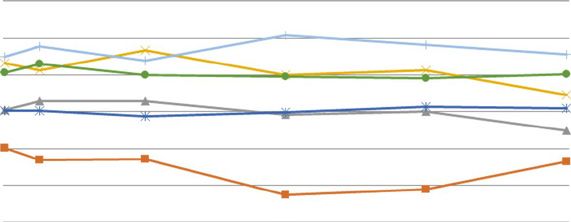
a                                                             b

                    Fig. 6. Example of ultrasound measurement at baseline (a) and after 12 weeks of treatment with MPA (b). No
                    dermal thinning was assessed. One black and white line on the scale on the left side of the image and at the bot-
                    tom corresponds to 1 mm. MPA, methylprednisolone aceponate.

throughout the observation period. Neither telangiecta-           Lehmann et al. [26] induced a steroid atrophy of the skin
sia formation nor increased skin transparency was de-             on the forearms by continuous, occlusive application of
tected.                                                           clobetasol propionate (CP) for 6 weeks. By scanning elec-
   Regarding TEWL and skin hydration, no significant              tron micrographs and histology, they were able to dem-
changes were found in BMV, MPV, HC, PIM, and pla-                 onstrate a progressive thinning of the epidermis with a
cebo between days 0 and 84. Only DBC significantly in-            reduction of the horny layer to a few wispy layers. The
creased skin hydration on the forearm (from 32.42 ± 7.81          basal cell polarity shifted from columnar to round or cu-
au to 36.07 ± 7.93 au, p = 0.029) and on the back of the          boidal. The dermal-epidermal interface was flattened,
hand (from 25.86 ± 8.9 au to 31.56 ± 5.23 au, p = 0.016).         and the dermis showed a great decrease in the ground
                                                                  substance. The compression of the fibers and their re-
                                                                  alignment parallel to the surface were noticeable. In a
     Discussion/Conclusion                                        comparable experimental setup, the same research group
                                                                  observed an exuberant hyperplasia of the epidermis with
    One of the main side effects of TCSs is skin atrophy,         a 4-fold maximal increase of viably epidermal thickness
which is characterized by a thin, transparent skin; a ciga-       within 4 days poststeroid. The dermal restitution was
rette-paper-like surface; increased fragility; and tearing        similarly rapid. Fibroblasts appeared very active and
and bruising of the skin [1, 3, 4]. In the epidermis, TCSs        within 14 days the ground substance increased continu-
may reduce the size of keratinocytes and inhibit their pro-       ously to normal levels [27].
liferation [3]. Moreover, TCSs may have a negative im-               Looking at the epidermis, Barnes et al. [24] showed
pact on the intercellular lipid layers of the upper epider-       that when used short term on the epidermis, the most at-
mis, contributing to skin barrier dysfunction [21–23]. Di-        rophogenic TCS was the very potent CP (−26%), followed
rect and indirect effects of TCS on fibroblasts and the           by BMV (−18%), which is a class III TCS according to
formation of collagen I and III and hyaluronic acid lead          Niedner et al. [28] and MPA (−8%), which is a class II
to a decrease in dermal thickness [24]. The extent of the         TCS. Even the less potent TCS, that is, HC 1%, which was
skin atrophy depends on the strength of the TCS, fre-             applied twice daily for 4 weeks on the uninvolved fore-
quency and duration of application, anatomical area               head skin of patients with AD, demonstrated a 6.1% re-
treated, and the vehicle of the emollient used [3, 24, 25].       duction in epidermal thickness according to the highly

46                  Skin Pharmacol Physiol 2022;35:41–50                                 Aschoff/Lang/Koch
                    DOI: 10.1159/000518214
sensitive OCT technique, and the thickness recovered al. [37] during continuous use of methylprednisolone
within 4 weeks after the end of treatment [11]. aceponate for 3 months on the limbs or on the trunk as
Regarding dermal thickness, the results are compara- measured by reflectance confocal microscopy. It is
ble. Korting et al. [29] investigated the influence of BMV, known that highly proliferating cells are seen in the epi-
mometasone furoate, and prednicarbate on total skin dermis as early as 2 days after alleviation of TCS, and the
thickness over a treatment period of 6 weeks measured by viable epidermis is increased in thickness with larger cells
20-MHz sonography; they found that the decrease in skin of irregular sizes and shapes [27]. Thus, it might be that
thickness was strongest by BMV and least by prednicar- a balance between atrophogenic effect of TCS and flare-
bate. Overall, dermal thinning is proportionally less than up of epidermal proliferation has been achieved in our
epidermal thinning [30, 31]. study at the back of the hand. Within 4 weeks after the
Considering that TCSs have a negative impact on skin end of the therapy, the epidermal thinning regressed.
integrity and that chronic diseases such as AD require re- Meanwhile, the dermal thickness reduced nonsignifi-
peated use of anti-inflammatory drugs, a proactive ther- cantly by −1.75% and −3.6% on the back of the hand and
apy has currently become the standard of care in AD [8]. volar forearm, respectively. MPA and HC revealed no
In clinical randomized controlled trials, fluticasone pro- changes in skin thickness, particularly epidermal thick-
pionate 0.05% cream, fluticasone propionate 0.005% ness, despite that the epidermis is known to change fast-
ointment, and MPA cream used 2 days per week for 16 er and more strongly than the dermis [38]. Using the skin
weeks reduced the risk of relapses of AD and extended the compression and thickness method, Lubach et al. [39]
time of remission [32–35]. Clinically, skin atrophy was also demonstrated skin atrophy induced by continuous
not evident in these investigations. topical application of CP, which is a highly potent TCS,
In our study, we applied TCSs of different strengths followed by intermittent application. They found that
and therapeutic indexes on the volar forearm and back continuous CP application resulted in −15% dermal
of the hand of healthy volunteers only once daily twice thinning. When continued every 5th or 7th day, no any
weekly for 12 weeks and measured the epidermal and improvement was observed; however, when CP was ad-
dermal thickness by using the highly sensitive OCT and ministered every 10th day, the skin recovered to a more
ultrasound, respectively. At baseline, the epidermal or less normal level.
thickness on the volar forearm was comparable to the Topical TCIs are also approved for treating AD and
results of Czekalla et al. [36], who measured the epider- are unlikely to cause skin atrophy. Even when PIM was
mal thickness on 5 different body areas using 2-photon applied twice daily for 4 weeks on the uninvolved fore-
microscopy and OCT. In addition to the decrease in epi- head skin of patients with AD, epidermal thinning did
dermal thickness with age, they showed that the thick- not occur according to OCT measurement [11]. There-
ness of the epidermis on the gluteal region, which is ex- fore, TCIs are preferred in sensitive locations such as the
posed to high mechanical stress, was higher than the epi- face, genital area, axilla region, or inguinal folds [1, 23,
dermal thickness on the dorsal forearm, the volar forearm, 40]. They also showed significant superiority over pla-
the sural region, and the abdomen. The epidermal thick- cebo in the proactive treatment of AD. When tacrolimus
ness in sun-expressed skin areas was higher than on ointment was applied twice weekly as a proactive ap-
those that are sun-protected. Similarly, in our study, the proach, disease exacerbations and the number of flares
epidermal thickness on the back of the hand, which is significantly declined, and the quality of life was im-
also exposed to high mechanical stress and UV radiation, proved in both adults and children [13, 14]. In our study,
was found to be thicker than the epidermis on the volar as expected, PIM showed no epidermal thinning on the
forearm. Although BMV was only applied intermittently volar forearm or back of the hand. Using ultrasound, we
for 84 days, the epidermal thickness continued to de- noticed dermal thinning on the back of the hand at
crease significantly by −6.56% on the back of the hand −2.2%, whereas that on the volar forearm remained un-
and −7.96% on the volar forearm. However, the effect changed. Interestingly, the decrease in dermal thinning
was lower than the 20% assumption in sample size calcu- on the back of the hand was statistically significant, al-
lation; the study was still able to demonstrate this sig- though the standard deviation was approximately 11%
nificant difference. We found a continuous decrease of and the decrement was less than that of the dermis on the
the epidermal thickness for the forearm but not for the forearm under BMV treatment (−3.6%), which was not
back of the hand, where the thickness stayed stable after significant. In Kyllönen et al. [41] study, the administra-
day 56. Such a plateau was also observed by Jiráková et tion of the TCI tacrolimus for over 12 months did not
Intermittent Treatment of AD with Skin Pharmacol Physiol 2022;35:41–50 47
Corticosteroids DOI: 10.1159/000518214cause skin thinning, as measured by ultrasound. How- Acknowledgments
ever, the skin thickness increased, as determined by the
The Department of Clinical Sensoring and Monitoring (Tech-
mean of 8 anatomic locations independently of their AD- nische Universität Dresden, Dresden, Germany) provided the
presenting status, and was accompanied by an increase technical knowledge and the equipment for the testing.
in collagen synthesis. The other group treated with con-
ventional steroid-based therapy showed a significant de-
crease of 8% in skin thickness, supporting our investiga- Statement of Ethics
tion on BMV.
In another study by Hofmann et al. [42], the applica- The study was approved by the institutional review board of the
tion of PIM once daily for 3 weeks did not change the skin Technische Universität Dresden (Approval No: EK 145052012)
and the German Federal Institute for Pharmaceuticals and Medical
thickness, as measured by ultrasound; in contrast, mo- Products (EudaCT-No. 2011-004953-17, Bundesinstitut für Arz-
metasone furoate (1 mg/g) and CP 0.05% cream resulted neimittel und Medizinprodukte). The study was conducted in
in skin thinning. Hoffman also discovered that signs of compliance with the ethical principles laid down in the Declara-
skin atrophy could be detected by Dermaphot® only after tion of Helsinki and posted in clinicaltrial.gov. All volunteers gave
treatment with a TCS, but not with PIM. In our study, their written informed consent to participate in the study.
skin thinning was not detected by Dermaphot®. In a
study in which PIM was administered under semiocclu-
sion 6 h for 13 consecutive days to skin of domestic pigs, Conflict of Interest Statement
there were no atrophogenic effects seen on skin thickness The authors have declared no conflicting of interests.
as measured by ultrasound and histomorphometry [43].
Moreover, PIM did not affect the expression of cytokines,
chemokines or adhesion molecules in optimally stimu- Funding Sources
lated human fibroblasts [44]. A decrease of dermal thick-
ness of 2% in our study is most likely not clinically rele- This study was financially supported by the Department of Der-
vant, but interestingly statistically significant. Therefore, matology, Technische Universität Dresden, Dresden, Germany.
PIM does not appear to have a negative effect on complete
skin thickness as repeated before.
In the study of Chittock et al. [45] involving patients Author Contributions
with quiescent AD treated with Betnovate® 0.1% cream Roland Aschoff performed the research, designed the research
twice per week for 8 weeks, TEWL and capacitance did study, and wrote the manuscript. Awena Lang also performed the
not change, but treatment with tacrolimus significantly research, analyzed the data, and drafted parts of the manuscript.
increased skin hydration. In another study, PIM was tre- Edmund Koch contributed experimental tools. All the authors
mendously effective in reducing TEWL and improving critically revised and approved the submitted manuscript.
skin hydration in patients with mild-to-moderate AD on
the affected skin [46].
In conclusion, the use of the TCSs MPA and HC in the Data Availability Statement
intermittent long-term therapy may be recommended All data generated or analyzed during this study are included
because they did not result in skin atrophy over a period in this article. Further enquiries can be directed to the correspond-
of 3 months. Considering the potential thinning of the ing author or can be requested via https://katalog.slub-dresden.de/
epidermis, BMV should rather be avoided. Especially in id/0-1551128357.
sensitive regions such as the face and intertrigines, PIM
is a good alternative in the proactive therapy because it
also does not cause atrophy. Given that epidermal thin-
ning is more pronounced and may occur earlier than der-
mal atrophy, OCT may be a valuable tool for the early
detection of skin atrophy.
However, no statement can be made as to whether
TCSs lead to skin atrophy when used over a prolonged
period of time. Therefore, further and longer studies, es-
pecially in patients with AD, are required.
48 Skin Pharmacol Physiol 2022;35:41–50 Aschoff/Lang/Koch
DOI: 10.1159/000518214References 1 Siegfried EC, Jaworski JC, Kaiser JD, Hebert 13 Wollenberg A, Reitamo S, Girolomoni G, La- 26 Lehmann P, Zheng P, Lavker RM, Kligman AA. Systematic review of published trials: hfa M, Ruzicka T, Healy E, et al. Proactive AM. Corticosteroid atrophy in human skin. A long-term safety of topical corticosteroids treatment of atopic dermatitis in adults with study by light, scanning, and transmission and topical calcineurin inhibitors in pediatric 0.1% tacrolimus ointment. Allergy. 2008 Jul; electron microscopy. J Invest Dermatol. 1983; patients with atopic dermatitis. BMC Pediatr. 63(7):742–50. 81(2):169–76. 2016 Jun;16(16):75. 14 Thaçi D, Reitamo S, Gonzalez Ensenat MA, 27 Zheng PS, Lavker RM, Lehmann P, Kligman 2 Broeders JA, Ahmed Ali U, Fischer G. Sys- Moss C, Boccaletti V, Cainelli T, et al. Proac- AM. Morphologic investigations on the re- tematic review and meta-analysis of random- tive disease management with 0.03% tacroli- bound phenomenon after corticosteroid-in- ized clinical trials (RCTs) comparing topical mus ointment for children with atopic der- duced atrophy in human skin. J Invest Der- calcineurin inhibitors with topical corticoste- matitis: results of a randomized, multicentre, matol. 1984;82(4):345–52. roids for atopic dermatitis: a 15-year experi- comparative study. Br J Dermatol. 2008 Dec; 28 Niedner R. [Therapy with systemic glucocor- ence. J Am Acad Dermatol. 2016 Aug; 75(2): 159(6):1348–56. ticoids]. Hautarzt. 2001 Nov; 52(11): 1062–4; 410–9.e3. 15 Fitzpatrick TB. The validity and practicality of quiz 1072. 3 Schoepe S, Schäcke H, May E, Asadullah K. sun-reactive skin types I through VI. Arch 29 Korting HC, Unholzer A, Schäfer-Korting M, Glucocorticoid therapy-induced skin atro- Dermatol. 1988;124(6):869–71. Tausch I, Gassmueller J, Nietsch KH. Differ- phy. Exp Dermatol. 2006 Jun;15(6):406–20. 16 Koch P, Boller D, Koch E, Welzel J, Hüttmann ent skin thinning potential of equipotent me- 4 Shlivko IL, Kamensky VA, Donchenko EV, G. Ultrahigh-resolution FDOCT system for dium-strength glucocorticoids. Skin Pharma- Agrba P. Morphological changes in skin of dermatology. Proc SPIE. 2005;5690:24–30. col Appl Skin Physiol. 2002;15(2):85–91. different phototypes under the action of 17 Popp A, Wendel M, Knels L, Knuschke P, 30 Josse G, Rouvrais C, Mas A, Haftek M, Delal- topical corticosteroid therapy and tacroli- Mehner M, Koch T, et al. Common-path Fou- leau A, Ferraq Y, et al. A multitechnique eval- mus. Skin Res Technol. 2014 May; 20(2): rier domain optical coherence tomography of uation of topical corticosteroid treatment. 136–40. irradiated human skin and ventilated isolated Skin Res Technol. 2009 Feb;15(1):35–9. 5 Luger T, Loske KD, Elsner P, Kapp A, Ker- rabbit lungs. Proc SPIE. 2005;5861:145–53. 31 Kolbe L, Kligman AM, Schreiner V, Stoude- scher M, Korting HC, et al. [Topical skin ther- 18 Frosch PJ, Behrenbeck EM, Frosch K, Macher mayer T. Corticosteroid-induced atrophy and apy with glucocorticoids: therapeutic index]. E. The Duhring chamber assay for corticoste- barrier impairment measured by non-inva- J Dtsch Dermatol Ges. 2004 Jul;2(7):629–34. roid atrophy. Br J Dermatol. 1981;104(1):57– sive methods in human skin. Skin Res Tech- 6 Schmitt J, von Kobyletzki L, Svensson A, Ap- 65. nol. 2001;7(2):73–7. felbacher C. Efficacy and tolerability of proac- 19 Rogiers V; EEMCO Group. EEMCO guidance 32 Hanifin J, Gupta AK, Rajagopalan R. Inter- tive treatment with topical corticosteroids for the assessment of transepidermal water loss mittent dosing of fluticasone propionate and calcineurin inhibitors for atopic eczema: in cosmetic sciences. Skin Pharmacol Appl Skin cream for reducing the risk of relapse in atop- systematic review and meta-analysis of ran- Physiol. 2001 Mar–Apr;14(2):117–28. ic dermatitis patients. Br J Dermatol. 2002 domized controlled trials. Br J Dermatol. 2011 20 Berardesca E. European Group for Efficacy Sep;147(3):528–37. Feb;164(2):415–28. Measurements on Cosmetics and Other Top- 33 Berth-Jones J, Damstra RJ, Golsch S, Livden 7 Wollenberg A, Bieber T. Proactive therapy of ical Products (EEMCO). EEMCO guidance JK, Van Hooteghem O, Allegra F, et al. Twice atopic dermatitis: an emerging concept. Al- for the assessment of stratum corneum hydra- weekly fluticasone propionate added to emol- lergy. 2009 Feb;64(2):276–8. tion: electrical methods. Skin Res Technol. lient maintenance treatment to reduce risk of 8 Wollenberg A, Barbarot S, Bieber T, Christen- 1997 May;3(2):126–32. relapse in atopic dermatitis: randomised, Zaech S, Deleuran M, Fink-Wagner A, et al. 21 Sheu HM, Lee JY, Chai CY, Kuo KW. Deple- double blind, parallel group study. BMJ. 2003 Consensus-based European guidelines for tion of stratum corneum intercellular lipid la- Jun 21;326(7403):1367. treatment of atopic eczema (atopic dermati- mellae and barrier function abnormalities af- 34 Van Der Meer JB, Glazenburg EJ, Mulder PG, tis) in adults and children: part I. J Eur Acad ter long-term topical corticosteroids. Br J Eggink HF, Coenraads PJ. The management of Dermatol Venereol. 2018 May;32(5):657–82. Dermatol. 1997;136(6):884–90. moderate to severe atopic dermatitis in adults 9 Holm EA, Wulf HC, Thomassen L, Jemec GB. 22 Jensen JM, Pfeiffer S, Witt M, Bräutigam M, with topical fluticasone propionate. The Neth- Instrumental assessment of atopic eczema: Neumann C, Weichenthal M, et al. Different erlands Adult Atopic Dermatitis Study Group. validation of transepidermal water loss, stra- effects of pimecrolimus and betamethasone Br J Dermatol. 1999 Jun;140(6):1114–21. tum corneum hydration, erythema, scaling, on the skin barrier in patients with atopic der- 35 Peserico A, Städtler G, Sebastian M, Fernan- and edema. J Am Acad Dermatol. 2006 Nov; matitis. J Allergy Clin Immunol. 2009 Sep; dez RS, Vick K, Bieber T. Reduction of relaps- 55(5):772–80. 124(33 Suppl 2):R19–28. es of atopic dermatitis with methylpredniso- 10 Proksch E, Fölster-Holst R, Jensen JM. Skin 23 Jensen JM, Weppner M, Dähnhardt-Pfeiffer lone aceponate cream twice weekly in addi- barrier function, epidermal proliferation and S, Neumann C, Bräutigam M, Schwarz T, et tion to maintenance treatment with emollient: differentiation in eczema. J Dermatol Sci. al. Effects of pimecrolimus compared with tri- a multicentre, randomized, double-blind, 2006 Sep;43(3):159–69. amcinolone acetonide cream on skin barrier controlled study. Br J Dermatol. 2008 Apr; 11 Aschoff R, Schmitt J, Knuschke P, Koch E, structure in atopic dermatitis: a randomized, 158(4):801–7. Bräutigam M, Meurer M. Evaluation of the double-blind, right–left arm trial. Acta Derm 36 Czekalla C, Schönborn KH, Lademann J, atrophogenic potential of hydrocortisone 1% Venereol. 2013 Sep 4;93(5):515–9. Meinke MC. Noninvasive determination of cream and pimecrolimus 1% cream in unin- 24 Barnes L, Kaya G, Rollason V. Topical corti- epidermal and stratum corneum thickness in volved forehead skin of patients with atopic costeroid-induced skin atrophy: a compre- vivo using two-photon microscopy and opti- dermatitis using optical coherence tomogra- hensive review. Drug Saf. 2015 May; 38(5): cal coherence tomography: impact of body phy. Exp Dermatol. 2011 Oct;20(10):832–6. 493–509. area, age, and gender. Skin Pharmacol Physi- 12 Queille-Roussel C, Graeber M, Thurston M, 25 Maubec E, Laouénan C, Deschamps L, Nguy- ol. 2019;32(3):142–50. Lachapelle JM, Decroix J, de Cuyper C, et al. en VT, Scheer-Senyarich I, Wackenheim-Ja- 37 Jiráková A, Rob F, Sečníková Z, Koblová K, SDZ ASM 981 is the first non-steroid that cobs AC, et al. Topical mineralocorticoid re- Džambová M, Rajská L, et al. Topical cortico- suppresses established nickel contact derma- ceptor blockade limits glucocorticoid-in- steroids but not calcineurin inhibitors in- titis elicited by allergen challenge. Contact duced epidermal atrophy in human skin. J duced atrophy after four weeks. Biol Regul Dermatitis. 2000 Jun;42(6):349–50. Invest Dermatol. 2015 Jul;135(7):1781–9. Homeost Agens. 2015;29(3):701–6. Intermittent Treatment of AD with Skin Pharmacol Physiol 2022;35:41–50 49 Corticosteroids DOI: 10.1159/000518214
38 Cossmann M, Welzel J. Evaluation of the at-        41 Kyllönen H, Remitz A, Mandelin JM, Elg P,          44 Wolff B, Herzig G, Stuetz A. Pimecrolimus
   rophogenic potential of different glucocorti-         Reitamo S. Effects of 1-year intermittent             does not affect cytokine and chomekine secre-
   coids using optical coherence tomography,             treatment with topical tacrolimus monother-           tion and adhesion molecule expression in pri-
   20-MHz ultrasound and profilometry; a dou-            apy on skin collagen synthesis in patients with       mary human keratinocytes and dermal fibro-
   ble-blind, placebo-controlled trial. Br J Der-        atopic dermatitis. Br J Dermatol. 2004 Jun;           blasts. J Invest Dermatol. 2003;121(1):1243.
   matol. 2006 Oct;155(4):700–6.                         150(6):1174–81.                                    45 Chittock J, Brown K, Cork MJ, Danby SG.
39 Lubach D, Rath J, Kietzmann M. Skin atrophy        42 Hofmann M, Salgo R, Aschoff R, Luger TA,              Comparing the effect of a twice-weekly tacro-
   induced by initial continuous topical applica-        Meurer M, Bräutigam M, et al. Validation of           limus and betamethasone valerate dose on the
   tion of clobetasol followed by intermittent ap-       Dermaphot(®) for the assessment of steroid-           subclinical epidermal barrier defect in atopic
   plication. Dermatology. 1995;190(1):51–5.             induced skin atrophy. Arch Dermatol Res.              dermatitis. Acta Derm Venereol. 2015 Jul;
40 Reitamo S, Ortonne JP, Sand C, Cambazard F,           2013 Apr;305(3):215–21.                               95(6):653–8.
   Bieber T, Fölster-Holst R, et al. A multicentre,   43 Meingassner JG, Grassberger M, Fahrngruber         46 Aschoff R, Schwanebeck U, Bräutigam M,
   randomized, double-blind, controlled study            H, Moore HD, Schuurman H, Stütz A. A nov-             Meurer M. Skin physiological parameters
   of long-term treatment with 0.1% tacrolimus           el anti-inflammatory drug, SDZ ASM 981, for           confirm the therapeutic efficacy of pimecroli-
   ointment in adults with moderate to severe            the topical and oral treatment of skin diseases:      mus cream 1% in patients with mild-to-mod-
   atopic dermatitis. Br J Dermatol. 2005 Jun;           in vivo pharmacology. Br J Dermatol. 1997;            erate atopic dermatitis. Exp Dermatol. 2009;
   152(6):1282–9.                                        137(4):568–76.                                        18(1):24–9.

50                         Skin Pharmacol Physiol 2022;35:41–50                                             Aschoff/Lang/Koch
                           DOI: 10.1159/000518214
You can also read