Enterovesical Fistula and Bladder Calculus caused by a Migrated Intrauterine Device: A Case Report
←
→
Page content transcription
If your browser does not render page correctly, please read the page content below
Case Report Frontiers in Medical Case Reports, ISSN: 2582-8142
DOI: http://dx.doi.org/10.47746/FMCR.2021.2509
Enterovesical Fistula and Bladder Calculus caused by a Migrated Intrauterine
Device: A Case Report
Niurka Wallace1* | Shenelle Wilson2
*Correspondence: Niurka Wallace
Address: 1Rowan University School of Osteopathic Medicine, Medical Center Drive, Stratford NJ; 2Georgia Urology, Atlanta,
Georgia
e-mail : wallac13@rowan.edu
Received: 13 September 2021; Accepted: 21 September 2021
Copyright: © 2021 Wallace N. This is an open-access article distributed under the terms of the Creative Commons Attribution
License, which permits unrestricted use, distribution, and reproduction in any medium, provided that the original work is
properly cited.
ABSTRACT
Intrauterine devices (IUD) have proven to be one of the most reliable methods of contraception. However, as it is a foreign
body, it can migrate from the intended position in the uterus, perforating surrounding structures and creating fistulas. We
report a case of a 42-year-old woman who presented with abdominal pain, recurrent urinary tract infections, and chronic
pelvic pain for eight years. Imaging revealed an intravesical IUD with attached bladder calculus, and a transurethral
cystolitholapaxy subsequently uncovered an enterovesical fistula. Conservative urethral catheter management did not resolve
the fistula, which was then repaired via robotic excision. The patient had an uncomplicated postoperative course, and voiding
cystourethrogram confirmed water-tight repair of the fistula tract after two weeks.
Keywords: Intrauterine Device, Enterovesical Fistula, Bladder Calculi, Chronic Pelvic Pain
Introduction
The intrauterine device (IUD) is a reversible, economical, highly effective contraceptive, with one of
the lowest failure rates and the highest continuation rates in the general population (Krashin et al., 2015).
However, IUDs can migrate, causing severe but rare complications days to years after insertion, such as
uterine perforation, colonic perforation, intestinal obstruction, and fistula formation (Almarhabi, 2020).
These complications are often unnoticed as they carry a low index of suspicion, thus making diagnosis
difficult. Additionally, some patients experience nonspecific inflammatory symptoms such as cystitis,
frequent urination, urinary urgency, dysuria, and discomfort of the lower abdomen, thus further causing a
delay in the relevant examination, diagnosis, and treatment (Wan et al., 2021). Another rare complication
of IUD migration is the formation of vesical calculus or bladder stone. Bladder calculi are usually a
manifestation of underlying pathology, including urinary stasis, infection, urinary tract obstruction,
voiding dysfunction, or foreign body retention (Schwartz et al., 2000). The foreign body can be self-
inflicted or iatrogenic. It may enter the bladder from the genitourinary tract, bowel, or perivesical tissues.
Pelvic ultrasonography and plain X-ray are diagnostic of bladder stones and radio-opaque foreign body
Cite this article: Wallace N, et al. Enterovesical Fistula and Bladder Calculus caused by a Migrated Intrauterine 1
Device: A Case Report. Front Med Case Rep 2021; 2(5): 1-05.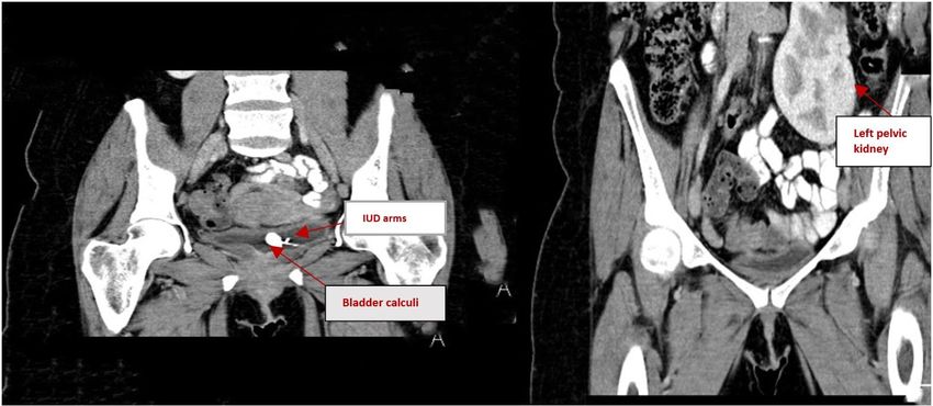
Case Report Wallace N et al., 2021; 2(5): 1-05
DOI: http://dx.doi.org/10.47746/FMCR.2021.2509
and can be used for early detection (Abdulwahab‑Ahmed and Ogunleye, 2013). We report the case of a
woman found to have an enterovesical fistula, an abnormal connection between the bladder and the
intestine caused by a migrating IUD, as well as a bladder stone.
Case Presentation
Our patient is a 42-year-old woman who presented with an 8-year history of vague abdominal
discomfort exacerbated by physical activity, chronic pelvic pain, and recurrent urinary tract infections
(UTIs). She had a history of multiple copper IUDs placed for contraception in her home country Eritrea in
2012. After the first IUD was inserted, she developed intractable abdominal pain that lasted a few weeks.
When she returned to her gynecologist three weeks after IUD placement, she reports being told that the
IUD had fallen out because the string was not visible on pelvic exam. A second IUD was placed at that time
without incident or post-procedure pain. Over the next few years, she reports intermittent, vague lower
abdominal pain that resolved with rest. In 2019, the second IUD was removed, causing subsequent
worsening persistent abdominal pain. In December 2020, the patient underwent computerized
tomography (CT) scan, which showed the first IUD in the bladder with a 12x12 mm stone attached (Fig.
1) and a left pelvic kidney. In January 2021, the IUD and stone were removed via a transurethral
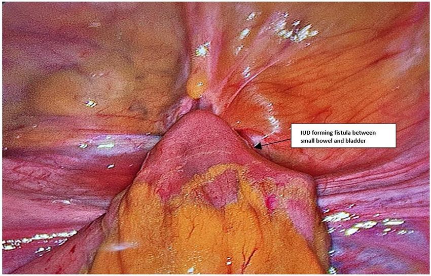
cystolitholapaxy, revealing a vesical fistula where the IUD was attached (Fig. 2). A catheter was left in
place for passive fistula closure. However, a retrograde cystogram performed after one month
demonstrated a persistent fistula to the small bowel (Fig. 3). The patient also reported seeing food
particles within her catheter bag.
Figure 1: CT images showing IUD and bladder calculus.
Front Med Case Rep, ISSN: 2582-8142 https://www.jmedicalcasereports.org/ 2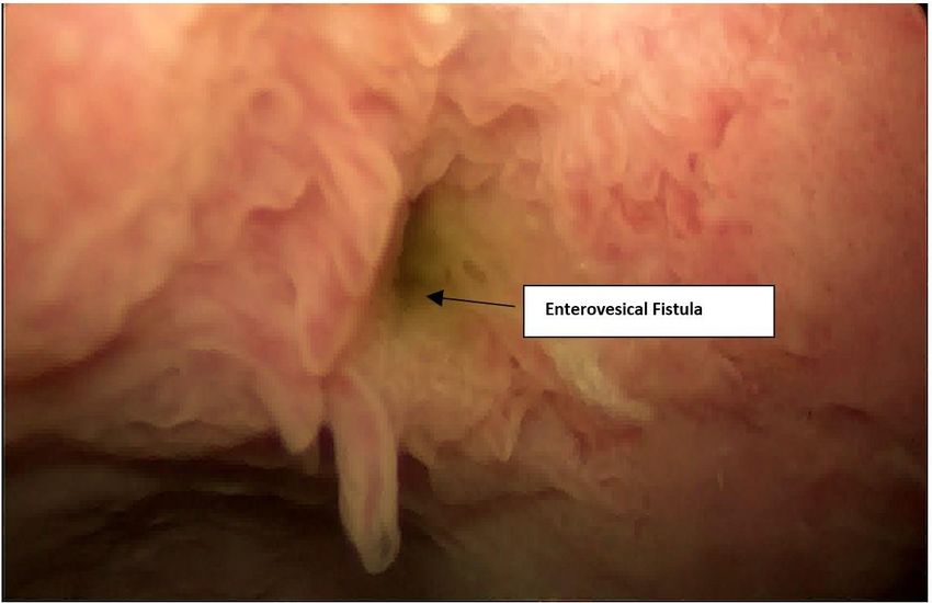
Case Report                                                                                 Wallace N et al., 2021; 2(5): 1-05
DOI: http://dx.doi.org/10.47746/FMCR.2021.2509

                   Figure 2: Vesical fistula visualized after removal of IUD and attached bladder stone.

                 Figure 3: Enterovesical fistula visualized one month post transurethral cystolitholapaxy.

      The patient was taken for definitive surgical treatment. She underwent cystoscopy with stent
placement within the fistulous tract, then robotic enterovesical fistula excision and small bowel resection.
The patient was discharged four days later after the resolution of postoperative ileus. Voiding
cystourethrogram then confirmed a water-tight repair at the two-week postoperative visit, and the
patient reported resolution of all pre-and peri-operative symptoms after six weeks.

Discussion

      Migration of IUD into surrounding organs is a rare but severe complication after uterine
perforation (Cheung et al., 2018). It is often asymptomatic but may be associated with vague pelvic or
abdominal pain (Almarhabi, 2020).

 Front Med Case Rep, ISSN: 2582-8142                                               https://www.jmedicalcasereports.org/          3
Case Report Wallace N et al., 2021; 2(5): 1-05
DOI: http://dx.doi.org/10.47746/FMCR.2021.2509
Likely migration sites include the bladder, rectosigmoid colon, peritoneum, appendix, small bowel,
and adnexa (Cheung et al., 2018). Once the IUD penetrates the uterus and bladder wall, it can act as a
nidus for calculus formation. However, the incidence of an IUD migration to the bladder and stone
formation is about 1 per 1000 insertions (Cheung et al., 2018), making this a rare complication of IUD
placements. Our patient had an extremely rare case as the IUD migrated to the bladder forming a stone
and one arm of the device created a fistula between the bladder and small bowel.
When an IUD string is not visualized on a gynecological examination, this should prompt further
detailed investigation. Diagnostic methods include urine examination, abdominal ultrasound, a
transvaginal ultrasound, KUB x-ray, and pelvic CT or MRI (Chai et al., 2017). However, CT is most useful
for identifying the exact location of the IUD and diagnosing whether it is penetrating surrounding organs
(Madden et al., 2016). If the IUD migrates intraperitoneally, minimally invasive techniques or an open
approach may be used to solve any associated complications as well as to remove the device, whereas, in
incidents of intravesical translocation and stone formation, cystolitholopaxy of the bladder calculus
should be employed along with the removal of the IUD (Madden et al., 2016).
This case presentation is unique for several reasons. First, our patient reported an atypical finding
of increased abdominal pain after the removal of the visualized IUD. Second, unlike prior published case
reports, the second IUD migrated to our patient’s bladder, creating a vesical fistula and a subsequent
enterovesical fistula. Finally, the closure of the first fistula tract was complicated by food particles
repeatedly clogging our patient’s foley because of the additional perplexing enterovesical fistula. Due to
the lack of similar reported cases seen in urogynecology practice, there is no consensus on surgical
approach and management. We hope that this case report will serve as a reference when cases of similar
complexity are encountered.
Conclusion
Intrauterine devices are one of the safest and most reliable methods of contraception. However, like
all foreign objects placed in the body, the IUD is not without complications. Although severe
complications are rare, they may include bladder calculi, fistula formation between the bladder, and small
bowel, all of which were seen in this patient. Fortunately, our patient had a positive outcome and has not
reported any complications since her last follow-up.
Abbreviations
IUD: Intrauterine device
CT: Computed Tomography Scan
UTI: Urinary Tract Infection
Front Med Case Rep, ISSN: 2582-8142 https://www.jmedicalcasereports.org/ 4Case Report Wallace N et al., 2021; 2(5): 1-05
DOI: http://dx.doi.org/10.47746/FMCR.2021.2509
References
Abdulwahab‑Ahmed A and Ogunleye OO. Vesical calculus 10 years post missing intrauterine contraceptive device. J Surg
Tech Case Rep 2013; 5: 48–50.
Almarhabi Y. Asymptomatic cecal perforation and ileocecal fistula after intrauterine device migration: a case report. J Surg
Case Reports 2020; 2020: 1-3.
Chai W, Zhang W, Jia G, Cui M, Cui L. Vesical transmigration of an intrauterine contraceptive device. Medicine (United
States) 2017; 96: 4-6.
Cheung ML, Rezai S, Jackman JM, Patel ND, Bernaba BZ, Hakimian O, Nuritdinova D, Turley CL, Mercado R, Takeshige T,
Reddy SM. Retained Intrauterine Device (IUD): Triple Case Report and Review of the Literature. Case Rep Obstet Gynecol 2018;
2018: 1-8.
Krashin J, Tang JH, Mody S, Lopez LM. Hormonal and intrauterine methods for contraception for women aged 25 years
and younger. Cochrane Database Syst Rev 2015; (8).
Madden A, Aslam A, Nusrat NB. A Case of Migrating “Saf-T-Coil” Presenting With a Vesicovaginal Fistula and Vesicovaginal
Calculus. Urol Case Reports 2016; 7: 17-19.
Schwartz BF, Stoller ML. The vesical calculus. Urol Clin North Am 2000; 27: 333–346.
Wan L, Wang Y, Xiao C, Li X, Cao J, Wang S, Wei X, Liu X. Four cases of heterotopia of an intrauterine device embedded in
the bladder muscular layer causing cystolithiasis: case report and review of the literature. J Int Med Res 2021; 49:
300060520979444.
Front Med Case Rep, ISSN: 2582-8142 https://www.jmedicalcasereports.org/ 5You can also read