Evaluation of Antibacterial Potency of Citrus Limon (Lemon) Juice Against Some Pathogenic Organisms as Alternative Source of Chemotherapy
←
→
Page content transcription
If your browser does not render page correctly, please read the page content below
ORIGINAL ARTICLE
European Journal of Biology and Biotechnology
www.ejbio.org

 Evaluation of Antibacterial Potency of Citrus Limon
 (Lemon) Juice Against Some Pathogenic Organisms as
 Alternative Source of Chemotherapy
 J. U. Ewansiha

 ABSTRACT

 The development of antibiotic resistance by pathogenic microorganisms
have necessitated the quest for alternative drug therapy. The efficacy and
safety of extract from Citrus limon for the development of alternative Published Online: April 01, 2020
antibacterial drug using the cold-pressing extraction methods and column DOI : 10.24018/2020.1.1.12
chromatography to obtain crude juice extracts and fractions respectively
while the agar well diffusion and tube dilution methods were used to screen
the juice extract and fractions for antibacterial activity against Salmonella
enterica, Klebsiella pneumoniae, Streptococcus pneumoniae and J.U Ewansiha *
Streptococcus pyogenes. A total of 277.5mL (12.313%) of juice was obtained Modibbo Adama University of Technology,
from 2253.8g of fruit while the phytochemical analysis revealed the Yola, Nigeria.
presence of 10 phytocomponents namely tannins, flavonoids, (e-mail: ewansihajoel@gmail.com)
anthraquinones, alkaloids, steroids, phenols, cardiac glycoside, terpenes,
 *Corresponding Author
resins and saponins. One hundred percent (100%) of the juice crude extract
exhibited the highest activity with mean inhibition zones (MIZ) ranged from
25.00±0.57mm to 32.33±0.33mm while 25% (6.67±1.15mm to
10.00±1.00mm) exhibited the least activity. The minimum inhibitory
concentration and minimum bactericidal concentration (MIC and MBC)
for the juice crude extract ranged from 25% to 12.5% and 100%
respectively while S. paratyphi C, S. typhi, K. pneumoniae and S. pneumoniae
were still viable at 100%. Out of the three fractions eluted, only one
(JEtOAc) was active against all the test organisms with MIZ ranged from
14.00±0.33mm to 22.33±1.20mm while 27 compounds were identified in the
fractions by GCMS. Notable group of compounds identified include fatty
acids, terpenes, aliphatic and aromatic hydrocarbons. The MIC and MBC
for the fractions ranged from 30mg/mL to 15mg/mL and >120mg/mL to
30mg/mL. Based on these findings, it can therefore be concluded that the
juice extract and ethyl acetate fraction of Citrus limon possess antibacterial
activity due to the abundant presence of secondary metabolites and it is
recommended that toxicity test be carried out on the crude extract and
fraction for the development of alternative drugs for the control and
treatment of infections caused by resistant organisms.
 Key Words: Citrus limon, Juice, Antibacterial, Cold pressing, Fractions,
Chemotherapy.

 the world population rely on traditional herbal medicine to
 I. INTRODUCTION meet their primary health care needs. It has been reported
Medicinal plants are increasingly gaining acceptance and that a substantial percentage of modern chemotherapeutic
well acknowledged even among the rural and urban drugs contained one or more of the natural products which
settlements, due to the increasing development of drug are of plant origin. [4] Suck (1989) reported that more than
resistance, inefficacy of many modern chemotherapeutic 75 pure compounds derived from higher plants are used in
drugs used for the control of microbial infections as well herbal medicine but most of those applied in modern
as side effects by several antibiotics and the increasing medicine are now produced synthetically. Citrus limon is
cost of drugs prescription, for the maintenance of personal an evergreen plant native to Asia. It is popularly utilized
health [1]. Increases also in human population has made it for its juice, pulp & also the peels. It is used worldwide in
impossible for modern health facilities to meet health cooking dishes because its juice provides a unique sour
demands all over the world, thus causing more demands taste. It is rich in citric acid which gives a pH of 2-3.
on the use of natural herbal health remedies. Medicinal Lemon is used to make lemonades, cocktails, beverages,
plants are used in traditional health care systems since etc. It is also a plant of interest because of its medicinal
prehistoric times and are still the most important health value [5]. It exhibits antimicrobial properties thus, its
care source for the vast majority of the population around potentials to inhibit microbial growth should be explored.
the world [2]. [3] reported that an estimate of 70-80% of Since it is easily available and common in use, its extracts

 DOI: http://dx.doi.org/10.24018/ejbio.2020.1.1.12 Vol 1 | Issue 1 | April 2020 1
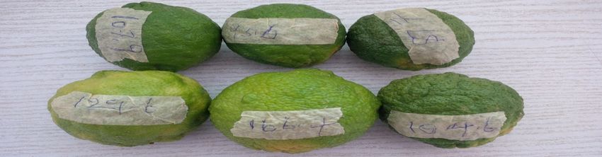
ORIGINAL ARTICLE
European Journal of Biology and Biotechnology
www.ejbio.org

can serve as medicines. Lemon is an important medicinal #TM050) (www.promega.com) and their identities and
plant of the family Rutaceae. It is cultivated mainly for its accession numbers were determined by BLAST
alkaloids, which are having anticancer activities and the (comparison of the extracted GENE sequence with the
antibacterial potentials in crude extracts of different parts known sequence from the GENE bank)
(viz., leaves, stem, root, juice and flower) of Lemon (www.ncbi.nlm.nih.gov).
against clinically significant bacterial strains has been Extraction of Fruit Juice.
reported [6]. Citrus flavonoids have a large spectrum of
biological activity including antibacterial, antifungal, Lemon fruit were washed with distilled water and weighed
antidiabetic, anticancer and antiviral activities on a digital weighing balance and the weight was
 recorded. The peels were removed with newly purchased
 razor blade, to prevent contamination of the juice with the
[7] [8]. The aim of this study was to evaluate the contents of the peel during extraction. The fruits were
antibacterial potential of lemon fruit juice crude extracts massaged gently to release the juice within the pulp and
 again they were washed with distilled water and then
against some pathogenic bacteria and its toxicological
 wiped with cotton soaked in 70% alcohol. The juice were
properties using standard routine antibacterial assay
 extracted by cold pressing method, which was achieved by
techniques.
 dividing the fruits into two halves with a sterile laboratory
 knife after which the juice were squeezed out gently and
 II. MATERIALS AND METHODS aseptically. The juice was then filtered through a whatman
A Collection, identification and processing of Plant filter paper with pore size of 20µm. The volume of the
sample juice was measured in a graduated cylinder while the
 Fruits of Citrus limon (Plate I) were collected from percentage yield was determined by dividing the total
Bosso estate Minna, Nigeria and identified/authenticated volume of the extracted juice by the total original weight
at the Herbarium Department of the National Institute of of the fruit juice before extraction.
Pharmaceutical Research, and Development, Idu, Abuja
where voucher specimens were deposited with voucher ( / ) =
 !"#$%& "( ()$*+ ,$*-& (%#)
number: NIPRD/H/6780. 100/1 (1)
 0&*12+ "( 32"#& ()$*+ ,$*-& (1)

 V DETERMINATION OF THE PH OF CITRUS LIMON
 FRUIT JUICE

 The pH (power of hydrogen) of the fruit juice extract of
 Citrus limon was determined using a digital pH meter. The
 pH meter was first calibrated using buffer 4, 7 and 9 pH,
 this was achieved by insertion of the pH electrode in the
 buffer solutions while the calibration control nub was
 Fig. 1. Citrus limon adjusted to the desired pH value. After calibration, the pH
 of the fruit juice extract was then taking by the insertion of
 the PH electrode in the sample and the pH values
B Test Organisms. recorded.
The test organisms, Salmonella enteric subs. enterica
serotype typhi, Salmonella enteric serotype paratyphi A, B VI. PHYTOCHEMICAL SCREENING
& C, Klebsiella pneumoniae, Streptococcus pneumoniae
 Phytochemical analyses was performed using the method
and Streptococcus pyogenes were obtained from stock
 as described by [10] to screen the extracts for the presence
cultures in the Microbiology laboratory Federal University
 of the following active principles: alkaloids, tannins,
of Technology, Minna. The microorganisms were
 saponins, flavonoids, anthraquinones, cardiac glycosides,
reconstituted by sub-culturing onto freshly prepared
 volatile oils, terpenoids, resins, steroids and phenol.
nutrient agar and then incubated at 370C for 24hours, after
 Standardization of Inoculums
which their identities were confirmed using gram staining
and molecular identification. Zero point two millilitre (0.2ml) of overnight cultures of
 III. GRAM STAINING the test organism was transferred into 20ml of sterile
 nutrient broth and the culture was incubated for 3 – 5h at
An overnight culture of the test organisms were Gram 370C to standardize the culture to 106 CFU/mL
stained according to the method described by [9], Gram McFarland. A loopful of the standardized inoculum was
positive and Gram negative organisms were recorded. A used for the antibacterial assay [11].
control smear of known Gram positive organism
 VII. PREPARATION OF EXTRACT
(Staphylococcus aureus) and a known gram negative
organism (Escherichia coli) was stained simultaneously to CONCENTRATION
confirm the accuracy of the procedure. One hundred percent of the lemon juice (100%) was
 VI. MOLECULAR IDENTIFICATION. directly used against the test organisms, while 75%, 50%
 and 25% were prepared by transferring 7.5, 5, and 2.5mL
The organism’s identity were molecularly authenticated of the juice crude extract into tubs containing 2.5, 5 and
according to Promega Protocol (Technical Manual

 DOI: http://dx.doi.org/10.24018/ejbio.2020.1.1.12 Vol 1 | Issue 1 | April 2020 2
ORIGINAL ARTICLE
European Journal of Biology and Biotechnology
www.ejbio.org

7.5mL distilled water respectively for the antibacterial CONCENTRATION (MIC AND MBC) OF THE JUICE
susceptibility test. EXTRACTS
 A Serial Dilution of Juice Extracts
 VIII. ANTIBACTERIAL SUSCEPTIBILITY TEST The tube dilution method as described by [16] [17] with
 slight modification using spectrophotometer was used to
The antibacterial activity of the fruit juice extract was determine the minimum inhibitory concentration. 100%
carried out using the agar-well diffusion method as and 75% as described above was used directly while two
described by [12]. Muller-Hinton agar was prepared fold serial dilutions of the lemon juice were prepared to
according to manufacturer instructions and seeded with the give a decrease in concentration ranging from 50, 25, 12.5,
standardized test organisms by the spread plate method 6.25, 3.125, 1.563 & 0.78%/ml of lactose broth.
using a sterile rod spreader to obtain uniform microbial
growth. Wells were made in the inoculated media using B Determination of MIC And MBC
sterile cork-borer (6 mm diameter) after which a little
molten media was used to seal the base of the wells to All the prepared dilutions were properly shaken to obtain a
prevent unwanted spread of the extracts. 100µl each of the homogenous mixture and they were next inoculated with
prepared juice extract equivalent to the desired 100μl of the test organisms appropriately. Positive and
concentrations per millilitre was transferred into the wells negative control tubes were also maintained for each test
with a sterile micropipette and it was well labelled, while batch of extract concentrations and test organisms
100µl of water (free of juice extract) was transferred into respectively [16] [17]. For the positive control, sterile
wells to serve as the negative control. Ciprofloxacin nutrient broth was inoculated with 100μl of the test
(1mg/ml) was used as the positive control. This was done organisms without the addition of the extract while for the
by transferring 100µl of the prepared standard antibiotics negative control, the prepared serially diluted juice were
into the well and the cultures were allowed to stand for incubated without the test organisms. The test tubes were
30min after which the seeded plates were incubated at all incubated at 370C for 24 h. At the end of the incubation
37°C for 24 hours. The experiment was carried out in period, the optical density of the cultures in the test tubes
triplicate and the mean values with the corresponding were read using spectrophotometer at a wavelength of
standard deviation of the inhibition zone diameters (IZD) 600nm while the spectrophotometer was adjusted to zero
were calculated. The performed agar well diffusion using sterile lactose broth void of extracts and test
susceptibility test was based on the modified methods of organism. The MIC was determined by subtracting the
the Science Laboratory Standards Institute [13]. absorbance of the negative control from the absorbance of
 the test and comparing the result with the absorbance of
 IX. THIN LAYER CHROMATOGRAPHY OF CRUDE the positive control (see below formulae). The
 EXTRACTS concentration/test tube where significant reduction in
The Analytical thin layer chromatographic technique was absorbance was observed, was recorded as the MIC.
done according to the method described by [14] to spot,
separate and determine the Rf (Retension factors) values Absorbance of Test (T) minus absorbance of negative
and a suitable solvent systems for fractionation of the control (C0) equal to the absorbance of positive control
phytochemical coponents by column chromatography on (C1):
the crude extracts. This was achieved by using the TLC T-C0 = C1 (2)
silica gel 60 F254 Aluminium sheet made by Merck KGaA,
Millipore Corporation Germany was used as the stationary The minimum bactericidal concentration (MBC) was
phase for the most active limon juice. The solevent system determined by subculturing the cultures with the lowest
that gave the best separation based on the Rf values were optical density beginning with the test tube containing the
used to fractionate the crude extracts by column minimum inhibitory concentration and above onto a
chromatography, while the number of spots seen was freshly prepared nutrient agar medium. The cultures were
recorded. incubated for 24 hours at 370C, after incubation, the
 culture concentration without visible growth was regarded
 X. COLUMN CHROMATOGRAPHY (PARTIAL as the minimum bactericidal concentration [16] [17].
 PURIFICATION) OF CRUDE EXTRACT
The micro scale column chromatographic method
according to [15] was used to separate the fractions of the
fruit juice extract. The column (40mm diameter width and XII. QUANTITATIVE ANALYSIS AND
150mm length) was prepared by packing it with 150g IDENTIFICATION OF COMPOUNDS
silica gel (0.015-0.04mm mesh size) desolved in 500ml n- The determination of the identity of components in the
hexane to make a slorry using the wet method. The most active fraction (JEtOAc) were done by GC-MS
fractions were collected in test tubes according to their analysis using GC-MS-QP 2010 Plus, Shimadzu system
colour development, bulked using thin layer (SHIMADZU, JAPAN) as described by [18]. The gas
chromatography and the eluting solvents were allowed to chromatograph interface to a mass spectrometer (GC-MS)
vapourise until a constant weight was obtained. instrument was used while the Column elite-1 was fused
 with silica capillary column (30m x0.25mm 1D x µL df,
 composed of 100% dimethyl polysiloxane). An electronic
 XI. DETERMINATION OF THE MINIMUM ionization system with ionization energy of 60eV was used
 INHIBITORY AND BACTERICIDAL for the GC-MS detection while Helium gas (99.99%) was
 used as the carrier gas at a flow rate of 1ml/min and

 DOI: http://dx.doi.org/10.24018/ejbio.2020.1.1.12 Vol 1 | Issue 1 | April 2020 3
ORIGINAL ARTICLE
European Journal of Biology and Biotechnology
www.ejbio.org

injection size of the fraction was 2µl (0.002ml with split B ± 1.15a ± ± ±
ratio of 1:40 and film thickness of 0.20μm). Total GC 1.00b 0.57c 0.33de 0.88d
 S. paratyphi 9.33± 12.33 14.00 29.00 24.66 NA
running time was 28.00minutes. Relative percentages and C 0.57a ± ± ± ±
amount of each components were deduced by comparing 0.57c 1.00a 0.00b 1.45a
individual average peaks area to the total areas. Turbomass S. typhi 10.00 13.33 15.33 30.00 25.33 NA
was used for the mass spectra and chromatogram while the ± ± ± ± ±
 1.0b 0.57c 0.57a 0.00b 0.33a
detection of compounds was done using the database from K. 8.67± 13.67 14.33 25.00 25.66 NA
the library of National Institute of Standard and pneumoniae 0.57ab ± ± ± ±
Technology (NIST) NIST Ver. 2.0 year 2009. 1.53c 0.57a 0.57a 0.88a
 S. 6.67± 11.00 14.33 30.00 25.33 NA
 pneumoniae 1.15ab ± ± ± ±
 XIII. RESULTS AND DISCUSSION 1.00b 1.53a 0.00b 0.88a
 S. pyogenes 8.67± 13.33 14.67 29.66 30.00 NA
 A. Results 1.53b ± ± ± ±
 TABLE I: IDENTITY AND ACCESSION NUMBER OF TEST 2.08c 1.15a 0.33b 1.00b
 ORGANISMS Key: Cpx: ciprofloxacin, D: distilled water, NA: no activity,
 *Specification for Cpx and E are: ≤15 (resistance), 16-20 (intermediate),
 Test Organisms Gram Total Identity Accession ≥21 (susceptible) [13]. Values on the same column with different
 s Score number superscript are significantly different (p
ORIGINAL ARTICLE
 European Journal of Biology and Biotechnology
 www.ejbio.org

 100% methanol - 5.0 - -
 Key: DMS= distance moved by the solvent (mobile phase), DMF=
 distance moved by fraction, Rf = Retention factor, NS = Number of spot.

 TABLE VI: PERCENTAGE YIELD AND RETENTION FACTOR OF
 CITRUS LIMON FRUIT JUICE FRACTIONS (50ML)
 TABLE VIII: MINIMUM INHIBITORY CONCENTRATION (MIC)
 Fractio Solvent Description Percenta N Rf AND MINIMUM BACTERICIDAL CONCENTRATION
 n system ge S (c (MBC) OF CITRUS LIMON JUICE FRACTIONS
 &Volume Yield g m) JEtOAc (mg/mL)
 (ml) (%w/v) Organisms MIC MBC
 JCHCl3 100% CHCl3 Yellow semi 1g (2) 1 0.8
 (500) solid 6 S. paratyphi A 30 120
 S. paratyphi B 15 60
 JEtOAc 100% EtOAc Brown semi 5g (10) 1 0.7
 (700) solid 6 S. paratyphi C 30 >120
 S. typhi 30 >120
 JH2O 100% water White pellet 9g (18) N N K. pneumoniae 30 >120
 (400) A A
 S. pneumoniae 15 120
 S. pyogenes 15 60
 TABLE VII: MEAN ZONES OF INHIBITION OF CITRUS LIMON
 JUICE FRACTIONS (MM) Key: JEtOAc = Ethyl acetate juice fraction,
 Juice fractions Control
 Organism JCHCL3 JEtOAc JH2O Cpx D
 (40mg (40mg (40mg (1mg (100µL)
 /mL) /mL) /mL) /mL)
S. paratyphi A 0.00a 15.00±1. 0.00a 23.50±1.50a
 00a
S. paratyphi B 0.00a 22.33±1. 0.00a 26.66±0.88a 0.00a
 20b
S. paratyphi C 0.00a 17.00±1. 0.00a 24.66±1.45a 0.00a
 a
 00
 S. typhi 0.00a 17.00±0. 0.00a 25.33±0.33a 0.00a
 57a
K. pneumoniae 0.00a 15.00±0. 0.00a 25.33±0.33a 0.00a
 00a
S. pneumoniae 0.00a 14.00±0. 0.00a 25.33±0.88a 0.00a
 33a
 S. pyogenes 0.00a 16.65±0. 0.00a 30.00±1.00b 0.00a
 63a
 Key: JEtOAc = Juice ethyl acetate fraction, JCHCL3 = Juice chloroform
 fraction, JH2OC = Juice aqueous fraction, Cpx = ciprofloxacin, D =
 dimethyl sulfoxide, Values on the same column with different
 superscript are significantly different (p
ORIGINAL ARTICLE
European Journal of Biology and Biotechnology
www.ejbio.org

 10 9.690 2.93 172.26 C10H20O2 p-Menthane-1,8-diol

 11 13.036 0.73 196.37 C14H28 1-Tetradecene

 12 15.527 0.76 210.4 C15H30 1-Pentadecene

 13 17.889 0.57 270.45 C17H34O2 Methyl 14-methylpentadecanoate

 14 18.773 1.23 336.38 C18H24O6 Phthalic acid, butyl ester, ester with
 butyl glycolate

 15 19.010 5.98 256.42 C16H32O2 n-Hexadecanoic acid

 16 19.248 0.92 224.43 C16H32 1-Hexadecene

 17 19.655 1.01 124 C8H12O 3-Isopropyl-2-cyclopenten-1-one

 18 20.911 0.46 294.47 C19H34O2 Linolelaidic acid, methyl ester

 19 21.000 0.91 296 C19H36O2 11-Octadecenoic acid, methyl ester

 20 21.360 0.29 298.5 C19H38O2 Octadecanoic acid, methyl ester

 21 21.818 10.74 282.46 C18H34O2 Oleic Acid

 22 22.081 2.38 284.48 C18H36O2 Octadecanoic acid
 23 22.255 0.64 266.50 C20H37F3O2 Octadecyl trifluoroacetate

 24 23.485 0.20 310 C20H38O2 Methyl 5-(2-
 undecylcyclopropyl)pentanoate

 25 24.487 0.32 308.59 C22H44 1-Docosene

 26 25.961 0.48 390.56 C24H38O4 Di-n-octyl phthalate

 27 26.331 0.16 396.73 C27H56O 1-Heptacosanol

Total - 100.00 - - - -
 19 21.000 0.91 296 C19H36O2 11-Octadecenoic acid, methyl ester

 20 21.360 0.29 298.5 C19H38O2 Octadecanoic acid, methyl ester

Key: RT = Retention time; PA = Peak area; MW = Molecular weight; MF = Molecular formula

 their quantity. The presence of most of these constituents
 B. Discussion
 has been reported in several research findings and have
The extraction result reveals a considerable percentage of been linked to antimicrobial potency of several medicinal
the juice (277.5mL (10.08) from 2257.8g of whole lemon plants. Tannins are known to be astringent, plants
juice. It is reported that extraction yield of medicinal plant polyphenols that bind to protein, precipitate and shrink
has a direct relationship with the extracting solvent and them [20]. Flavonoids is a potent antioxidant which
methods employed [19], but in this case the cold pressing increases with increase in hydroxyl groups and reduces in
method which is a direct method was used and therefore glycosylation [21], this could be responsible for the
was not affected by any external factor. Of the 11 antibacterial activity of the juice. The antioxidant activity
phytochemical constituents analysed for, 10 namely has been reported to be concomitant with the development
tannins, flavonoids, anthraquinones, alkaloids, steroids, of reducing power [22]. Tannins also possess the ability to
phenols, cardiac glycoside, terpenes, resins and saponins chelate metal ions such as Fe (II) and interact with one of
were present while volatile oil was absent. The qualitative the process in the Fenton reaction and retard oxidation
phytochemical analysis method was used which limits the [23]. The presence of these constituents are responsible
research work to determine only their presence and not for the potent antibacterial activity of the extract.

 DOI: http://dx.doi.org/10.24018/ejbio.2020.1.1.12 Vol 1 | Issue 1 | April 2020 6
ORIGINAL ARTICLE
European Journal of Biology and Biotechnology
www.ejbio.org

Notable compounds where identified in the fractions of [16] O. A. Kabir, O. Olukayode, E. O. Chidi. C. I. Christopher and A. F.
 Kehinde, Screening of crude extracts of six medicinal plants
the active extract by GCMS, compounds such as Oleic
 used in South-West Nigerian unorthodox medicine for anti-
Acid, Octadecanoic acid, Linolelaidic acid etc. these are methicillin resistant Staphylococcus aureus activity, BMC
groups of lipids which are reported to possessing Complementary and Alternative Medicine; 5(6): 1-7, 2005.
antimicrobial potency in different capacity. Free fatty [17] K. O. Akinyemi, O. K. Oluwa and E. O. Omomigbehin,
 Antimicrobial activity of crude extracts of three medicinal
acids are reported to having the ability to kill or inhibit the plants used in south-west Nigerian folk medicine on some food
growth of bacteria, owing to the fact that they are used by borne bacterial pathogens. African Journal of Traditional
many microorganisms to defend themselves against Complementary and Alternative Medicines; 3 (4): 13 – 22,
parasitic or pathogenic bacteria [24]. The antibacterial 2006.
 [18] N. O. Sarswati, S. N. Kuldeep, M. T. Lalit, S. M. Puran, C. R. Jai,
properties of fatty acids are well recognized and they are P. Veena, S. Sandeep and P. Amit, Variation in essential oil
reported to act through different mechanisms to most composition and anti-microbial activity of Indian Oregano
conventional antibiotics they offer potential for (Origanum vulgare L.) population from Indian Himalayan
commercial exploitation. They can also be unstable and Region (IHR). Journal of Medicinal Plants Research; 7(46):
 3375-3384, 2013.
have the tendency to bind non-specifically to proteins [19] J.D. Maruti, B. J. Chidamber, S.G. Jai and D. S. Kailash, Study
[25]. [26] reported that most free fatty acids possess an Antimicrobial Activity of Lemon (Citrus lemon L.) Peel
exciting potential applications as topical antibacterial Extract. British Journal of Pharmacology and Toxicology; 2(3):
medicine used for the prevention and treatment of 119-122, 2011.
 [20] T. A. Rana, M. Nasreen, V.M. Atheer and M. Sabri, Antibacterial
bacterial diseases. The crude extract was found to be more Activity of Tannins Extracted from Some Medicinal Plants in
active than the fractions, therefore it could be standardized vitro. Department of Biochemistry, Medicine College, Al-Anbar
for direct use provided it is safe. University, Ramadi, IRAQ; 6(1): 1-7, 2008.
 [21] S. Frankel, G.E, Robinson and M R. Berenbaum, Antioxidant
 capacity and correlated characteristics of 14 unifloral honeys.
 REFERENCES
 Journal of Apicultural Research, 37: 27–31, 1998.
[1] M.S. Smolinski, M.A. Hamburg and J. Lederberg, (eds) (2003).
 [22] A. C. Akinmoladun, E O. Ibukun,, E. Afor, B L. Akinrinlola, T R.
 Microbial threats to health: Emergence, detection, and
 Onibon, A O. Akinboboye, E M. Obutor, E O. Farombi.
 response. Washington, DC: Institute of Medicine, National
 Chemical constituents and antioxidant activity of Alstonia
 Academies Press. pp 203-210, 2003.
 boonei. African Journal of Biotechnology, 6:1197-1201, 2007.
[2] Y. Uprety, H. Asselin, E.K. Boon, S.Yadav, K.K. Shrestha,
 [23] M. Karamac, A. Kosinska, and R. Amarowicz, Phytochemical
 Indigenous uses andbio-efficacy of medicinal plants in the
 Screening and Assessment of Invitro Antioxidant Activities of
 Rasuwa district, Central Nepal. Journal of Ethnobiology and
 Calpurnia aurea Seeds and Leaves. Bromate Chem Toksykol,
 Ethnomedicine; 2: 34-49, 2010.
 39: 257–260, 2006.
[3] WHO, World Health Organization traditional medicine strategy
 [24] P. Andrew, Desbois and J. S. Valerie. Antibacterial free fatty acids:
 Geneva, 2005.
 Activities, Mechanisms of Action and Biotechnological
[4] D. Suck, Higher plants as a source of drugs. 2nd Edition, Macmillan
 potentials. Applied Microbiology and Biotechnology; 85(6):
 publishing Company limited, London. pp. 15-65, 1989. 1629-1642, 2009.
[5] B. Jitu Buragohain, K. Konwar and M. J. Bordoloi. Der Pharmacia [25] J.L. Guil-Guerrero, A, Giménez- Giménez, Robles-Medina, A., Del
 Sinica, 2 (6): 149-152. 2001. Mar, Rebolloso-Fuentes, M., E-H, Belarbi, L. Esteban-Cerdán
[6] S. T. Kawaii, K.Yasuhiko, K. Eriko, O. Kazunori, Y. Masamichi, K. and E. Molina-Grima, Hexane reduces peroxidation of fatty
 Meisaku, ChihiroIto and F. Hiroshi, Quantitative study of acids during storage. European Journal of Lipid Science and
 flavonoids in leaves of Citrus plants, Journal of Agriculture and Technology 103: 271-278, 2001.
 Food Chemistry., 48: 3865-3871, 2000. [26] A.P. Desbois,, A. Mearns-Spragg and V.J. Smith, A fatty acid from
[7] S.A. Burt. Essential oils: Their antibacterial properties and potential the diatom Phaeodactylum tricornutum is antibacterial against
 applications in foods: A review. International Journal of Food diverse bacteria including multiresistant Staphylococcus aureus
 Microbiology; 94: 223-253, 2004. (MRSA). Mar Biotechnol 11: 45-52, 2009.
[8] A.A. Ortuno, P. Baidez, M.C. Gomez, I. Arcas, A.G. Porras and J.A.
 Del Rio, Citrus paradise and Citrus sinensis flavonoids: Their
 influence in the defence mechanism against Penicillium
 digitatum. Food Chem., 98(2): 351-358, 2006.
[9] M. Cheesbrough, District Laboratoy Practice in tropical Countries
 (part 2). University press. Cambridge pp: 180 — 300, 2002.
[10] B. A. Hajir, I. M. Alaa, A. A. Khansa, I. A. Naga, A. Wdeea and N.
 H. Monier, Evolution of Antimicrobial, Antioxidant Potentials
 and Phytochemical Studies of Three Solvent Extracts of Five
 Species from Acacia Used in Sudanese Ethnomedicine.
 Advances in Microbiology, 6: 691-698, 2016.
[11] H I. Babayi, J. I. Kolo, Okogun and U.J.J. Ijah, The antimicrobial
 activities of methanolic extracts of Eucalyptus camadulensis
 and Terminalia catapa against some pathogenic
 microorganisms. Journal of Biokemistri; 16: 106-111, 2004.
[12] M. Perez, M. Paul and P. Bazerque, An Antibiotic assay by the agar
 well diffusion method. Acta Biologiae et Medicine
 Experimentals. 15:113-115, 1990.
[13] Clinical and Laboratory Standard Institute-CLSI, Performance
 standards for antimicrobial susceptibility testing; seventeenth
 information supplement (ed.), 27(1); M100-S17, 2012.
 [14] J.K. Oloke, D.O. Kolawole and W.O. Erhun, The Antibacterial and
 Antifungal Activities of Certain Components of Aframomum
 melegueta, Roscoe fruits. Fitoterapia lix: pp. 383-389, 1988.
[15] J.D. Fair, and C.M. Kormas, Thin Layer Chromatography. Journal
 on Chromatography, 1211(1-2), 49-54, 2008.

 DOI: http://dx.doi.org/10.24018/ejbio.2020.1.1.12 Vol 1 | Issue 1 | April 2020 7
ORIGINAL ARTICLE
European Journal of Biology and Biotechnology
www.ejbio.org

 J. U. Ewansiha was born at Kaduna State,
 Nigeria on 11th August, 1978. He holds PhD
 Microbiology (pharmaceutical microbiology),
 M.Tech Microbiology (pharmaceutical
 microbiology) and B.Tech microbiology, all
 degrees were obtained at the Federal University of
 Technology, Minna Niger State, Nigeria in 2019,
 2012 and 2006 respectively.
 He has worked as a quality control personnel in
 a pharmaceutical company between 2007 and 2012
and presently he is a lecturer in one of the federal university i.e.
Modibbo Adama University of Technology, Yola Nigeria from 2013 till
date. He has also published a number of good number articles both
internationally and locally, few of which include:
 1. J. U. Ewansiha, S. A. Garba, J. D. Mawak, O. A. Oyewole,
 Antimicrobial Activity of Cymbopogon Citratus (Lemon Grass)
 and its Phytochemical Properties. Frontiers in Science 2012,
 2(6): 214-220, 2012.
 2. J. U. Ewansiha, S. A. Garba, M. Galadima, S.Y. Daniyan,
 M.B. Busari, J.H. Doughari, Bioactivity of Chromatographic
 Fractions from Eucalyptus citriodora Leaf against Some
 Bacterial Pathogens. International Journal of Clinical Medicine
 Research; 4(1): 1-14, 2017.
 3. J. U. Ewansiha, B. Sekinat and J.S. Obida, Microorganisms
 associated with nursing mother’s breast nipple and their
 corresponding antibiotic susceptibility pattern. Basic Research
 Journal of Microbiology. Vol. 2(1) pp. 01-04, 2015.
 Dr. Ewansiha. Memberships of the Nigeria Association for
Microbiology.

 DOI: http://dx.doi.org/10.24018/ejbio.2020.1.1.12 Vol 1 | Issue 1 | April 2020 8
You can also read