Every Diagnosis Counts - CytoPower Imaging and Analysis for Cytogenetic Samples
←
→
Page content transcription
If your browser does not render page correctly, please read the page content below

Every
                                               Diagnosis
                                               Counts

CytoPower
Imaging and Analysis for Cytogenetic Samples
A Complete Cytogenetics Workflow
Digitize slides, analyze chromosomes and score probes all-in-one

        1 2
Welcome to your comprehensive Cytogenetics testing solution:

 Scan                                        View
 & Capture                                   & Analyze

Digitize your slides for onscreen    All in one platform to analyze
analysis and case management.        chromosomes and classify cells
Capture both metaphase and
interphase cells for analysis
3 4
        Scan        Analyze         Review    Report       Complete!

     Review                                   Report
     Results

Click of a button karyotype and real-        Submit comprehensive case details
time statistical results on signals
Cutting edge scanning, imaging and analysis
instrumentation for karyotyping and FISH
All in one system

 "CytoPower doubled our lab productivity in bone marrow karyotyping and tripled our productivity
                                in blood sample karyotyping."
                             Nettie Rietema, University Medical Center Groningen, NL

 Benefits to your lab:
     Higher diagnostic confidence with more cell analysis
     Automatic metaphase and interphase finder for better results
     Validated analysis tools for standardization across users
     Data management for performance metrics
     And so much more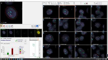
Digital Karyotyping & FISH Analysis
Manual and Automated Scanning
An all-in-one imaging & analysis solution for digital karyotyping and FISH diagnostics.
Automated workflows, powerful algorithms and feature-rich software provide optimized lab
productivity and greater confidence in patient assessment for cytogenetic labs.

Fully Automated

    Patient Database        Automatic Barcode Reader          Unattended Scanning           Oil Dispenser     Image Acquisition

     Immediate Access        Onscreen Analysis     Advanced Supervisor Review   Customized Reporting           LIS Connectivity

Cytogenetic Clinical Applications to visualize, assess and identify:
    Chromosomal Abnormalities
    Structural and Numerical Changes
    Prenatal Amniotic Fluid
    Postnatal Blood
    Bone Marrow Cancer Genetics
    And much more…

Hematology Fish Flow:

                                                                                                            Diagnostics
                                                                                                                 &
                                                                                                            Treatment
  Karyotyping                                    FISH
  Bright-Field Microscopy                        Fluorescence Microscopy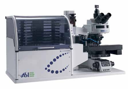
Digital Chromosome Analysis You Can Rely On
Efficiency, Precision, Versatility

State of the Art Image Quality
Start & Walkaway Scanning
                                                         Broad Staining and Sample Menu

                                                                  G-Band                 Q-Band                   R-Band

High resolution 5MP camera sensor combined with a high             FISH                  Spectral                 R-Band
quality 100x immersion oil objective
                                                                 Blood         Bone Marrow       Amniotic Fluid      CVS

Increased Lab Productivity
66% Technologist Time Savings (Hours / Month)
       4,532
                                                         1,352
                                   624

                 -65%                            -68%                -65%

                 1,548                                                   468
                                                 204
                                                                                             Traditional Karyotyping

                                                                                             CytoPower
         Bone Marrow                     Amnio               Blood

    Free technologist time to where their expertise is needed most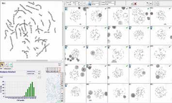
Comprehensive Working Platform

 Feature Rich Review & Analysis

              Count                                                 Analyze                                   Karyotype
        Accurate automatic                               Fast and easy chromosome                High accuracy of automated karyotyping,
       chormosome counting                                indexing and classification             with auto ISCN, auto bands estimation
                                                                                                          and auto overlap score

Easy Separation and Boundary Editing
“Magic Tool” combining 12 operations in a simple mouse click

                                                                                                             Saves hundreds of
                                                                                                             mouse-clicks per
                                                                                                                karyotype!
       Overlap                    Split                    Extend                  Combine

Advanced Onscreen Supervisor or Director Review

Image Gallery                                      Chromosome Compare                         Aberrant Ideogram
Display of all case metaphases                     All captured cells and chromosomes         Chromosomes, cells, ideograms and
and karyotypes                                     side by side                               annotations in an image generator

* FDA cleared for BandView, FISHView, SpotScan for CEP XY, UroVysion, ALK and HER2/neu FISH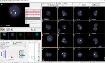
Digital FISH Analysis
Accuracy, Consistency, Ease-of-Use

Image Quality Driven to the Max!
                                   Wide Application Coverage
                                   HER2/neu (Breast)                ALK (Lung)                   UroVysion (Bladder)

                                                Hematology Fusion                Hematology Enumeration

   Any Sample
   Any Probe

“Digital FISH analysis provides more efficient and accurate results and better patient care in comparison
                                      to traditional FISH methods.”
                          Liew M, Rowe L, Clement PW, Miles RR, Salama ME., J Pathol Inform.

Workflow Efficiencies for Increasing Test Volumes
Over 55% Time Savings (Minutes / Case)

                                                           50
       45                     45

                  -78%                   -71%                         -58%
                                                                                            Manual FISH
                                                                       21
                                          13
                                                                                            CytoPower
                   10

            ALK                    CLL                          AML
In-depth Cell Analysis

Increased Diagnostic Confidence

                                                                                                  Digital
                                                                                                  Z-Stacking

                                                                                                  Multi Layer
                                                                                                  Display

Accuracy                                                                                          Signal
Automated cell/ signal detection and                                                              Detection
classification for consistent reliable results

Easy Classification & Review
                                                                                  No More     Double Blinded
                                                                                 Dark Room!      Analysis

High image quality
with digital Z-stack of
each cell

                                                                                              Cell gallery for
                                                                                              fast and easy
Multi-layer view for
                                                                                              classification review
each signal

Real time statistics

* FDA cleared for BandView, FISHView, SpotScan for CEP XY, UroVysion, ALK and HER2/neu FISH
CytoPower for Research Applications

FISH Research

Circulating Tumor Cells                        Immuno Fluorescence                                  Sperm Cells

clgFISH                                        Successive Staining                                  Telomeres

Scanning Protocols for All Probes, Samples and Preparation Techniques
            High Cellular                    High Cellular                            Low Cellular                        Low Cellular
              Density                          Density                                  Density                             Density

User                         User pre-                               DensityScan™                        Dual scan
selected                     configured                              automatic                           automatic
Fields of                    scanning                                detection and                       detection &
View                         pattern and                             ranking of                          classification
                             stop criteria                           Fields of View                      of metaphases
                                                                                                         & interphases

            Semi Automated                   Fully Automated                          Fully Automated                     Fully Automated

                                                               Quality Control of Probe Assays
                                                               Cell phenotyping and signal classification
                                                               Measurment and display of multiple cellular and signal properties
*HiSKY for Test Assurance
*HiSKY
Gold Standard Spectral Karyotyping
Multi-Color FISH Analysis for Result Verification
HiSKY Probe Kit
Automatic identification of translocations and chromosomal origins Chromosome
paints for human,
Simultaneous detection of chromosomal aberrations in one hybridization mouse and rat
Measurement of entire spectrum at each point
Brain Tumor Ewing Sarcoma
*HiSKY is an add on application for research purposes
MN Score
Micronuclei imaging, scoring and analysis for measurement of
DNA damage, cytostasis and cytotoxicityData Management and Connectivity
Modern Paperless Workflow
Central Portal and Database
Easily Integrates with Lab LIS
Efficient
Comprehensive
Eliminates human error
Case Data Management (CDM)
Work from Home!
GenASIs AnyWhere™ for GLOBAL
ACCESS
Remote Access Supervisor
easy access
Director sign
off from any
location
Lab Connectivity Anytime, Anywhere
Review, analyze and sign off case information from any location Remote access,
via a secured network review and analysis
Advanced Reporting 1D/2D Barcode Reader LIS Connectivity
Performance
Security
Data Integrity
HIPAA CompliantBecome a Data-Driven Lab with NEW
LabLife
Generate lab performance statistics
LabLifeTM for Lab Management
NEW
Benchmarks Optimization Growth Annual analysis
and review
Calculate performance Identify best practices Justify investment Compare performance
benchmarks and to increase ROI per case in additional capital year on year and make
track your KPIs. Meet and focus improvement equipment for the lab data driven decisions
certification and efforts
regulatory requirements
Click for Atlas NEW
Reference Atlas for higher assurance
Atlas provides the expert support you need to investigate, research, and confirm
challenging abnormalities.
This new feature is located in BandView’s toolbar for easy access during the
analysis process.
http://atlasgeneticsoncology.org/About Applied Spectral Imaging
Applied Spectral Imaging (ASI) is a global leader in biomedical imaging with a comprehensive product
portfolio and a global distribution footprint.
Founded in 1993, ASI markets, services and supports its products in nearly 50 countries. With a wide FDA
clearance portfolio, you can rest assured that ASI applications have been rigorously tested for compliance
and clinical use.
The Company's technology, powered by GenASIs, enables pathology, cytogenetics and research
laboratories to provide advanced diagnostics to patients through superior digital diagnostic tools.
ASI has a wide portfolio of dedicated solutions for brightfield, fluorescence and spectral imaging and
analysis, including HiPath Pro, PathFusion, HiBand, HiFISH, CytoPower and Rainbow.
The Company has offices in the US and Asia and a global network of distribution partners.
Global Presence
3,500 54 63
Systems installed Countries through direct Third party distribution
worldwide & indirect sales forces partners
ASI Inc
USA ASI Ltd
Asia
Direct sales
Indirect salesThe Company’s Product Portfolio
Exceptional Imaging & Analysis Solutions for Laboratories
Cytogenetics Pathology Research
HiBand HiFISH CytoPower HiPath Pro PathFusion Rainbow
Diverse platforms to accommodate all laboratory needs
99-Slide Tray Loader 9-Slide Scanning System 1-Slide Capture System
HyperSpectral System Review & Analysis Station AnyWhere Remote ConnectivitySystem Specifications
Manual 1 Slide 9 Slide Motorized Stage 99 Slide Tray Loader HyperSpectral 1 Slide
Microscope BF and FL upright OLYMPUS BX61 BF + FL OLYMPUS BX61 BF + FL BF and FL upright
Support microscopes OLYMPUS BX63 BF+ FL OLYMPUS BX63 BF+ FL microscopes
ZEISS AxioImager Z2 BF+ FL ZEISS AxioImager Z2 BF+ FL
Objectives Olympus ZEISS Olympus ZEISS Olympus ZEISS 10x/0.3
10x/0.3NA 10x/0.3NA 1.25x/0.04NA 1.25x/0.03NA 1.25x/0.04NA 1.25x/0.03NA 60x/1.42 or 63x/1.25
60x/1.25NA 63x/1.25NA 4x/0.16NA 5x/0.16NA 4x/0.16NA 5x/0.16NA 100x/1.3
100x/1.3NA 100x/1.3NA 10x/0.3NA 10x/0.3NA 10x/0.3NA 10x/0.3NA
40x/1.4NA 40x/1.3NA 40x/1.4NA 40x/1.3NA
60x/1.25NA 63x/1.25NA 60x/1.25NA 63x/1.25NA
100x/1.3NA 100x/1.3NA 100x/1.3NA 100x/1.3NA
Camera 5MP CMOS Monochrome 5MP CMOS Monochrome 5MP CMOS Monochrome Spectral 1.3MP Monochrome
Slide Capacity 1 slide (Manual or Motorized) 9 slides 99+ slides 1 slide (Manual or Motorized)
Barcode Reader Handheld 1D/2D Handheld 1D/2D Integrated 1D/2D Handheld 1D/2D
Automated Oil
N/A Optional Integrated N/A
Dispenser
Dimensions According to clients 61cm x 69cm x 85cm 100cm x 90cm x 90cm According to clients
[WxDxH] microscope (24” x 27.2” x 33.5”) (39.4” x 35.5” x 35.5”) microscope
Weight According to clients 45kg 80kg According to clients
microscope 99.2lb 176.4lb microscope
SCAN ME
North America
Applied Spectral Imaging Inc.
Tel: +1 760 929 2840
sales-inc@spectral-imaging.com
Headquarters
Applied Spectral Imaging Ltd.
Tel: +1 817 886 6031
sales@spectral-imaging.com www.spectral-imaging.com
DOC000349 Rev. EYou can also read