Evolution of the liver biopsy and its future - Translational ...
←
→
Page content transcription
If your browser does not render page correctly, please read the page content below
Review Article

Evolution of the liver biopsy and its future
Dhanpat Jain1, Richard Torres2, Romulo Celli1, Jeremy Koelmel3, Georgia Charkoftaki3, Vasilis Vasiliou3
1
    Department of Anatomic Pathology, 2Department of Laboratory Medicine, Yale University School of Medicine, New Haven, CT, USA;
3
Department of Environmental Health Sciences, Yale School of Public Health, New Haven, CT, USA
Contributions: (I) Conception and design: D Jain; (II) Administrative support: None; (III) Provision of study material or patients: None; (IV)
Collection and assembly of data: None; (V) Data analysis and interpretation: None; (VI) Manuscript writing: All authors; (VII) Final approval of
manuscript: All authors.
Correspondence to: Dhanpat Jain, MD. Professor of Pathology, Yale University School of Medicine, New Haven, CT, USA.
Email: dhanpat.jain@yale.edu.

                 Abstract: Liver biopsies are commonly used to evaluate a wide variety of medical disorders, including
                 neoplasms and post-transplant complications. However, its use is being impacted by improved clinical
                 diagnosis of disorders, and non-invasive methods for evaluating liver tissue and as a result the indications of
                 a liver biopsy have undergone major changes in the last decade. The evolution of highly effective treatments
                 for some of the common indications for liver biopsy in the last decade (e.g., viral hepatitis B and C) has
                 led to a decline in the number of liver biopsies in recent years. At the same time, the emergence of better
                 technologies for histologic evaluation, tissue content analysis and genomics are among the many new
                 and exciting developments in the field that hold great promise for the future and are going to shape the
                 indications for a liver biopsy in the future. Recent advances in slide scanners now allow creation of “digital/
                 virtual” slides that have image of the entire tissue section present in a slide [whole slide imaging (WSI)].
                 WSI can now be done very rapidly and at very high resolution, allowing its use in routine clinical practice.
                 In addition, a variety of technologies have been developed in recent years that use different light sources
                 and/or microscopes allowing visualization of tissues in a completely different way. One such technique
                 that is applicable to liver specimens combines multiphoton microscopy (MPM) with advanced clearing and
                 fluorescent stains known as Clearing Histology with MultiPhoton Microscopy (CHiMP). Although it has
                 not yet been extensively validated, the technique has the potential to decrease inefficiency, reduce artifacts,
                 and increase data while being readily integrable into clinical workflows. Another technology that can provide
                 rapid and in-depth characterization of thousands of molecules in a tissue sample, including liver tissues, is
                 matrix assisted laser desorption/ionization (MALDI) mass spectrometry. MALDI has already been applied
                 in a clinical research setting with promising diagnostic and prognostic capabilities, as well as being able
                 to elucidate mechanisms of liver diseases that may be targeted for the development of new therapies. The
                 logical next step in huge data sets obtained from such advanced analysis of liver tissues is the application of
                 machine learning (ML) algorithms and application of artificial intelligence (AI), for automated generation
                 of diagnoses and prognoses. This review discusses the evolving role of liver biopsies in clinical practice over
                 the decades, and describes newer technologies that are likely to have a significant impact on how they will be
                 used in the future.

                 Keywords: Liver biopsy; matrix assisted laser desorption imaging (MALDI); Clearing Histology with
                 MultiPhoton Microscopy (CHiMP); whole slide imaging (WSI)

                 Received: 12 January, 2020; Accepted: 19 March, 2020; Published: 05 April 2021.
                 doi: 10.21037/tgh.2020.04.01
                 View this article at: http://dx.doi.org/10.21037/tgh.2020.04.01

© Translational Gastroenterology and Hepatology. All rights reserved.   Transl Gastroenterol Hepatol 2021;6:20 | http://dx.doi.org/10.21037/tgh.2020.04.01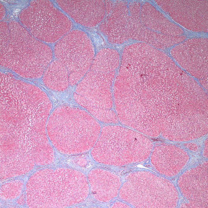
Page 2 of 21                                                                              Translational Gastroenterology and Hepatology, 2021

Introduction                                                                  lesion is difficult to approach by other means. Liver biopsies
                                                                              (needle or wedge) can also be obtained intra-operatively
Since its original description by Ehrlich in 1883, the
                                                                              whenever liver disease is suspected or suspicious lesions
clinical role of liver biopsy has changed dramatically over
                                                                              are identified. While needle biopsies provide a core of
the years (1). Initially, it was used for determination of
                                                                              liver parenchyma, wedge biopsies provide a “wedge” of
glycogen stores in the livers of diabetic patients while in
                                                                              liver tissue taken from it surface and are much larger.
the ensuing years, its indications have become more diverse
                                                                              In many instances, an abnormal appearance of the liver
and numerous (2). It took a few decades for the liver biopsy
                                                                              during surgery for an unrelated procedure (most often
to be accepted as a standard medical procedure by the
                                                                              cholecystectomy) is the first indication of an underlying
medical community. Only after Menghini described “one-
                                                                              liver disease. Liver biopsies harvested intra-operatively
second needle biopsy of the liver” in 1958 did the procedure
                                                                              have the added advantage of obtaining tissue samples from
come into more widespread clinical use (2). While liver
                                                                              grossly visible/suspicious lesions. However, these wedge
biopsies are commonly used to evaluate a wide variety of
                                                                              biopsies are often suboptimal for assessment of liver fibrosis
medical disorders, including neoplasms and post-transplant                    and/or inflammation due to the preponderance of Glissen’s
complications, its use is being impacted by improved                          capsule, wider portal tracts in subcapsular area, and frequent
clinical diagnosis of disorders, and non-invasive methods for                 (but inconsequential) surgically-induced hepatitis (8).
evaluating liver tissue. Nevertheless, liver biopsies continue                Therefore, needle biopsy should be the technique of choice
to provide highly valuable information for clinical and                       at laparotomy or be used in addition to a wedge biopsy.
research purposes. This review considers the evolving role                        Once obtained, liver tissue needs to be triaged for
of liver biopsies in clinical practice over the decades, and                  various special procedures e.g., electron microscopy,
describes technologies that are likely to have a significant                  microbiological cultures, immunofluorescence studies or
impact on how they will be used in the future.                                flow cytometry, based on the clinical differential diagnosis.
    Liver tissue can be obtained for clinical analysis in                     Thus, it is very important that critical clinical details (e.g.,
several ways, including percutaneous, transjugular,                           liver enzymes, history of medications/toxin exposure,
laparoscopic, and intra-operative approaches. Each of these                   viral and autoimmune serology) and a clinical differential
have advantages and disadvantages. The percutaneous liver                     diagnosis be communicated to the pathologist so that
biopsy technique remains the standard practice in most                        appropriate tissue triage and special procedures can be
medical centers. Whenever a percutaneous needle biopsy                        undertaken without loss of time.
is contraindicated (e.g., presence of significant ascites or                      Histological examination is an essential tool for the
coagulopathy), a transjugular needle biopsy is preferred.                     evaluation of patients with liver disease, particularly those
An important advantage of the transjugular approach is the                    with persistent unexplained liver function abnormalities.
ability to obtain intrahepatic portal pressures that aid in                   Most pathology labs will routinely perform trichrome,
the diagnosis and management of select groups of patients,                    hematoxylin and eosin (H&E), reticulin, iron and D-PAS
particularly those with cirrhosis (3). However, transjugular                  (periodic acid-Schiff stain after diastase digestion)
biopsies often render smaller and fragmented tissue                           stains on liver biopsies for clinical evaluation of a liver
samples, making pathologic assessment somewhat difficult.                     disorder. Under certain circumstances, special sample
Percutaneous or transjugular biopsies are used to obtain                      preparation and fixation may be required, such as placing
samples from patients suffering from disorders that involve                   the tissue in appropriate culture medium (for suspected
a diffuse change in the liver (3,4). By contrast, image-guided                infections), processing in metal-free containers (for metal
needle biopsies are necessary for focal diseases/lesions (5,6).               quantification), fixation in glutaraldehyde (for electron
Laparoscopic liver biopsy enables the gross features of                       microscopy), using frozen tissue (for Oil red O stain for the
the liver to be assessed prior to taking the biopsy. During                   estimation of microvesicular fat), or simply “RUSH/STAT”
laparoscopy apart from needle core biopsy, a wedge of liver                   processing in critically ill patients (e.g., those with fulminant
tissue (wedge biopsy) can be obtained. Due to the nature of                   liver failure or the post-transplant situation) (2). Routine
surgery involved, laparoscopic liver biopsy has added costs                   histological evaluation of liver biopsies involves assessment
and the potential for more complications (7). As such, it is                  of inflammation, cellular changes involving hepatocytes and
used in select settings at most centers e.g., when laparoscopy                bile ducts, abnormal accumulation of various substances,
is being anyway performed for another indication or the                       the extent of the liver injury, and/or fibrosis. The liver

© Translational Gastroenterology and Hepatology. All rights reserved.   Transl Gastroenterol Hepatol 2021;6:20 | http://dx.doi.org/10.21037/tgh.2020.04.01
Translational Gastroenterology and Hepatology, 2021                                                                                        Page 3 of 21

conditions in which a biopsy is most commonly performed                       Pérez-Tamayo first provided evidence in both animal
include viral hepatitis, non-alcoholic fatty liver disorder                   models and human disease showing reversal of fibrosis and
(NAFLD), autoimmune disorders, drug/toxin-induced liver                       cirrhosis (18). Subsequently, serial biopsies from a patient
injury (including alcoholic liver disease, ALD), cholestatic                  with hepatitis B following antiviral treatment showed
diseases, metabolic/inherited disorders, and hepatic masses                   regression of fibrosis from fully developed cirrhosis to
(2,3,5,9-14). In the post-transplant setting, liver biopsies                  incomplete septal cirrhosis. In this landmark paper based
are used for the evaluation of organ rejection, infections,                   on livers removed at transplantation having cirrhosis
surgical complications, and disease recurrence. However,                      or incomplete septal cirrhosis, Wanless and colleagues
the indications for liver biopsy in all of these conditions                   reported histologic parameters reflective of progression
continues to evolve as other approaches to monitoring liver                   or regression of fibrosis (19). Subsequent studies reported
health are developed or diseases that previously necessitated                 biopsy-documented regression of cirrhosis associated with
liver biopsies are more effectively treated (2).                              hepatitis C, hepatitis B and autoimmune hepatitis following
   Historically, liver biopsies have been used in hepatitis B                 disease-specific therapy (20). Given that liver fibrosis may
and C because it permitted evaluation of the stage and severity               be reversed by therapeutic intervention, a renewed focus
of the disease, its response to therapy (15,16). Due to high                  has been placed on tools to monitor fibrosis (and therefore
specificity, their less invasiveness, faster turn-around and cost,            therapy effectiveness). However, assessment of changes
serological diagnostic tests are now used rather than liver                   in the severity of fibrosis remains challenging. Most
biopsies to establish the diagnosis of hepatitis B or C infection             investigations have employed fibrosis staging systems that
(13,14). A biopsy may be used as an adjunct to serological tests              have been in use for several decades; these works well when
for the assessment of the degree of liver injury and fibrosis,                there is a definite numerical change in the fibrosis stage
presence of other concomitant liver disease or, under some                    despite issues of sampling variability. Most common fibrosis
circumstances, the response to antiviral therapy. The efficacy                scoring systems, such as the Ishak, Batts and Ludwig or
of direct-acting antiviral (DAA) therapies for HCV infection                  METAVIR staging systems, were developed before the idea
(>90% cure) make liver biopsies redundant in this condition.                  of fibrosis regression gained importance and, as such, are
Thus, it is not surprising that the use of liver biopsies in                  not equipped for assessing this aspect of fibrosis (21,22). In
clinical practice is declining worldwide.                                     essence, these systems can detect a change as progression or
   Another major liver disorder in clinical practice is                       regression only when there is definite numerical change in
NAFLD. With better control of viral hepatitis, it is                          fibrosis stage assignment. However, they are not reliable for
expected that NAFLD will likely become the most common                        evaluating changes within a given stage, especially cirrhosis.
liver disease worldwide. The need for a liver biopsy in this                  Historically, cirrhosis has been considered to be “end-stage”.
setting is limited as existing imaging techniques are excellent               It is now recognized that cirrhosis is a spectrum that varies
for the evaluation of fat in the liver. However, current                      from mild to severe disease (23). Several systems have been
methodologies are sub-optimal for assessing inflammations                     developed to sub-classify cirrhosis, but even these systems
and fibrosis. In the presence of other concomitant liver                      fail to predict the course of fibrosis based on a given single
diseases, the relative contribution from each disease also can                liver biopsy obtained at a given point of time (24,25).
be difficult to evaluate without a biopsy. On the other hand,                 However, recent studies focusing on fibrosis regression have
given the limitations of treatment options and ability to                     shown a variety of histopathologic changes that at any given
predict the disease course based on histology, the biopsies                   time point suggest fibrosis regression without requiring
are performed only in select settings in NAFLD. Further,                      sequential biopsies (19). Cirrhosis regression is associated
advances in the non-invasive methods (serologic or imaging                    with a net decrease in the amount of fibrous tissue, thinning
based) for assessing fibrosis in the liver make the need to                   or loss of fibrous septa, disappearance of shunting neo-vessels
liver biopsy even less (13).                                                  (that develop as a result of angiogenesis associated with
   While a variety of histologic changes are evaluated                        fibrogenesis), disappearance of ductular proliferation and an
in a liver biopsy for medical disorders, one of the most                      overall decrease in the cellularity of the fibrous septa (26).
important parameters is fibrosis which can be indicative                      Regression of fibrosis is associated with a partial or full
of disease progression or response to any treatment. Once                     restoration of the lobular organization (27). These various
a controversial topic, it is now well-accepted that fibrosis                  features have been taken into account to develop a novel
can regress, and even cirrhosis can regress (17). In 1979,                    “progressive-indeterminate-regressive (or PIR) system” to

© Translational Gastroenterology and Hepatology. All rights reserved.   Transl Gastroenterol Hepatol 2021;6:20 | http://dx.doi.org/10.21037/tgh.2020.04.01
Page 4 of 21                                                                              Translational Gastroenterology and Hepatology, 2021

                         A                                                 B

                        C                                                  D

Figure 1 Fibrosis in liver represents an important feature of liver injury and characterization of fibrosis forms an important aspect of
evaluation of hepatic disorders. Representations of liver fibrosis showing key features suggesting “progression” and “regression”. A case
of hepatitis C cirrhosis (A) with predominantly progressive pattern of fibrosis characterized by many broad fibrous septa. Compare this
with predominantly regressive pattern of fibrosis (B) post therapy with sustained viral response in a case of hepatitis C cirrhosis showing
many thin fibrous septa (A&B, 40×, Trichrome stain). Higher power images showing increased thickness, more inflammation and increased
cellularity in progressive septum (C) compared to those undergoing regression characterized by relatively hypocellular, less vascular and thin
septum (D) (C&D, 200×, H&E stain).

assess the dynamic change in fibrosis that can be applied                      will have to adapt to incorporate this aspect of fibrosis
to routine liver biopsies (Figure 1A,B,C,D). This system                       assessment.
takes several histologic characteristics into account and                          Thus, over the years, the role of the liver biopsy has
provides insight into the quality or directionality of fibrosis—               been changing from a diagnostic modality to a prognostic
progressive or regressive—in patients with cirrhosis based                     evaluation. It is anticipated that liver biopsy will continue to be
on a single liver biopsy. This system can be easily applied on                 used in: (I) cases of viral hepatitis with unusual presentation;
H&E and trichome stained slides obtained from formalin-                        (II) patients with more than one concomitant disorder; (III)
fixed paraffin-embedded tissue. Importantly, it has already                    liver disease with unclear etiology; and (IV) liver diseases
been shown to correlate with liver-related morbidity and                       in which the results of various non-invasive measurements
mortality in the setting of treated chronic hepatitis B (28,29).               are discrepant with each other or inconsistent with clinical
As this and/or similar systems are validated for routine                       findings. However, the use of liver biopsy in the future is
clinical practice, the evaluation and reporting of liver biopsies              likely to be eroded as new technologies are developed that are

© Translational Gastroenterology and Hepatology. All rights reserved.   Transl Gastroenterol Hepatol 2021;6:20 | http://dx.doi.org/10.21037/tgh.2020.04.01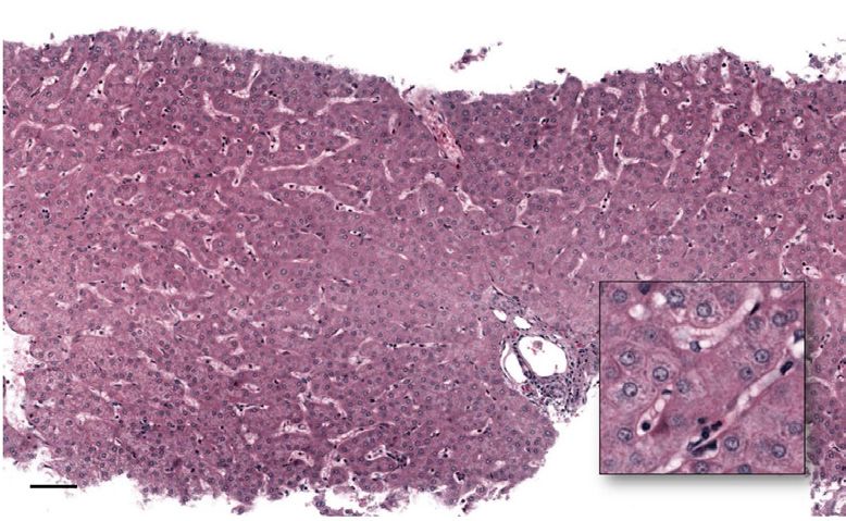
Translational Gastroenterology and Hepatology, 2021                                                                                        Page 5 of 21

less invasive, and able to identify changes in liver structure or             or Scheuer systems (32-36). In the setting of cirrhosis,
function with higher resolution/fidelity.                                     DIA techniques correlate with sub-classification staging
   It is also important to realize that technologies are evolving             indices, such as the Laennec or Jain-Garcia systems (37).
that allow visualization of liver tissue in three-dimensions,                 They have also been shown to correlate well with certain
digital morphometry, and analysis of the content of liver cells               clinical parameters, such as the Hepatic Venous Pressure
that go far beyond what immunohistochemistry or other                         Gradient (33), and with the progression or regression of
current molecular techniques can accomplish. The application                  fibrosis in the setting of treated chronic viral hepatitis
of artificial intelligence (AI) and machine learning (ML)                     (33,38,39).
further enhances these technologies by automating their                           Other areas that lend themselves to quantitation by DIA are
analysis and interpretation.                                                  assessment of steatosis and inflammation (40,41). The degree
                                                                              of fat in liver tissues has been associated with worse prognosis
                                                                              and the development of steatohepatitis in NAFLD (42).
Role of digital pathology
                                                                              In addition, fat in the liver has been associated with worse
The application of digital technology has had a significant                   graft outcomes (43). These represent a few of many reasons
impact on medicine and pathology has been at the forefront                    justifying comparison of digital morphometric methods for
of this ‘digital revolution’. Analysis of images obtained from                assessing the degree of hepatic fat content by more objective
select parts of histologic tissue sections has been in practice               methods (41,44-46). DIA algorithms have recently been
for decades. Only recently development of advanced slide                      designed to be incorporated in large scale research studies,
scanners has allowed creation of “digital/virtual” slides                     including clinical trials for NAFLD (47).
that have image of the entire tissue section present in a                         Despite the advantages and potential of WSI, its adoption
slide [whole slide imaging (WSI)]. This can now be done                       in clinical practice has been slow. Slide scanners are very
very rapidly and at very high resolution, allowing its use                    expensive and storing of large quantities of digital data
even in routine clinical practice for making diagnoses and                    remains an issue. In addition, scanners require trained
evaluating prognostic biomarkers. Digitally-scanned tissue                    technicians to operate, and good informatics support is
sections also permit rapid access and sharing of image data.                  required on the back end. Thus, a major barrier to adoption
In addition, it permits the development and application                       of WSI has been the associated increase in cost and labor
of novel, computer-based methods for analyzing tissue                         to the already labor-intensive and slow process of standard
parameters. To this end, AI is being leveraged to automate                    tissue formalin fixation, paraffin wax-embedding, slice cutting
tissue analyses and enhance processes and accuracy in                         and staining. A second barrier has been the reduction in
pathological diagnoses (30). One notable example of this                      image quality compared to the currently used transmitted
new approach is the increased accuracy and efficiency of                      light microscopy (48-50). Although artifacts from focusing
AI-assisted diagnosis of lymph node metastasis detection                      error in WSI continue to be reduced, WSI systems will
compared to human pathologists (31).                                          remain limited by artifacts, such as tissue tears, folds and
   Application of digital pathology to the liver has                          stain variability that are inherent in the process of physically
overwhelming been focused on establishing quantitative                        cutting thin tissue slices. None the less, the slide scanners are
measurements of fibrosis. Routine human-based histologic
                                                                              getting cheaper and more versatile in terms of their speed
examination of fibrosis has been notoriously fraught with
                                                                              and DIA applications. Storage capacity of digital data is
inter- and intra-observer variability and inter-laboratory
                                                                              improving. With these advances, it is expected that the use of
staining variability. Digital image analysis/morphometry
                                                                              DIA will continue to expand as new algorithms are developed
(DIA) techniques increase the level of objectivity and
                                                                              that allow objective assessment of variety of other histologic
reproducibility of these analyses. While DIA can overcome
                                                                              features in liver biopsies, besides fibrosis and steatosis.
some of the limitations of tissue artifacts or staining
variability, it cannot address the issue of sampling error, as
this occurs before the tissue is received in the pathology lab.               Evolving technologies for histologic evaluation
Variations of this technique have been used for more than                     of liver tissues
thirty years now, and have been shown to correlate with
                                                                              In vivo multi-photon and 3-dimensional microscopy
previously established histology-based semi-quantitative
measures of fibrosis, such as the Ishak, Metavir, Knodell                     For over a century, diagnostic histology has relied on

© Translational Gastroenterology and Hepatology. All rights reserved.   Transl Gastroenterol Hepatol 2021;6:20 | http://dx.doi.org/10.21037/tgh.2020.04.01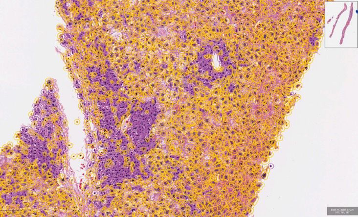
Page 6 of 21                                                                              Translational Gastroenterology and Hepatology, 2021

thinly cut tissue slices mounted on slides, stained with dyes                 adoption. Slow imaging time is a critical limitation of
(most commonly H&E), and examined in 2-dimensions                             several of these approaches (especially SRS and the standard
using an ocular microscope using white light. A variety                       implementations of confocal and MPM) which may
of technologies developed in recent years use different                       take many minutes or hours to generate a single biopsy
light sources and/or microscopes that allow visualization                     optical slice, and thus are unable to achieve adequate
of tissues in a completely different way. For example,                        throughput for clinical use. Some (e.g., SPIM) are limited
multiphoton microscopy (MPM) (described in more detail                        by the computationally-intensive image processing. This
below) allows visualization of tissues in 3-dimensions based                  may be addressable by more computing power, but this
on fluorescence excitation and detection (51-53). It has                      adds to analysis cost and complexity. The perpendicular
been applied widely in research and is being developed                        optical setup of SPIM also makes specimen orientation
for clinical use. Other exciting advances such as new                         incompatible with clinical workflow in its standard
techniques for improvement of deep imaging of tissue via                      configuration, for which specialized approaches have been
chemical clearing and advanced image analysis methods                         devised that permit placing a specimen on a flat surface
such as convolutional neural networks (CNNs) (see below),                     and imaging at an angle (61). Finally, those methods that
when combined with MPM microscopy, portend dramatic                           produce data that are not directly convertible to images
improvements in the efficiency and precision of histologic                    using standard histologic stains, e.g., H&E, require
evaluation of liver tissues.                                                  pathologist retraining; this represents an additional barrier
    Advances in optics over the past few decades have                         to adoption. The overarching limitation of these so-
generated an array of potential alternatives to formalin-                     called ‘ex vivo’ imaging approaches is that all (except the
fixed, paraffin-embedded tissue (FFPE) and WSI for digital                    confocal and MPM techniques) produce images that are
image analysis of specimens without thin physical sectioning                  of inferior quality relative to standard manual transmitted
or wax-embedding, including liver. The approaches                             microscopy. As such, they should only be considered as
can generally be categorized into the following: optical                      an adjunct to existing methods for definitive diagnosis.
coherence tomography (OCT) (54), stimulated Raman                             Given the irreversible and often more marked distortion
spectroscopy (SRS) (41,55), selective plane illumination                      of morphology associated with the use of frozen sections,
(light-sheet) microscopy (SPIM) (56), deep UV surface                         several of these new approaches may offer a viable
imaging [e.g., microscopy with ultraviolet surface excitation                 alternative for intraoperative evaluation, even in their
(MUSE)] (57), confocal microscopy (58), and MPM                               current state of development. Nevertheless, only those
(59,60). Other modalities exist but generally involve higher                  methods with the highest quality imaging, such as confocal
complexity, making their near-term applicability to routine                   and MPM, are likely to be able to move histology beyond
clinical use less likely.                                                     standard physical slide use.
    It is important to appreciate that all of these new                          The image quality delivered by many of these new
methods require less processing than the standard                             approaches can be increased by incorporating another
histological tissue preparation, and they are intrinsically                   recent advance in imaging called “clearing”. This involves
digital. Several require very little to no tissue processing                  making a tissue transparent through chemical treatment.
time (i.e., OCT, SRS, MUSE), of particular interest for                       A clearing step has been a part of standard histology
time-sensitive applications such as intraoperative evaluation.                processing for nearly a century, and it referrs to the partially
Others enable three-dimensional visualization (i.e., SPIM,                    cleared appearance that the use of xylene imparts in its role
confocal, MPM), an approach of still theoretical interest for                 as an intermediary reagent with solubility in both alcohol
clinical evaluation of tissue samples. All of these approaches                and molten paraffin wax. With the advent of confocal
avoid tissue cutting artifacts and generate morphologic                       microscopy, MPM, and SPIM for deep imaging of uncut,
information without consuming valuable tissue. As such,                       un-embedded tissue specimens, there has been a resurgent
they improve the yield of informative data that is extractable                interest in the development and use of chemicals that can
from a given sample relative to the commonly-used physical                    match the optical properties of proteins and membranes (i.e.,
slices.                                                                       the high refractive index of these components), thereby
    Despite these benefits, these alternative imaging                         improving the quality and extending considerably the depth
techniques, often grouped under the misnomer of                               within tissue at which fluorescent images can be obtained.
‘ex vivo’ imaging, face challenges of their own for clinical                  The past few decades have engendered a host of approaches

© Translational Gastroenterology and Hepatology. All rights reserved.   Transl Gastroenterol Hepatol 2021;6:20 | http://dx.doi.org/10.21037/tgh.2020.04.01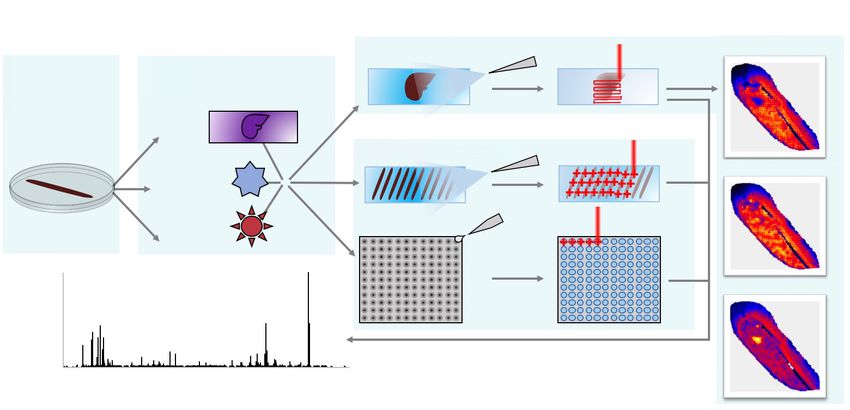
Translational Gastroenterology and Hepatology, 2021                                                                                        Page 7 of 21

and chemical compositions for improving clearing of tissues                   multiphoton-based in vivo endoscopes for diagnostic liver
which allows better light penetration (62). The approaches                    assessment, these modalities have seen the greatest use in
can be divided into those that involve (I) electrophoretic                    ex vivo liver samples (58,63,64). In unstained liver specimens,
removal of lipids (e.g., CLARITY and its extensions), or                      confocal microscopy and MPM allow visualization of
(II) the replacement of the water with a high refractive                      overall cellular features using intrinsic fluorescence derived
index fluid, e.g., BABB, 3DISCO, etc. While of potential                      primarily from nicotinamide adenine dinucleotide hydride
value for investigative use due to high transparency, which                   (NADH), its phosphorylated form (NADPH), and flavin
improves light collection at depth, electrophoretic removal                   adenine dinucleotide (FAD) (59,64,65). Such unstained
of lipids presents particular challenges for clinical use in                  approaches have been applied to the identification and
that it is more labor intensive and more complex than pure                    characterization of liver cancer and steatosis using standard
chemical clearing. By removing membrane and membrane                          H&E stained sections as the reference (41,66).
bound molecules, it may also limit the reliability of certain                     Added information can be derived by exploiting the
immunohistochemical (IHC) stains in routine clinical use,                     finding that the typical length of time a fluorophore
making it incompatible with the current diagnostic standard.                  spends in an excited state before releasing its light energy
On the other hand, chemical clearing is a processing step                     (known as the fluorescence lifetime) varies depending on
that can allow fluorescence microscopy to achieve contrast,                   the fluorophore and its microenvironment. Specialized
clarity, and resolution surpassing that of transmitted light                  detection systems can be combined with MPM excitation
microscopy. This offers an opportunity for improvement in                     in a modality called fluorescence lifetime imaging (FLIM)
the reliability of diagnostic interpretation of liver.                        to produce image patterns with enhanced biological
   The quality of digital images ties into the emerging                       information, such as recognition of certain cell types,
advances in computerized image analysis. Just as                              reduction-oxidation state, and apoptosis/necrosis (67).
pathologists require quality images for interpretation,                       Unfortunately, this detection process requires more
computerized classification depends on high quality image                     extended imaging times and expensive equipment, reducing
data. The introduction of CNNs has significantly advanced                     its clinical potential for cost-effective routine analysis.
automated image pattern recognition. The method involves                          A somewhat more established role for MPM in liver
mathematically combining (convolving) known patterns,                         analysis is measurement of collagen fibrosis (53,60,68-70).
such as repeating black and white columns adopting varying                    Although multiphoton laser excitation systems are more
frequencies and orientations, with portions of an image.                      expensive than confocal laser systems, MPM has several
The resulting nodes are then convolved further with other                     advantages over confocal for clinical (and investigative) liver
patterns to form layers of a network. Deep networks refer                     imaging. Specifically, it (I) can image deeper into tissue
to algorithms that involve many layers, each progressively                    (typically ≈200 µm, depending on fixation and clearing
representing higher order patterns and from which the                         state), (II) has lower risk of damaging tissue or nucleic acids
training process can select the most relevant to a given                      from high intensity light, and (III) can access the collagen-
histologic feature recognition, biological process, or cell                   specific signal derived from second harmonic generation
type. In the past few years, CNN methods have begun to                        (SHG) (Figure 2). Collagen is nearly the only liver molecule
perform as well as humans at image pattern recognition for                    that, due to its non-centrosymmetric properties, can
specific tasks. Given this rapid progress, it is expected that                combine two photons arriving nearly simultaneously into
such digital image interpreting tools will aid pathologists                   one photon of exactly twice the energy. This SHG signal
in reproducibly grading and accurately recognize disorders.                   differs from fluorescence signals and occurs at exactly half
In as much as they provide higher quantity and potentially                    the wavelength of the excitation wavelength so it can be
higher quality of digital data, techniques such as MPM                        readily separated during imaging, producing a digital map
and confocal can be combined with the new image analysis                      of collagen fibrosis.
developments to augment the efficiency and precision of                           SHG produces a relatively reproducible digital collagen
microscopic diagnostic assessment further.                                    distribution signal. As such, it has been touted as a potential
                                                                              means to improve upon the recognized low concordance
                                                                              of fibrotic scoring by visual estimation, which occurs even
New imaging: applications to liver disease
                                                                              when collagen specific stains, such as Masson’s, Picrosirius
Owing perhaps to the use of commercial confocal and                           Red, and van Gieson, are used (71). Underscoring this

© Translational Gastroenterology and Hepatology. All rights reserved.   Transl Gastroenterol Hepatol 2021;6:20 | http://dx.doi.org/10.21037/tgh.2020.04.01
Page 8 of 21 Translational Gastroenterology and Hepatology, 2021
Figure 2 Second harmonic generation for liver collagen assessment using CHiMP multiphoton microscopy in a human cirrhotic liver.
Red—Autofluorescence. Blue—SHG. Scale bar =0.5 mm. CHiMP, Clearing Histology with MultiPhoton Microscopy.
standard collagen stains, a process that does not require a
specialized instrument.
MPM, clearing, and CNNs in liver disease
Implementation of new histological techniques in clinical
practice is most likely to succeed only if it can satisfy
several key requirements: (I) it must be fast enough to be
compatible with the expected clinical volume; (II) it must
reduce (rather than increase) labor requirements; (III) it
Figure 3 Multiphoton microscopy image of normal human liver
should not create an undue burden at other steps of the
tissue without need for wax-embedding or physical sectioning.
overall process (such as surgical collection); (IV) it must
Image collected by CHiMP processing and imaging scope
improve diagnostic yield; and (V) it must be cost-effective
technique. Fluorescence image has been converted to a pseudo-HE
to perform. MPM has been combined with advanced
coloration. Scale bar =100 μm. CHiMP, Clearing Histology with
clearing and fluorescent stains that mimic H&E in an
MultiPhoton Microscopy.
approach that satisfies the requirements for practical clinical
implementation. Although it has not yet been extensively
contention, a commercial instrument has been developed validated, the technique known as Clearing Histology with
MultiPhoton Microscopy (CHiMP) is also applicable to
that uses SHG derived via MPM on unstained slides for
liver specimens and is capable of producing images that
liver fibrosis assessment (71). It also employs algorithms
look like standard H&E preparation that may be used for
analyzing fiber distribution for additional fibrosis
primary diagnostic assessment (Figure 3) (60,72). It has
characterization. It is important to appreciate, however,
the potential to decrease inefficiency, reduce artifacts, and
that SHG is relatively specific for the detection of type I
increase data while being readily integrable into clinical
and II collagen. Given that Masson’s stain detects type I and workflows.
large type III collagen fibers, and Picrosirius Red detects Using a prototype specialized MPM with high speed
both type I and III fibers, SHG patterns may differ from acquisition, CHiMP can image with sufficient speed to
these standard stains. Nevertheless, overall studies have adequately match clinical volumes, taking ≈1 minute per
shown good correlation. Still, when using physical slides, optical slice. Multiple levels can be evaluated remotely
SHG does not save on labor, adds to cost and time, and it is within hours of biopsy. Since no tissue is consumed,
unclear whether the analysis has clinical significance beyond specimens may be subsequently processed for standard
image analysis assessment that can be derived from WSI of histologic stains or nucleic acid analysis, as dictated by
© Translational Gastroenterology and Hepatology. All rights reserved. Transl Gastroenterol Hepatol 2021;6:20 | http://dx.doi.org/10.21037/tgh.2020.04.01Translational Gastroenterology and Hepatology, 2021 Page 9 of 21
the morphology. A sample preparation method developed information, biopsies will continue to be able to provide
for detailed 3-dimensional collagen fibrosis assessment diagnostic data beyond what in vivo analysis can yield.
in liver samples in the research environment (52) Together with the techniques described in the section
could be extended to clinical samples. The additional that follows, the new microscopic imaging modalities,
clinical validation required would be worthwhile because particularly those based on MPM coupled with the
the technique offers considerable improvements over refinement of rapidly evolving ML approaches, are poised
the current histology standard and the real possibility of to usher in a new era of liver disease diagnostics.
clinical use in the near future. Like other ‘ex vivo’ imaging
modalities, a feature of CHiMP that is particularly
Role of matrix assisted laser desorption/
significant for its application in diagnostic liver pathology
ionization (MALDI) in evaluation of liver tissues:
is that it is intrinsically digital and devoid of the artifacts
successes, challenges, and future development
created by physical slicing and standard transmitted light
microscopy and WSI. This means clearer images for remote Clinical application of MALDI
interpretation by expert liver pathologists, and improved
data for computerized image analysis. In addition, MPM Obtaining a liver biopsy is an invasive procedure.
can produce very thin optical slices, and thereby provide Consequently, it is of paramount importance that the
clarity beyond what is achieved with routine histologic tissue sample yield clinically-relevant information that
sections. As a result, it is anticipated that the data generated cannot be obtained by non-invasive means. MALDI mass
by CHiMP will be particularly well suited for use with ML spectrometry is a technology that can provide rapid and in-
algorithms, including CNNs. depth characterization of thousands of molecules in a tissue
CNNs and other image analysis tools have been applied sample. Briefly, laser pulses are directed onto the surface
to H&E slides (73,74), collagen stains (75), and, less of a tissue coated with a crystalline matrix that is used to
commonly, MPM of liver tissue for the identification of extract biological molecules from the tissue. The laser
clinically-relevant findings (76). Published results thus pulses cause ions to be emitted from both the matrix and
far show considerable promise for efficient, reproducible, biological molecules; the ions enter a mass spectrometer
and quantitative assessment for a variety of tasks, such as where the entire mass range of molecules of interest can
assessment of lymphocytic inflammation and ploidy. The be measured (mass spectra) or the ions can be further
use of CNN has engendered tools that are already being fragmented for structural confirmation. Therefore, from a
offered for clinical use in radiology. Unfortunately, the single point on a liver tissue sample (usually in the range of
story is different for histology. This is, in part, due to the 30 to 500 µm), hundreds to thousands of molecules can be
fact that microscopic slide data are an order of magnitude identified.
more complex than radiologic images. In addition, there From a clinical perspective, this approach can be
is considerable variability stemming from stain variability, applied in two manners (Figure 4). First, MALDI can
cutting artifacts, and WSI system imagers. As a result, be used for high throughput screening (77,78). High
general applicability is a challenge for CNNs. In this regard, throughput screening is accomplished by rapidly obtaining
high quality MPM images (such as those derived using measurements of biological molecules across a large number
CHiMP) could enable reliable clinical implementation. of samples via selecting specific tissue regions, placing cell
There is a pressing need for studies that can demonstrate cultures in well plates, and/or increasing the diameter of the
the utility of CNNs as clinical diagnostic aids in MPM laser. Second, in a more time intensive experiment (hours
images of liver disease. Along with operationalized cost- to days), the approach can be used to generate a molecular
effective instrumentation, these data could stimulate rapid image. While the time to obtain MALDI images may be
adoption of these new imaging techniques as a new standard prohibitive in the context of diagnostics, except for cases
for microscopic liver assessment. where no other sufficient tool exists, MALDI imaging can
Radiologic imaging techniques have demonstrated be used to determine a region of interest where diagnostic
significant improvements in diagnostic precision that have markers may be found, and then a higher throughput
reduced the clinical need for standard histologic analysis. approach as described above can be developed.
Given that ‘ex vivo’ tissue can be manipulated in ways that A molecular image in the case of MALDI, is an image
yield far more detailed morphologic and compositional showing the spatial distribution of individual molecules (for
© Translational Gastroenterology and Hepatology. All rights reserved. Transl Gastroenterol Hepatol 2021;6:20 | http://dx.doi.org/10.21037/tgh.2020.04.01Page 10 of 21 Translational Gastroenterology and Hepatology, 2021
Mount on slide Apply Raster across Mass spectral
(if not already) matrix tissue image
3a) Tissue imaging Docosahexaenoic acid
1) Collect liver 2) Sample preservation
biopsy/tissue and storage
FFPE* Select ablation spots
3b) Rapid diagnostics
Arachidonic acid
Flash
Freeze
Heat
Treatment
Well Plate
100
Relative abundance
80 Oleic acid
60
40
20 4) Analyze mass spectrum
0
(ldentify compounds, determine diagnostic signals, etc.)
200 400 600 800 1000
m/z
Figure 4 A summary of the MALDI workflow for both rapid diagnostics and tissue imaging. Final data (both tissue images and spectra) are
actual experimental data from mouse liver. *Note that for formalin-fixed paraffin-embedded (FFPE) tissues other steps may be necessary
including deparaffinization and antigen retrieval or direct application of trypsin digestion. MALDI, matrix assisted laser desorption imaging.
example proteins, peptides or metabolites) at micrometer of liver molecular biology. In these regards, MALDI
resolution. The image is obtained by rastering the laser has been applied in a clinical research setting with
across a tissue region, generating mass spectra across the promising diagnostic and prognostic capabilities (including
tissue at every laser pulse. The resulting data contains differentiating disease states that closely resemble each
“molecular images” often for hundreds to thousands of other and differentiating stages of the disease), as well as
molecules: the clinician or researcher can use software being able to elucidate mechanisms of liver diseases that
to select molecules of interest and see the distribution may be targeted for the development of new therapies.
of numerous molecules from a single MALDI analysis. Clinically-relevant applications of MALDI include gastric
Depending on the setup of the mass spectrometer and cancer spread to the liver (80), liver metastasis of pancreatic
choice of matrix, MALDI mass spectrometry can be cancer (81), liver cancer (82), colorectal cancer (83), liver
designed to investigate a diverse range of compounds, infection (84,85), and non-alcoholic fatty liver disease (86).
including metabolites, lipids, proteins, peptides, and Its application in other areas of liver diseases, such as toxin-
exogenous molecules, e.g., drugs (79). or drug-induced injury, metabolic disorders, or infiltrative
disorders (e.g., amyloid), have yet to be explored.
The most promising and well-studied areas for the
Applications of MALDI to liver biopsies
application of MALDI to liver disease are currently liver
As discussed earlier, non-invasive procedures are replacing cancer and NAFLD. Liver cancer is expected to be the
liver biopsies for assessment of a variety of parameters; 3rd leading cause of cancer-related mortality in 2030, and
however, liver biopsies continue to play an essential role its prevalence is increasing at a rate of 3.7% in men and
in clinical practice. In all cases, it is advantageous to have 2.9% in women annually (87). Despite extensive research,
in-depth, spatially-resolved molecular measurements hepatocellular carcinoma (HCC) remains a cancer with
that can both describe the current condition of the liver poor prognosis and very limited treatment options.
and predict outcomes based on a detailed understanding Serum alpha-fetoprotein is the only clinically-used tumor
© Translational Gastroenterology and Hepatology. All rights reserved. Transl Gastroenterol Hepatol 2021;6:20 | http://dx.doi.org/10.21037/tgh.2020.04.01Translational Gastroenterology and Hepatology, 2021 Page 11 of 21
biomarker for screening and surveillance of HCC. However, favored HCC growth. This led LPCAT1 to be suggested
it suffers from low specificity and sensitivity (88). Surgical as a potential drug target for HCC treatment (96). These
resection or transplantation are the only curative options, studies illustrate the capacity of MALDI to identify a
but the outcomes are highly variable (88). In patients with single molecule (or molecular signature) that is a sensitive
an established diagnosis of HCC, the need for better tools and specific diagnostic or prognostic marker. In addition,
for prognosis, distinguishing it from similar cancers, and MALDI promotes a greater understanding of the molecular
assessment of margins during surgical resection cannot be mechanisms involved in the development of HCC and
overemphasized. other liver diseases.
Currently, the assessment of surgical margins for HCC The incidence of fatty liver disease (and associated
resections is based on histological examination of the tissues (89). cirrhosis) is increasing globally (97). In the United States,
Even with the use of this methodology, recurrence rates it has been associated with increasing incidence of HCC,
remain high and prognosis is still very poor (89). It is and has become the leading cause of liver disease-related
anticipated that more refined assessments of margins based mortality (97). Two of the most common fatty liver diseases
on molecular signatures of involved and uninvolved tissues are alcoholic fatty liver disease (AFLD) and NAFLD.
could lead to better outcomes. In this context, MALDI Both are characterized by steatosis of the liver, which can
technology has been applied to differentiate tumor from be complicated by steatohepatitis, progressive fibrosis,
non-tumor tissues at a molecular level, and it appears to be cirrhosis and/or HCC. While NAFLD and AFLD have
very promising. Han and colleagues conducted MALDI similar mortality rates, the underlying cause of mortality
imaging of peptides in flash frozen liver biopsies to better and associated risk factors differ significantly (98). It is
understand the disease progression and to identify potential clinically very important to differentiate between these
targets for treatment (89). Using their classification models, two diseases because the clinical management and options
the authors were able to distinguish tumor from non-tumor for transplants differ (98). However, such differentiation is
tissues and to study the tumor borders. Unfortunately, not always easy, and sometimes these diseases may coexist
they did not identify the specific peptides distinguishing in the same patient (99). In clinical practice, liver biopsy
these tissues. Another factor important in the prognosis of and/or various non-invasive imaging techniques (including
patients with HCC is microvascular invasion. Its assessment ultrasonography, computed tomography, and magnetic
by routine pathologic evaluation is fraught with many issues, resonance imaging) are used to diagnose fatty liver disease,
including sampling and subjectivity in interpretation (90). identify the cause, and evaluate disease progression
Using MALDI imaging, Poté and coworkers determined (100,101). However, these techniques have significant
modified forms of histone H4 that could be used to identify limitations for differentiating NAFLD from AFLD in the
regions of microvascular invasion. Importantly, they were able clinical setting (101).
to validate their findings in a second cohort of patients (91). Lipids play a key role in fatty liver disease pathology
An additional factor important in prognosis and the choice (86,102-104). As such, it is anticipated that lipid profiles are
of treatment is the accurate diagnosis of HCC in relation likely to be different based on the etiology. Since MALDI
to the stage of progression and its distinction from other allows rapid high-throughput analysis of tissue samples for
similar neoplasms or healthy adjacent liver. MALDI has lipids, it could be a very useful approach for differentiating
been applied to liver biopsies to distinguish (I) HCC from the different etiologies of NAFLD and AFLD. Lipids can be
cirrhotic nodules using monomeric ubiquitin in cancerous directly analyzed by MALDI. In AFLD, ethanol increases
regions (92), (II) hilar cholangiocarcinoma from peripheral lipogenesis by up-regulation of sterol regulatory element
cholangiocarcinoma using neutrophil peptides (93), (III) binding protein 1 (105). In addition, ethanol promotes the
fibrotic liver from HCC using classification models of formation of lipid droplets in the liver, and reduces the
unknown peptides (94), and (IV) HCC from metastatic secretion of very-low-density lipoprotein (VLDL) (105).
colon cancer using protein-based classification models (95). Other important lipid changes associated with fatty liver
MALDI has also been used to study progression of disease are those involved in inflammatory responses, e.g.,
HCC using the ratio of lysophosphatidylcholine to an increase in sphingolipids (106). There are also more direct
phosphatidylcholine (96). In this latter study, the enzyme changes related to cell damage, e.g., generation of oxidized
lysophosphatidylcholine acyltransferase 1 (LPCAT1) was lipid species (107). Following binge drinking, ethanol levels
shown to modulate phospholipid composition such that it in the liver can attain high millimolar concentrations (108),
© Translational Gastroenterology and Hepatology. All rights reserved. Transl Gastroenterol Hepatol 2021;6:20 | http://dx.doi.org/10.21037/tgh.2020.04.01Page 12 of 21 Translational Gastroenterology and Hepatology, 2021
and lead to the generation of lipid-ethanol adducts, well and compare these spectra against libraries of spectra
specifically phosphatidylethanol species. The presence (and unique to various organisms for identification.
levels) of these lipid molecules represent a potential indicator MALDI has been effectively applied in the clinical
of harmful and prolonged exposure to ethanol (109), laboratory to identify various aerobic (113), gram-positive
especially given that many of them are only produced (114,115) and gram-negative bacteria (116), and yeasts (117).
in the presence of ethanol (110). MALDI, which can Extension of such approaches to liver tissue for the
identify thousands of lipids at low micrometer resolution diagnosis of hepatic infections in the clinical setting may be
in a tissue section, thus has great promise for elucidating a more accurate, comprehensive, and cost-effective tool as
subtle tissue region-dependent differences. This can be compared to conventional approaches.
used to identify specific changes in the liver during disease Due to the capacity to identify and measure specific
development and progression to an extent that is unrivalled proteins and lipids (and their derivatives) in specific
by any conventional approaches. MALDI has been applied regions of liver tissue, MALDI has great potential for the
previously to NAFLD with great success to determine identification of specific protein deposition disease (e.g.,
lipid heterogeneity, with the distinct distribution of lipids amyloidosis), drug-induced liver injury, and metabolic
in NAFLD aiding researchers understanding of disease liver disease. The application of MALDI in these areas for
pathology and its classification (86,102-104). Using imaging potential clinical use warrants further exploration.
mass spectrometry, various lipid molecules have been shown
to localize to specific regions of the liver and to correlate
Is MALDI ready for the clinical laboratory? Successes,
with progression from normal to NAFLD to non-alcoholic
limitations, and future directions
steatohepatitis (NASH) (102). For example, lipidomics, the
comprehensive measurement of lipids within a biological There have been a number of promising diagnostic and
substrate or system, was able to distinguish NAFLD from prognostic omics-based tools that have been explored in
NASH using specific glycerophospholipids, sphingolipids, the last decade; only a few have made it into the clinic. This
and sterols (111). MALDI analysis of lipids has been used is for a number of reasons that include: (I) no follow-up
to assess disease progression in NAFLD, showing loss of validation studies or failure during validation; (II) high costs
lipid zonation with disease progression (102). The ability of analysis and need for highly trained personnel; (III) low
to measure the density and size of steatotic regions using reproducibility; and/or (IV) rejection by regulatory agencies
specific lipid species [including certain phosphatidylglycerol or the medical community due to lack of familiarity
(PG) species: PG(18:1_20:4) and PG(18:2_22:6)] also helps (118-120). Early research using MALDI has shown
in this regard (86). tremendous potential for application to liver tissues for
Another potential application for MALDI in the clinical diagnosis and prognosis.
setting is the diagnosis of infections. The liver can be However, cost represents a significant factor that could
affected by a variety of infections, the most common being limit the adoption of MALDI in the clinical setting. The
viral hepatitis A, B and C (112). While diagnosis of these instrument alone can approach a million dollars (depending
common infections in clinical practice is seldom a problem, on the desired performance and service contracts), and
detection of other infections can be very expensive and personnel training adds to this cost. Surprisingly, the few
challenging. Each of these infections can contribute to studies that have done a cost analysis of applying MALDI
the development of acute liver failure, chronic hepatitis, in the clinical setting showed that its implementation for
fibrosis, and cirrhosis (112). The application of MALDI in identification of microbes in cell cultures of patients with
the clinical setting for detecting pathogenic microbes and infections not only improved patient outcomes but also led
differentiating related species (including viruses) has been to cost savings. For example, a study from the University
shown (85). While generally applied to microbial cultures of Michigan Health System showed that implementing
from blood, the technique could readily be used in liver MALDI for rapid diagnosis of blood stream infections
tissues. As currently used, cell cultures from patients are decreased thirty-day mortality compared to traditional
placed into MALDI wells, treated with formic acid, and methods (12% vs. 21%, respectively), and total hospital
a matrix is applied. Common MALDI well plates contain costs decreased per patient by ~5%, saving the hospital
98 wells, and once cultures are added to wells the ~$2 million dollars annually (118). Of the studies conducted
instrument can rapidly obtain spectra moving from well to thus far, this is the most thorough cost-effective analysis
© Translational Gastroenterology and Hepatology. All rights reserved. Transl Gastroenterol Hepatol 2021;6:20 | http://dx.doi.org/10.21037/tgh.2020.04.01You can also read