Extreme miniaturization of a new amniote vertebrate and insights into the evolution of genital size in chameleons - Nature
←
→
Page content transcription
If your browser does not render page correctly, please read the page content below
www.nature.com/scientificreports
OPEN Extreme miniaturization of a new
amniote vertebrate and insights
into the evolution of genital size
in chameleons
Frank Glaw1*, Jörn Köhler2, Oliver Hawlitschek3, Fanomezana M. Ratsoavina4,
Andolalao Rakotoarison4, Mark D. Scherz5 & Miguel Vences6
Evolutionary reduction of adult body size (miniaturization) has profound consequences for organismal
biology and is an important subject of evolutionary research. Based on two individuals we describe
a new, extremely miniaturized chameleon, which may be the world’s smallest reptile species. The
male holotype of Brookesia nana sp. nov. has a snout–vent length of 13.5 mm (total length 21.6 mm)
and has large, apparently fully developed hemipenes, making it apparently the smallest mature
male amniote ever recorded. The female paratype measures 19.2 mm snout–vent length (total
length 28.9 mm) and a micro-CT scan revealed developing eggs in the body cavity, likewise indicating
sexual maturity. The new chameleon is only known from a degraded montane rainforest in northern
Madagascar and might be threatened by extinction. Molecular phylogenetic analyses place it as
sister to B. karchei, the largest species in the clade of miniaturized Brookesia species, for which we
resurrect Evoluticauda Angel, 1942 as subgenus name. The genetic divergence of B. nana sp. nov.
is rather strong (9.9‒14.9% to all other Evoluticauda species in the 16S rRNA gene). A comparative
study of genital length in Malagasy chameleons revealed a tendency for the smallest chameleons
to have the relatively largest hemipenes, which might be a consequence of a reversed sexual size
dimorphism with males substantially smaller than females in the smallest species. The miniaturized
males may need larger hemipenes to enable a better mechanical fit with female genitals during
copulation. Comprehensive studies of female genitalia are needed to test this hypothesis and to better
understand the evolution of genitalia in reptiles.
Numerous vertebrate lineages have achieved extremely small body sizes, especially among the ectothermic fish,
amphibians, and reptiles. Extremely miniaturized animals are generally thought to face physiological challenges
that limit further size reductions1. Yet, miniaturization has independently evolved many times. The repeated
evolution of such an extreme phenotype suggests that selection can often favour its e mergence1,2, but currently
our understanding of miniaturization and the underlying evolutionary pressures is far from complete. Morpho-
logically, miniaturization is often associated with an evolutionary loss of phalangeal elements, with modifications
of the skull and other features like relatively larger eyes and braincases, which often might reflect functional
constraints and p aedomorphosis1–5. To improve the picture, it is essential to complete our basic knowledge of
the diversity of diminutive vertebrates.
Two clades of squamate reptiles have independently converged on what seems to be the minimum body
size for the order, and indeed for amniotes as a w hole3: Sphaerodactylus dwarf geckos from Central America
and Brookesia dwarf chameleons from Madagascar. The smallest of these are 14–15 mm in minimum body size
(snout–vent length, SVL) of a dults4,5, but other members of the genera are considerably larger (S. pacificus and
B. perarmata reach maximum male body sizes of 49 mm and 66 mm, respectively6). In both genera, the smallest
1
Zoologische Staatssammlung München (ZSM-SNSB), Münchhausenstr. 21, 81247 München,
Germany. 2Hessisches Landesmuseum Darmstadt, Friedensplatz 1, 64283 Darmstadt, Germany. 3Centrum für
Naturkunde, Universität Hamburg, Martin‑Luther‑King‑Platz 3, 20146 Hamburg, Germany. 4Mention Zoologie et
Biodiversité Animale, Université d’Antananarivo, BP 906, 101 Antananarivo, Madagascar. 5Institute of Biochemistry
and Biology, Universität Potsdam, Karl‑Liebknecht‑Str. 24–25, 14476 Potsdam, Germany. 6Zoologisches Institut,
Technische Universität Braunschweig, Mendelssohnstr. 4, 38106 Braunschweig, Germany. *email: glaw@snsb.de
Scientific Reports | (2021) 11:2522 | https://doi.org/10.1038/s41598-020-80955-1 1
Vol.:(0123456789)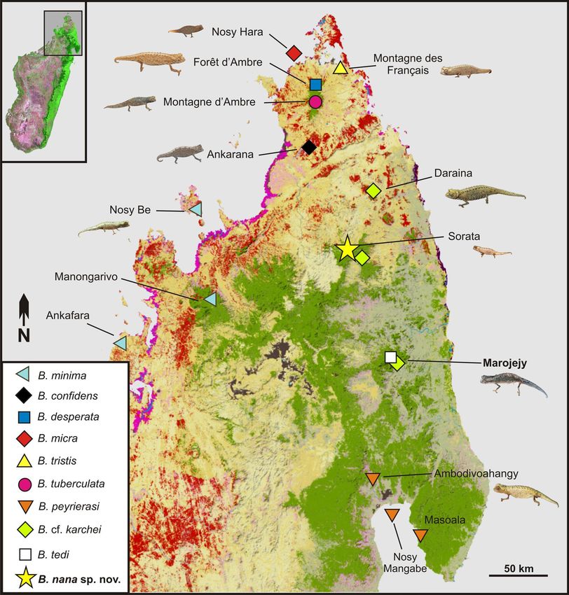
www.nature.com/scientificreports/
species are characterized by clear paedomorphism, a frequent feature of miniature a nimals1, often arising from
heterochrony, and particularly obvious by their relatively large heads and eyes.
The brookesiine chameleon genus Brookesia consists of predominantly terrestrial species divided in two major
lineages, which diverged from each other ca. 40–50 million years a go7–9. One of these lineages includes larger
species of 34–66 mm SVL, while the other contains only highly miniaturized species. At present, 12 described
species are known from this c lade5,10, none of which exceeds 30 mm SVL, with the smallest species B. micra reach-
ing a maximum adult female SVL of 19.9 mm5. A report of live B. micra reaching 23 mm SVL11 is unfortunately
not vouchered and cannot be verified.
Most miniaturized Brookesia are rainforest species, which inhabit mostly forests in lowlands (e.g. B. minima
on Nosy Be) and rarely at higher elevations > 1000 m a.s.l. (e.g. B. tedi on Marojejy). Other species prefer dry
nderground5,12. The majority of species exhibit very small ranges, with only few
forest, especially on karstic u
species being known from more than two locations. This microendemism may be related to the complex topog-
raphy in northern Madagascar where these and other Brookesia species are predominantly distributed13. Their
diminutive size combined with their small ranges have contributed to the fact that much of the diversity of this
clade has been overlooked until recently.
Here, we report on the discovery of a new species of Brookesia that is apparently still smaller than other
miniaturized species of the genus, measuring less than 14 mm SVL in an adult male and 19 mm in a female. We
describe this new species and discuss several aspects of miniaturization in these chameleons.
Results
Phylogenetic position of the new chameleon species. The Maximum Likelihood (ML) trees
obtained from analysis of two mitochondrial gene fragments (16S, ND2: Fig. 1) and one nuclear gene frag-
ment (CMOS: Fig. 2) suggested concordant relationships among species of Brookesia, similar to those previously
inferred5,10, which were based on a more limited taxon sampling. Among the new aspects of our analysis is the
confirmation of a specimen from the Masoala Peninsula as Brookesia peyrierasi. Also, the new samples of B.
karchei from Sorata cluster with other samples of this taxon from Marojejy and Daraina, and the new samples of
B. ramanantsoai from Tsinjoarivo and Vohimana cluster with other samples of this taxon from Mandraka. In all
these cases, the samples from the different locations show a substantial genetic divergence: uncorrected pairwise
distances (p-distances) in the 16S gene among localities were 3.2‒3.4% for B. peyrierasi, 3.4‒4.4% for B. karchei,
and 4.3‒6.3% for B. ramanantsoai.
The two specimens of our focal lineage from Sorata had identical 16S sequences, and in the two mitochondrial
trees they were placed sister to B. karchei (bootstrap support 58% and 66%), whereas in the CMOS tree they
formed the sister group of a clade containing B. karchei, B. peyrierasi, and B. tedi (bootstrap support 75%). 16S
genetic divergences of the Sorata lineage were 9.9‒11.3% to B. karchei, and 10.5‒14.9% to all other B. minima
group species. The Sorata lineage also showed a substantial divergence in the nuclear CMOS gene, which usually
is rather conserved among closely related reptiles; uncorrected pairwise distances were 5.1% to B. karchei, and
4.0‒7.3% to all other B. minima group species.
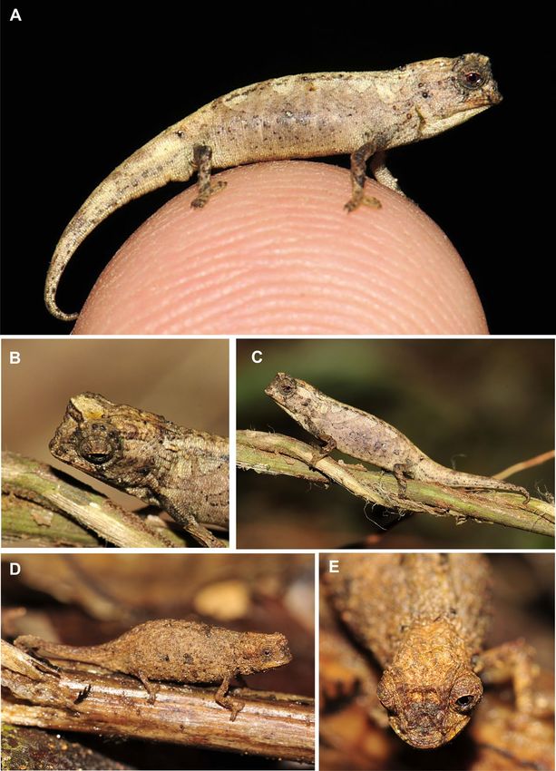
A map with genetically confirmed records of the B. minima group from northern Madagascar (Fig. 3), based
on data from previous p ublications5,10,14, indicates that species are reliably known from a maximum of three
localities.
Systematics
Order Squamata Oppel, 1 81115.
Family Chamaeleonidae Rafinesque, 181516.
Subfamily Brookesiinae Angel, 1 94217.
Genus Brookesia Gray, 186517.
Subgenus Evoluticauda Angel, 1942 (resurrected herein, justification below).
Brookesia nana sp. nov. Holotype. ZSM 1660/2012 (field number FGZC 3788), adult male, from near
a campsite in the Sorata massif, 13.6851° S, 49.4417° E, ca. 1280 m a.s.l., northern Madagascar, collected on 1
December 2012 by F. Glaw, O. Hawlitschek, T. Rajoafiarison, A. Rakotoarison, F. M. Ratsoavina, and A. Razafi-
manantsoa.
Paratype. UADBA-R/FGZC 3752, adult female, from near the pitfall site 1, Sorata massif, 13.6817° S, 49.4411°
E, 1339 m a.s.l., northern Madagascar, collected on 29 November 2012 by same collectors as the holotype.
Nomenclatural statement. A Life Science Identifier (LSID) was obtained for the new species (Brookesia nana)
from ZooBank: urn:lsid:zoobank.org:act: 37B38077-FA5D-48E9-BACF-723061B3921F and for this publication:
urn:lsid:zoobank.org:pub: 540F80C8-EC9F-49A9-B13F-77C75DED5962.
Diagnosis. A diminutive chameleon species assigned to the genus Brookesia on the basis of its small body
size, short tail, presence of rows of dorsolateral tubercles along vertebral column, presence of pelvic spine, and
molecular phylogenetic relationships. Brookesia nana sp. nov. is distinguished by the following unique suite of
morphological characters: (1) male SVL 13.5 mm, female SVL 19.2 mm; (2) male TL mm 21.6 mm, female TL
28.9 mm; (3) TaL/SVL ratio of 0.51 in male; (4) absence of lateral or dorsal spines on the tail; (5) absence of
Scientific Reports | (2021) 11:2522 | https://doi.org/10.1038/s41598-020-80955-1 2
Vol:.(1234567890)
www.nature.com/scientificreports/

                                  Figure 1.  Molecular phylogenetic trees of specimens in the subgenus Evoluticauda (known as Brookesia minima
                                  group), based on sequences of the mitochondrial (A) 16S (480 bp) and (B) ND2 (571 bp) genes, inferred under
                                  the Maximum Likelihood optimality criterion, and the GTR + G (16S) and HKY + I + G (ND2) substitution
                                  models. Values at nodes are support values from a bootstrap analysis in percent (500 replicates) and are shown
                                  only if > 50%. The two gene fragments were analysed separately and not concatenated because partly different
                                  samples were available for each of them. The trees were rooted with B. brygooi (removed for better graphical
                                  representation).

Scientific Reports |   (2021) 11:2522 |               https://doi.org/10.1038/s41598-020-80955-1                                              3

                                                                                                                                         Vol.:(0123456789)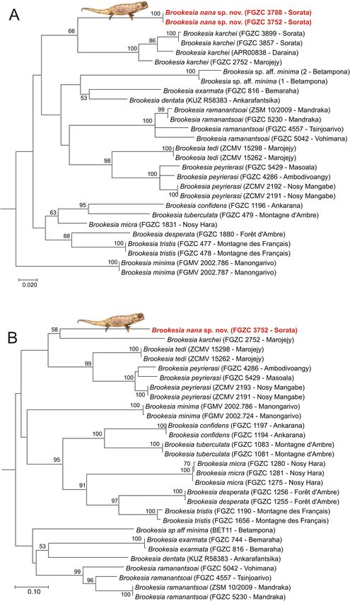
www.nature.com/scientificreports/

                                            Figure 2.  Molecular phylogeny of specimens in the subgenus Evoluticauda (known as Brookesia minima
                                            species group), based on the nuclear CMOS gene (alignment length 847 bp, but only about 400 bp available for
                                            all samples) and inferred under the Maximum Likelihood optimality criterion (K2P + G substitution model).
                                            Values at nodes are support values from a bootstrap analysis in percent (500 replicates) and are only shown
                                            if > 50%. The tree was rooted with B. brygooi (removed for better graphical representation).

                                            dorsal pelvic shield in sacral area; (6) presence of distinct pelvic spine; (7) pale brown dorsal colouration with
                                            slightly darker markings in life; (8) absence of apical spines on the hemipenis.
                                                Within the genus Brookesia, B. nana sp. nov. can easily be distinguished from all species that are not mem-
                                            bers of the B. minima species group by its diminutive size (SVL 13.5–19.2 mm vs. > 34 mm). Within the B.
                                            minima species group, it can be distinguished from most species by the smaller total length (TL). Based on TL
                                            (21.6–28.9 mm), both males and females are significantly smaller than all known specimens of B. desperata
                                            (39.7–47.6 mm), B. exarmata (39.8–40.1 mm), B. karchei (51.0 mm), and B. ramanantsoai (39.0–43.5 mm), and
                                            are slightly but distinctly smaller when compared to B. tristis (30.7–36.5 mm), B. confidens (29.2–36.2 mm),
                                            and B. peyrierasi (32.2–43.1 mm). Four species of the B. minima group are in an overall comparable size range:
                                            B. micra, B. minima, B. tedi, and B. tuberculata. Yet, the male of the new species (TL 21.6 mm) is the smallest
                                            adult Brookesia so far known, compared to the previously smallest specimen, a male of B. micra with 22.2 mm
                                            TL and 15.3 mm SVL.
                                                The very short tail of the male B. nana (TaL/SVL 0.51; 0.60 in the female) constitutes a difference to males of
                                            most species of the B. minima group: male TaL/SVL is 0.60–0.70 in B. confidens, 0.59–0.63 in B. desperata, 0.66
                                            in B. karchei, 0.65–0.73 in B. minima, 0.57–0.92 in B. peyrierasi, 0.8 in B. ramanantsoai, 0.74–0.92 in B. tedi,
                                            0.71–0.72 in B. tristis, and 0.68–0.88 in B. tuberculata.
                                                Given its tiny size, the new species is most similar to B. micra (SVL 15.3–15.8 and TL 22.5–23.6 in males; SVL
                                            18.7–19.9 mm and TL 26.9–28.8 in females), which has an even shorter relative tail length in males (TaL/SVL
                                            0.47–0.49). However, males of B. micra differ by a more robust habitus; by a flat surface distally forming a sym-
                                            metrical comb of six large, rounded papillae on the apex of the hemipenis (absent in the new species); and by life
                                            colouration, namely a dark brown body and a yellow-orange tail (versus pale brown body and tail with indistinct
                                            darker markings). Moreover, molecular data provide evidence for a distant relationship of B. nana and B. micra.

                                            Description of the holotype. Adult male in excellent state of preservation (Figs. 4A–C, 5). 21.6 mm TL
                                            and 13.5 mm SVL. For additional measurements, see Table 1. Lateral crest on head weakly developed, barely
                                            recognizable; weak orbital crest; transversal row of enlarged tubercles at the posterior edge of head lacking, no
                                            distinct border between head and body, posterior crest lacking; a pair of short curved parasagittal crests that
                                            start above the eyes and fade at midlevel of head; depression between the eyes lacking any further crests; one
                                            pointed tubercle on each side of head; few scattered, slightly enlarged tubercles on lateral surfaces of head; orbital
                                            crest slightly denticulated; distinct supraocular cone absent; supranasal cone very tiny, not projecting beyond
                                            tip of snout; head longer than wide; chin and throat without enlarged tubercles. Dorsal surface of body without
                                            vertebral ridge or keel; 5/5 (left/right) dorsolateral pointed tubercles that form an incomplete longitudinal line,

          Scientific Reports |   (2021) 11:2522 |                https://doi.org/10.1038/s41598-020-80955-1                                                     4

Vol:.(1234567890)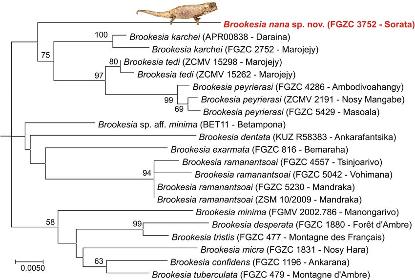
www.nature.com/scientificreports/
Figure 3. Map of northern Madagascar, showing the distribution of species of the subgenus Evoluticauda
(known as Brookesia minima group) in this region (only showing records verified by molecular data5,10,14).
Note that B. dentata, B. exarmata, and B. ramanantsoai occur further south and are not included in the map.
Orange (dry forest) and green (rainforest) show remaining primary vegetation in 2003–2006. Modified from the
Madagascar Vegetation Mapping Project; http://www.vegmad.org.
ending approximately at level of midbody; anteriormost pointed dorsolateral tubercle being largest; pointed dor-
solateral tubercles along vertebral column almost equally spaced; dorsal surface of tail lacking distinctly enlarged
tubercles; no dorsal pelvic shield in sacral area, but distinct small pelvic spine; lateral surface of body with few
irregularly spaced enlarged tubercles; venter without enlarged tubercles; no enlarged pointed tubercles on limbs;
no pointed tubercles around cloaca; longitudinal row of slightly enlarged tubercles lateral on anterior tail; no
dorsal, lateral, or ventral spines on tail; no enlarged tubercles on ventral surfaces of tail.
Left hemipenis fully everted, right hemipenis almost fully everted (Fig. 5). The fully everted hemipenis is
2.5 mm long, tubular, elongated, with a small flattened apical end with a clear lip around its circumference
(Fig. 5F,G). A pair of structures emerge from the apical surface, each of which consists of a fleshy lobe. The
truncus is smooth and lacks any trace of calyces.
Scientific Reports | (2021) 11:2522 | https://doi.org/10.1038/s41598-020-80955-1 5
Vol.:(0123456789)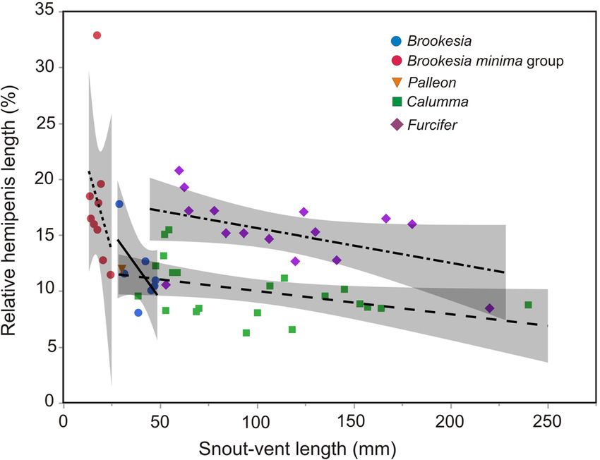
www.nature.com/scientificreports/

                                            Figure 4.  Brookesia nana sp. nov. in life. (A–C) male holotype (ZSM 1660/2012). (D, E) female paratype
                                            (UADBA-R/FGZC 3752).

                                                In life, overall dorsal ground colouration was pale brown, with some lighter blotches in the dorsal and dorso-
                                            lateral regions, partly extending to the flanks forming incomplete streaks, as well as numerous small dark brown
                                            and blackish tubercles and spots. A pattern of four diffuse beige streaks running obliquely from the dorsum to

          Scientific Reports |   (2021) 11:2522 |                https://doi.org/10.1038/s41598-020-80955-1                                                 6

Vol:.(1234567890)
www.nature.com/scientificreports/
Figure 5. Morphological characters of Brookesia nana sp. nov.: (A) preserved holotype (ZSM 1660/2012) in
lateral view, showing right everted hemipenis, (B) head in dorsal and (C) lateral (mirrored, indicated with
asterisk) views; head of female paratype (UADBA-R/FGZC 3752) in (D) dorsal and (E) lateral views; (F, G)
close-ups of everted left hemipenis of holotype photographed under different light conditions; (H) micro-CT
scan image of the female paratype in lateral view showing its skeleton. The inset image (I) shows the area
marked by the stippled square viewed at a different rendering threshold, showing two developing eggs in the
females’ ovaries.
Scientific Reports | (2021) 11:2522 | https://doi.org/10.1038/s41598-020-80955-1 7
Vol.:(0123456789)www.nature.com/scientificreports/
UADBA-R/FGZC
ZSM 1660/2012 3752
FGZC 3788 FGZC 3752
Holotype Paratype
Sex M F
TL 21.6 28.9
SVL 13.5 19.2
TaL 8.1 9.7
HW 2.5 3.0
HH 2.0 2.7
ED 1.3 1.5
FORL 3.9 5.1
Table 1. Morphometric measurements of holotype and paratype of Brookesia nana sp. nov. (all in mm). See
“Materials and Methods” for abbreviations.
the mid-flanks were recognizable (Fig. 4C). A beige patch was present on the anterior head (Fig. 4B). Two dark
streaks ran from the lower margin of the eye to the upper lip. The dorsolateral tubercles and the supraocular crest
were blackish. Exterior surfaces of forelimbs and hindlimbs were distinctly darker than flanks and mottled with
brown and grey. Darker radial streaks were present on the eyelid, and the iris was dark red (Fig. 4A).
After 6 years in ethanol, the body colouration is generally faded with less evident pattern. The ground coloura-
tion is pale brown, becoming distinctly lighter lateroventrally and ventrally. An interrupted dark brown mid-
dorsal line runs longitudinally on the dorsum. Head laterally with a diffuse pattern of different shades of brown,
grey, and white. Dorsolateral tubercles blackish, pelvic spines whitish. Flanks with dark brown to beige tubercles
and patches, including four nearly blackish circles. The dark radial streaks are more distinct than they were in life.
Variation. Female paratype is in very good state of preservation. Lateral crest on head present, starting at
midlevel of eye and stretching backwards to posterior crest; prominent orbital crests; transversal row of enlarged
tubercles at the posterior edge of head that separates the head from the body, forming posterior crest; a pair of
short curved parasagittal crests that start above the eyes and fade at posterior level of eyes; depression between
the eyes with short indistinct median crest and a pair of curved crests starting above eyes and converging to
midlevel of head; five pointed tubercles on each side of posterior crest; scattered, slightly enlarged tubercles
on lateral surfaces of head; orbital crest denticulated; distinct supraocular cone absent; supranasal cone dis-
tinct, small, not projecting beyond tip of snout; head longer than wide; chin and throat without enlarged tuber-
cles. Dorsal surface of body without vertebral ridge or keel; 5/5 (left/right) dorsolateral pointed tubercles along
vertebral column, barely recognizable, forming an incomplete longitudinal line; pointed dorsolateral tubercles
almost equally spaced; dorsal surface of tail lacking distinctly enlarged tubercles; enlarged tubercles on lateral
tail not recognizable; no dorsal pelvic shield in sacral area, but distinct pelvic spine; lateral surface of body with
few irregularly spaced enlarged tubercles; venter without enlarged tubercles; scattered enlarged and distinctly
pointed tubercles on limbs; no pointed tubercles around cloaca; no dorsal, lateral, or ventral spines on tail; no
enlarged tubercles on ventral surfaces of tail. In life, dorsal colour brown, with some darker coloured tubercles,
scattered flecks and spots, but generally lacking any conspicuous pattern. Dorsal surface of head slightly paler
(Fig. 4D,E). In preservative, the female paratype is generally darker than the holotype, with most enlarged lat-
eral tubercles and numerous small tubercles being dark brown. Dorsal side with a large dark brown patch in its
posterior part.
Etymology. The specific epithet is the Latin noun nana (meaning female dwarf) in the nominative singular.
Conservation status. Brookesia nana is known from just two specimens and a single location and thus
belongs to the ca. 14% of the world’s lizard species that are only known from the type locality18. This extremely
poor knowledge makes it difficult to reliably evaluate the distribution and the conservation status of this species.
However, given that most of the miniaturized Brookesia species are microendemic with limited elevational range
(Fig. 3), a small range might be also expected for B. nana. During our expedition in 2012 the natural habitats
of the Sorata massif were highly threatened. At lower elevations, the natural forest had been completely eradi-
cated and anthropogenic pressure at the existing edges was high, especially from deforestation, slash-and-burn
agriculture, and cattle. These threats were increasingly extending to higher altitude including the type locality
of B. nana. Recently, the Sorata massif has received official protection as part of the new protected area ‘Resé-
rve de Ressources Naturelles du Corridor Marojejy-Anjanaharibe Sud-Tsaratanàna partie Nord’, also known as
COMATSA Nord19. This new reserve may hopefully help to preserve the remaining forest habitats, but the cur-
rent threat situation around the type locality is unknown. However, according to the current state of knowledge,
we suggest that B. nana qualifies as Critically Endangered B1ab(iii) under the Red List Criteria of the I UCN20 as
the extent of occurrence is estimated to be less than 100 km2, all individuals occur in one threat-defined location,
and there is continuing decline in the extent and quality of its forest habitat in the Sorata massif. We recommend
that the extinction risk of this species be assessed officially for the IUCN Red List of Threatened Species as soon
Scientific Reports | (2021) 11:2522 | https://doi.org/10.1038/s41598-020-80955-1 8
Vol:.(1234567890)www.nature.com/scientificreports/
Figure 6. Relation between body size (snout–vent length, SVL) and relative hemipenis length (relative
HPL = HPL/SVL in percent), for genera of Malagasy chameleons. The graph shows a prevalent negative
correlation of relative HPL with SVL, but also clear differences among genera, especially among the two genera
of large-sized tree chameleons, Calumma and Furcifer. The species with the highest value of relative HPL is
Brookesia tuberculata.
as possible. This finding confirms the results of a previous study that Malagasy chameleons have a higher propor-
tion of threatened species compared to other species-rich reptile groups (geckos, skinks, and gerrhosaurids)21.
Genital organ size in Malagasy chameleons. Relative hemipenial length in Malagasy and Comoran
chameleons ranged over more than a half-order of magnitude, from a minimum of 6.3% of SVL in Calumma
capuroni to 32.9% in Brookesia tuberculata, and with an average of 13.1% over the 52 species for which data
were available. The value of B. tuberculata—with genitals of almost one-third of body length—is, however,
exceptional, with the next largest values being around 20% in B. peyrierasi, Furcifer cephalolepis, and F. later-
alis. A non-parametric Spearman rank test confirmed that relative hemipenial length was negatively correlated
to snout–vent length (Spearman’s R = − 0.358; P = 0.0085). This overall trend was consistent in all genera, but
among the tree chameleons, Calumma had distinctly and consistently shorter hemipenes than Furcifer (Fig. 6).
Although small-sized chameleons had relatively larger hemipenes, the very long genital organs of B. tuberculata
clearly stood out as an outlier (Fig. 6). The holotype of Brookesia nana had the fifth largest relative hemipenial
length (18.5%) of the 52 studied species and the third largest in the genus Brookesia, supporting that this tiny
chameleon is indeed an adult male.
Resurrection of Evoluticauda Angel, 1942 as subgenus of Brookesia Gray, 1865. Previous
molecular studies have revealed two major lineages in Brookesia, which separated from each other ca. 43 million
years ago9. This split is only slightly younger than the split between the genera Bradypodion/Nadzikambia, Trioc-
eros/Kinyongia and Calumma/Furcifer (all ca. 45–46 mya) and older than the split between Rieppeleon/Archaius
(ca. 35 mya) and between the different subgenera of Rhampholeon9. Most species of the large-bodied clade of
Brookesia show very distinct rows of bony lateral projections along the vertebral column, which are based on a
unique vertebral structure and might function as body armour to prevent predation22. These rows of projections
are either absent, incomplete, or very poorly developed in the lineage of small-bodied species, which are also
recognizable by their miniaturized adult size (22–51 mm versus 51–110 mm total length) and relatively larger
hemipenis length (Fig. 6). Due to the old divergence and the morphological distinctness of the two clades we
here suggest to consider them as different subgenera:
Subgenus Brookesia Gray, 1865 (large-bodied clade)
Type species: Chamaeleo superciliaris Kuhl, 1820.
Contents: Brookesia antakarana, B. bekolosy (attribution tentative), B. betschi, B. bonsi, B. brunoi, B. brygooi, B.
decaryi, B. ebenaui, B. griveaudi, B. lambertoni, B. lineata, B. perarmata, B. stumpffi, B. superciliaris, B. therezieni,
B. thieli, B. vadoni, B. valerieae.
Distribution: Madagascar.
Subgenus Evoluticauda Angel, 1942 (miniaturized clade, known as B. minima group)
Type species: Brookesia tuberculata Mocquard, 1894.
Contents: Brookesia confidens, B. dentata, B. desperata, B. exarmata, B. karchei, B. micra, B. minima, B. nana,
B. peyrierasi, B. ramanantsoai, B. tedi, B. tristis, B. tuberculata.
Scientific Reports | (2021) 11:2522 | https://doi.org/10.1038/s41598-020-80955-1 9
Vol.:(0123456789)www.nature.com/scientificreports/
Distribution: Northern half of Madagascar.
Discussion
Body size of Brookesia nana and B. micra. Brookesia nana sp. nov. is a remarkable addition to the diver-
sity of microendemic and miniaturized chameleons in northern Madagascar, and with a SVL of only 13.5 mm
the holotype represents once more a new record at the lower size limit of amniotes. Until now, the smallest
Brookesia was B. micra, with a confirmed minimal adult male size of 15.3 m m5. In a valuable ecological study,
Villeneuve observed 117 B. micra and measured the SVL of living individuals to the nearest 1 mm. In this paper,
11
a body size distribution graph was presented in which males and females as small as 9 mm SVL were reported,
but these data refer to juveniles, as adults were defined as ≥ 13 mm SVL and juveniles/sub-adults ≤ 13 mm SVL.
The largest body size of B. micra from this graph was 20 mm for a female, in line with data of Glaw et al.5, but two
males of 20 and 23 mm represent an important shift of maximal sizes. Unfortunately, these exceptional values
are not discussed in this s tudy11. We here consider them as in need of confirmation, although we are aware that
exceptionally large specimens are known from numerous amphibian and reptile species and may thus be found
in B. micra and other miniaturized species as well. Villeneuve11 also reported on the sexual size dimorphism of
B. micra and stated in the abstract that he ‘found adult males to have a significantly larger snout‒vent length
(SVL) than adult females’, whereas in the results (referring to a table with SVL measurements) it was reported
that ‘adult female B. micra tended to be larger than adult male individuals’. The latter result is typical for B. micra
and other species of the subgenus Evoluticauda5, and the former thus probably an error.
Evolution and consequences of miniaturized body size. Although all species of miniaturized Brooke-
sia belong to the subgenus Evoluticauda, important size differences are seen within the group. The mitochondrial
trees (Fig. 1) place B. nana sister to B. karchei, the largest species in Evoluticauda (30.7 mm SVL in females5).
A second extremely small species, B. micra (minimum male SVL 15.3 mm, maximum female SVL 19.9 mm) is
phylogenetically relatively close to B. desperata where females reach 30 mm S VL5. The available data do not allow
us to unambiguously distinguish in this case between convergent extreme miniaturization in B. micra and B.
nana, or convergent reversal to somewhat larger body sizes in B. desperata and B. karchei. The obvious presence
of homoplasy and/or reversal in the evolution of miniaturization agrees with the observations in other taxa, such
as the independent origin of morphologically similar miniaturized taxa in microhylid and other frogs23–26, and
the evolutionary lability of body size traits in other predominantly small-sized v ertebrates27–29.
The species of Brookesia that apparently have independently evolved their tiny sizes also share a number of
other morphological features, such as a general reduction of dorsolateral spines or tubercles along the vertebral
column, almost complete lack of head ornaments such as supraocular crests and cones, and short tails. Whether
these characteristics are allometric correlates of small body size, e.g. via p aedomorphism30,31 or may be driven by
32
convergence on a small-size body shape o ptimum cannot be decided without a substantial amount of further
data and analyses, including a greatly improved knowledge of the morphological variation of both sexes and the
ontogenetic development of these characters in juveniles.
Given possible functions of chameleon head ornaments in sexual selection33,34 and of the tail in Brookesia
for assisted walking35, it is likely that miniaturization in these lizards is linked to either functional causes, or
functional consequences, or both. For example, small chameleon species are known to outperform larger species
during ballistic tongue p rojection36, but none of the miniaturized Brookesia species has yet been studied in this
respect. Also, studies on the microhabitat requirements and ecology of Brookesia species are scarce and largely
restricted to the larger Brookesia species37,38, so that the behavioural and ecological consequences of the extreme
miniaturization of Evoluticauda species remain completely unknown.
Several miniaturized lizards, especially species of Sphaerodactylus, occur on islands4, as do the smallest spe-
cies of snakes39. It is appealing to relate this to the so-called island rule, much discussed especially for mammals
where small mammals tend to evolve larger sizes, and large mammals smaller sizes, compared to their mainland
conspecifics40,41 (but see ref.42). On the contrary, in lizards, it was found that small species on islands become
smaller than their mainland conspecifics, while large ones become larger still, opposite to predictions of the
island rule43. Whether the presence of miniaturized chameleons in Madagascar can be interpreted as supporting
this finding is uncertain given that Madagascar, with a surface of about 587,041 km2, qualifies more as a micro-
continent than an island. Comparing the distribution of the two most strongly miniaturized species, B. micra
appears to be restricted to the tiny 270 ha islet of Nosy Hara with an estimated population of 100,000 to 150,000
individuals11, which may have driven miniaturization. However, the new species B. nana occurs in a mountain
massif that can be considered rather as part of a major rainforest block of northern Madagascar and its small size
is unlikely to be related to specific insularity-related drivers. The elevational distribution of B. nana is, however,
remarkable in that it is only one of three species in Evoluticauda occurring at elevations above 1300 m a.s.l.
Patterns of fusion of fingers and toes. As all chameleons, Brookesia are characterized by a unique pat-
tern of fusion of fingers and toes: on the forelimbs, the outer two and inner three toes are fused, respectively,
whereas on the hindlimbs the pattern is reversed. This ‘chamaeleodactyl’ morphology is accompanied by numer-
ous modifications of the mesopodial elements, which however differ among chameleon genera44. The small-
sized genera of ground chameleons, including the Malagasy Brookesia and Palleon, but also the African Rham-
pholeon and Rieppeleon, were found to maintain the fewest independent carpal and tarsal elements as adults,
while the genera of larger-sized arboreal chameleons have a larger number of mesopodial elements, which may
be related to locomotor mode44. For Brookesia, these conclusions were drawn based on an analysis of B. stumpffi,
a species reaching over 50 mm SVL, while the truly miniaturized ground chameleons have not yet been studied
in detail for their hand and foot skeleton. A more comprehensive comparative analysis of skeletal anatomy across
Scientific Reports | (2021) 11:2522 | https://doi.org/10.1038/s41598-020-80955-1 10
Vol:.(1234567890)www.nature.com/scientificreports/
Brookesia of different sizes may reveal whether differences in hand and foot morphology are related to their split-
ting from a phylogenetically basal node among chameleons, or by-products of small size and miniaturization, or
functionally adaptive in relation to their forest floor habitat.
Size and evolution of male genitalia. One striking feature of miniaturized chameleons is the relatively
large size of their genital organs. This is particularly obvious in the very long hemipenes of Brookesia tuberculata,
but also the balloon-shaped hemipenes of B. minima and B. ramanantsoai attain an enormous volume and a
width much exceeding body width of these small lizards (photos in ref.5). The causes for this allometric relation-
ship are poorly understood. Sexual selection and communication in many chameleons relies on optical signals,
both related to colour and external ornaments such as crests, casques, spines, or snout p rotuberances33,34,45. Most
Malagasy ground chameleons of the genera Brookesia and Palleon stand out among other Malagasy chameleons
by being small-sized, by their dull colouration and lack of capacity for major colour changes, and their limited
amount of external ornamentation. In contrast to the situation in the larger-sized Calumma and Furcifer, in
Brookesia the females are typically larger than the males46, suggesting that male–male competition may play a
more limited role in their mate choice behaviour or that physiological constraints prevent further female size
reductions. Across the animal kingdom, extreme sizes of genitals occur. They can be similar to body length
in ducks, and up to eight times the body length in barnacles47, being usually related to functional necessities
(e.g., in sessile barnacles) or sexual conflict and male-male competition in waterfowl48. Sexual size dimorphism
can strongly influence the evolution of reproductive strategies and can lead to functional conflicts between the
sexes, e.g., an evolutionary mismatch between the absolute sizes of male and female genitalia within species,
as has been shown for orb-weaving spiders, where genital dimorphism increases with increasing sexual size
dimorphism49. The distinct differences in relative hemipenis length between large and small chameleon species
might be a consequence of the reversal of sexual size dimorphism, given that in larger-sized chameleon genera
like Furcifer and Calumma, males are generally larger than females, whereas the opposite is true in small-sized
genera, e.g. Brookesia and Rhampholeon46,50. In these miniaturized species, the smaller males may simply need
larger hemipenes to allow for a better mechanical fit that makes successful copulation with the much larger
females possible. To test this plausible hypothesis there is an obvious need for comprehensive studies of female
genitalia of chameleons and other squamates.
Although the current evidence (M.D. Scherz and collaborators in progress; and data herein) suggests that
hemipenial ornamentation in chameleons is predominantly determined by allometric factors related to body
size, it is obvious that additional factors play a relevant role causing for instance the distinct differences in hemi-
penial size among Calumma and Furcifer species of similar body sizes (Fig. 6). In-depth comparative studies of
the mating system of these genera as well as Brookesia, and especially of the miniaturized Brookesia species with
exaggerated genital sizes, emerges as an important priority for future research, in order to fully understand the
evolution of these highly specialized lizards, and the evolutionary consequences and drivers of miniaturization
in vertebrates.
Materials and methods
Fieldwork, permits and morphological measurements. Miniaturized Brookesia species were inten-
sively sought during the day on the ground and at night with torchlight. Vouchers were anaesthetised and subse-
quently euthanised by oral application of lidocaine. This method was carried out in accordance with all relevant
guidelines and regulations. No experiments were conducted with the living animals. After taking tissue samples
(stored in pure ethanol), vouchers were fixed with 90% ethanol and deposited in 75% ethanol for long-term
storage. Collection of specimens was conducted under permit No. 265/12/MEF/SG/DGF/DCB.SAP/SCB (dated
18 Oct. 2012) and exportation of specimens under permit No. 163N-EA12/MG12 (dated 17 Dec. 2012), both
issued by the Direction Générale des Forêts (Ministère de l’Environnement, des Eaux et Forêts de la République
de Madagascar). Import permits were issued by the German CITES authority (Bundesamt für Naturschutz).
Field numbers (FGZC) refer to the field series of F. Glaw. We deposited the vouchers in the collections of
the Mention Zoologie et Biodiversité Animale of the Université d’Antananarivo (UADBA-R) and Zoologische
Staatssammlung München (ZSM). Morphometric analysis and morphological descriptions follow a previous
study5. The following measurements were taken by MV to the nearest 0.1 mm using a digital calliper: TL (total
length); SVL (snout–vent length); TAL, tail length; HW, maximum head width; HH, maximum head height; ED,
eye diameter; FORL, forelimb length.
The X-ray micro-Computed Tomography (micro-CT) scan was produced using a phoenix|x nanotom m
cone beam scanner (GE Measurement & Control, Wunstorf, Germany), with details of the method as described
in ref.51. Scans were deposited in MorphoSource (https://www.morphosource.org/Detail/ProjectDetail/Show/
project_id/953).
Molecular analysis. For molecular analysis, we used DNA sequences of fragments of the mitochondrial
genes for 16S rRNA (16S) and NADH Dehydrogenase Subunit 2 (ND2), and the nuclear gene for oocyte matu-
ration factor mos (CMOS). Our dataset builds upon sequences from a previous study5, but with a reduced
representation (two sequences per species) for ND2, and also including sequences of one sample of B. peyrierasi
and two samples of B. tedi10. This data set was expanded by newly determined sequences of the two available
samples of the new species from Sorata, and of several additional samples of Brookesia karchei, B. peyrierasi,
and B. ramanantsoai. Because for some species the individual samples sequenced for ND2 differed from those
sequenced for 16S, we refrained from combining these two mitochondrial DNA fragments for analysis. Further-
more, in order to test for genealogical concordance between nuclear and mitochondrial DNA52, we also analysed
the CMOS sequences separately.
Scientific Reports | (2021) 11:2522 | https://doi.org/10.1038/s41598-020-80955-1 11
Vol.:(0123456789)www.nature.com/scientificreports/
We extracted genomic DNA and amplified the target gene fragments using standard protocols as described
reviously5, with the primers ND2F17 (5′-TGAC
p AAA
AAA CNCC-3′)53 and ALAR2 (5′-AAAA
TTG TRT
CTG
RG
54
TTGC ATTCAG-3′) for ND2, 16SA-L (5′-CGCCTGTTTATCAAAAACA T-3′) and 16S-BH (5′-CCGGTCT GA
ACTCAGATCACGT-3′) for 1 6S55, and CO8 (5′-GCTTGGTGTTCAATAGACTGG-3′) and CO9 (5′-TTGGGA
GCATCCAAAGTCTC-3′) for C MOS56. We purified PCR products with ExoSAPIT (Thermo Fisher Scientific,
Waltham, MA, USA) and sequenced them on an automated DNA sequencer (ABI 3130 XL; Applied Biosystems).
We checked, corrected, and trimmed sequences with the software CodonCode Aligner (CodonCode Corpora-
EGA755. Newly obtained sequences were submitted
tion), and aligned them using the Clustal algorithm in in M
to GenBank (accession numbers MK452380‒MK452387, MK45737‒MK457374, and MK457447‒MK457451);
for accession numbers of previously published sequences, see refs.5,10.
Sequences were analysed in M EGA757. We determined the most suitable substitution models determined
under the Bayesian Information Criterion, implemented in MEGA7 (16S: GTR + I + G; ND2: HKY + I + G; CMOS:
a K2P + G model), and conducted phylogenetic analyses under the Maximum Likelihood optimality criterion,
with nearest-neighbour interchange (NNI) branch-swapping, and with 500 heuristic bootstrap replicates.
Genital morphology. To understand patterns of allometry in hemipenes of chameleons, we measured
body size (snout–vent length) and everted hemipenis length (HPL) in a total of 97 adult males of 52 species
of the genera Brookesia, Palleon, Calumma and Furcifer from Madagascar and the Comoros (Supplementary
Table S1). Where several individuals per species were available, we calculated species averages for both charac-
ters for further analysis. Relative hemipenis length, i.e., the ratio between SVL and HPL, was plotted against SVL,
and non-parametric correlations calculated with Statistica 7.1 (Statsoft Inc.).
Data availability
All data generated or analysed during this study are included in this published article (and its Supplementary
Information files), MorphoSource, and GenBank.
Received: 9 August 2020; Accepted: 29 December 2020
References
1. Hanken, J. & Wake, D. B. Miniaturization of body size: Organismal consequences and evolutionary significance. Ann. Rev. Ecol.
Syst. 24, 501–519 (1993).
2. Rittmeyer, E. N., Allison, A., Gründler, M. C., Thompson, D. K. & Austin, C. C. Ecological guild evolution and the discovery of
the world’s smallest vertebrate. PLoS ONE 7, e29797 (2012).
3. Glaw, F., Vences, M., Ziegler, T., Böhme, W. & Köhler, J. Specific distinctness and biogeography of the dwarf chameleons Brookesia
minima, B. peyrierasi and B. tuberculata (Reptilia: Chamaeleonidae): evidence from hemipenial and external morphology. J. Zool.
247, 225–238 (1999).
4. Hedges, S. B. & Thomas, R. At the lower size limit in amniote vertebrates: A new diminutive lizard from the West Indies. Caribb.
J. Sci. 37, 168–173 (2001).
5. Glaw, F., Köhler, J., Townsend, T. M. & Vences, M. Rivaling the world’s smallest reptiles: Discovery of miniaturized and microen-
demic new species of leaf chameleons (Brookesia) from northern Madagascar. PLoS ONE 7, e31314 (2012).
6. Meiri, S. Traits of lizards of the world: variation around a successful evolutionary design. Glob. Ecol. Biogeogr. 27, 1168–1172 (2018).
7. Townsend, T. M., Vieites, D. R., Glaw, F. & Vences, M. Testing species-level diversification hypotheses in Madagascar: The case of
microendemic Brookesia leaf chameleons. Syst. Biol. 58, 641–656 (2009).
8. Townsend, T. M., Tolley, K. A., Glaw, F., Böhme, W. & Vences, M. Eastward from Africa: Palaeocurrent-mediated chameleon
dispersal to the Seychelles islands. Biol. Lett. 7, 225–228 (2011).
9. Tolley, K. A., Townsend, T. M. & Vences, M. Large-scale phylogeny of chameleons suggests African origins and Eocene diversifica-
tion. Proc. R. Soc. B 280, 20130184 (2013).
10. Scherz, M. D., Köhler, J., Rakotoarison, A., Glaw, F. & Vences, M. A new dwarf chameleon, genus Brookesia, from the Marojejy
massif in northern Madagascar. Zoosyst. Evol. 95, 95–106 (2019).
11. Villeneuve, A. R. Habitat selection and population density of the world’s smallest chameleon, Brookesia micra, on Nosy Hara,
Madagasar. Herpetol. Conserv. Biol. 12, 334–341 (2017).
12. Schimmenti, G. & Jesu, R. Brookesia exarmata sp. nov. (Reptilia, Chamaeleonidae): a new dwarf chameleon from the limestone
outcrops of western Madagascar. Ital. J. Zool. 63, 193–197 (1996).
13. Raxworthy, C. J. & Nussbaum, R. A. Systematics, speciation and biogeography of the dwarf chameleons (Brookesia; Reptilia,
Squamata, Chamaeleontidae) of northern Madagascar. J. Zool. 235, 525–558 (1995).
14. Penny, S. G. et al. Combining old and new evidence to increase the known biodiversity value of the Sahamalaza Peninsula, North-
west Madagasar. Contrib. Zool. 86, 273–1296 (2017).
15. Oppel, M. Die Ordnung, Familien und Gattungen der Reptilien als Prodrom einer Naturgeschichte Derselben (Joseph Lindauer,
München, 1811).
16. Speybroeck, J. et al. Species list of the European herpetofauna: 2020 update by the Taxonomic Committee of the Societas Europaea
Herpetologica. Amphib.-Reptil. 41, 139–189 (2020).
17. Glaw, F. Taxonomic checklist of chameleons (Squamata: Chamaeleonidae). Vertebr. Zool. 65, 167–246 (2015).
18. Meiri, S. et al. Extinct, obscure or imaginary: The lizard species with the smallest ranges. Divers. Distrib. 24, 262–273 (2018).
19. Goodman, S. M. et al. (eds) Les Aires Protégées Terrestres de Madagascar : Leur Histoire, Description et Biote/The Terrestrial Protected
Areas of Madagascar: Their history, description, and biota (Association Vahatra, Antananarivo, 2018).
20. IUCN. IUCN Red List Categories and Criteria: Version 3.1. (IUCN, Gland, 2012).
21. Jenkins, R. K. B. et al. Extinction risks and the conservation of Madagascar’s reptiles. PLoS ONE 9, e100173 (2014).
22. Schucht, P. J., Rühr, P. T., Geier, B., Glaw, F. & Lambertz, M. Armored with skin and bone: A combined histological and µCT study
of the exceptional integument of the Antsingy leaf chameleon Brookesia perarmata (Angel, 1933). J. Morphol. 281, 754–764 (2020).
23. Kraus, F. New genus of diminutive microhylid frogs from Papua New Guinea. ZooKeys 48, 39–59 (2010).
24. Matsui, M. Taxonomic revision of one of the Old World’s smallest frogs, with description of a new Bornean Microhyla (Amphibia,
Microhylidae). Zootaxa 2814, 33–49 (2011).
25. Lourenco-De-Moraes, R. et al. Diversity of miniaturized frogs of the genus Adelophryne (Anura: Eleutherodactylidae): A new
species from the Atlantic Forest of northeast Brazil. PLoS ONE 13, e0201781 (2018).
Scientific Reports | (2021) 11:2522 | https://doi.org/10.1038/s41598-020-80955-1 12
Vol:.(1234567890)www.nature.com/scientificreports/
26. Scherz, M. D. et al. Morphological and ecological convergence at the lower size limit for vertebrates highlighted by five new min-
iaturised microhylid frog species from three different Madagascan genera. PLoS ONE 14, e0213314 (2019).
27. Rüber, L., Kottelat, M., Hui Tan, H., Ng, P. K. L. & Britz, R. Evolution of miniaturization and the phylogenetic position of Paedo-
cypris, comprising the world’s smallest vertebrate. BMC Evol. Biol. 7, 38 (2007).
28. Blackburn, D. C. Biogeography and evolution of body size and life history of African frogs: Phylogeny of squeakers (Arthroleptis)
and long-fingered frogs (Cardioglossa) estimated from mitochondrial data. Mol. Phylogenet. Evol. 49, 806–826 (2008).
29. Zimkus, B. M., Lawson, L., Loader, S. P. & Hanken, J. Terrestrialization, miniaturization and rates of diversification in African
puddle frogs (Anura: Phrynobatrachidae). PLoS ONE 7, e35118 (2012).
30. Alberch, P., Gould, S. J., Oster, G. F. & Wake, D. B. Size and shape in ontogeny and phylogeny. Paleobiology 5, 296–317 (1979).
31. Klingenberg, C. P. Heterochrony and allometry: The analysis of evolutionary change in ontogeny. Biol. Rev. 73, 79–123 (1998).
32. Frédérich, B. et al. Body shape convergence driven by small size optimum in marine angelfishes. Biol. Lett. 13, 20170154 (2017).
33. Parcher, S. P. Observations on the natural histories of six Malagasy Chamaeleontidae. Zeitschr. Tierpsychol. 34, 500–523 (1974).
34. Karsten, K. B., Andriamandimbiarisoa, L. N., Fox, S. F. & Raxworthy, C. J. Social behavior of two species of chameleons in Mada-
gascar: Insights into sexual selection. Herpetologica 65, 54–69 (2009).
35. Boistel, R. et al. Assisted walking in Malagasy dwarf chamaeleons. Biol. Lett. 6, 740–743 (2010).
36. Anderson, C. Off like a shot: Scaling of ballistic tongue projection reveals extremely high performance in small chameleons. Sci.
Rep. 6, 18625 (2016).
37. Randrianantoandro, J. C. et al. Roost site characteristics of sympatric dwarf chameleons (genus Brookesia) from western Mada-
gascar. Amphib-Reptil. 28, 577–581 (2007).
38. Randrianantoandro, J. C. et al. Identifying important areas for the conservation of dwarf chameleons (Brookesia spp.) in Tsingy
de Bemaraha National Park, western Madagascar. Oryx 42, 578–583 (2008).
39. Hedges, S. B. At the lower size limit in snakes: Two new species of threadsnakes (Squamata: Leptotyphlopidae: Leptotyphlops) from
the Lesser Antilles. Zootaxa 1841, 1–30 (2008).
40. Van Valen, L. M. A new evolutionary law. Evol. Theory 1, 1–30 (1973).
41. Lomolino, M. V. Body size evolution in insular vertebrates: Generality of the island rule. J. Biogeogr. 32, 1683–1699 (2005).
42. Meiri, S., Cooper, N. & Purvis, A. The island rule: made to be broken?. Proc. R. Soc. B 275, 141–148 (2008).
43. Meiri, S. Size evolution in island lizards. Glob. Ecol. Biogeogr. 16, 702–708 (2007).
44. Diaz, R. E. J. & Trainor, P. A. Hand/foot splitting and the ‘re-evolution’ of mesopodial skeletal elements during the evolution and
radiation of chameleons. BMC Evol. Biol. 15, 184 (2015).
45. Stuart-Fox, D. M. & Moussalli, A. Selection for social signalling drives the evolution of chameleon colour change. PLoS Biol. 6,
e25 (2008).
46. Glaw, F. & Vences, M. A Field Guide to the Amphibians and Reptiles of Madagascar 3rd edn. (Vences & Glaw Verlag, New York,
2007).
47. Neufeld, C. J. & Palmer, A. R. Precisely proportioned: Intertidal barnacles alter penis form to suit coastal wave action. Proc. R. Soc.
B 275, 1081–1087 (2008).
48. Brennan, P. L. R., Gereg, I., Goodman, M., Feng, D. & Prum, R. O. Evidence of phenotypic plasticity of penis morphology and
delayed reproductive maturation in response to male competition in waterfowl. Auk 134, 882–893 (2017).
49. Ramos, M., Coddington, J. A., Christenson, T. E. & Irschick, D. J. Have male and female genitalia coevolved? A phylogenetic analysis
of genitalic morphology and sexual size dimorphism in web-building spiders (Araneae: Araneoidea). Evolution 59, 1989–1999
(2009).
50. Tilbury, C. R. Chameleons of Africa: An Atlas, Including the Chameleons of Europe the Middle East and Asia (Chimaira Buchhan-
delsgesellschaft mbH, London, 2018).
51. Scherz, M. D. et al. A review of the taxonomy and osteology of the Rhombophryne serratopalpebrosa species group (Anura: Micro-
hylidae) from Madagascar, with comments on the value of volume rendering of micro-CT data to taxonomists. Zootaxa 4273,
301–340 (2017).
52. Avise, J. C. & Ball, R. M. Principles of genealogical concordance in species concepts and biological taxonomy. In Oxford Surveys
in Evolutionary Biology (eds Futuyma, D. & Antonovics, J.) 45–67 (Oxford University Press, Oxford, 1990).
53. Macey, J. R. et al. Evaluating trans-tethys migration: An example using acrodont lizard phylogenetics. Syst. Biol. 49, 233–256 (2000).
54. Macey, J. R., Larson, A., Ananjeva, N. B., Fang, Z. & Papenfuss, T. J. Two novel gene orders and the role of light-strand replication
in rearrangement of the vertebrate mitochondrial genome. Mol. Biol. Evol. 14, 91–104 (1997).
55. Palumbi, S. R. et al. The Simple Fool’s Guide to PCR, Version 2.0. (Privately published, University of Hawaii, 1991).
56. Han, D., Zhou, K. & Bauer, A. M. Phylogenetic relationships among gekkotan lizards inferred from C-mos nuclear DNA sequences
and a new classification of the Gekkota. Biol. J. Linn. Soc. 83, 353–368 (2004).
57. Kumar, S., Stecher, G. & Tamura, K. MEGA7: molecular evolutionary genetics analysis version 7.0 for bigger datasets. Mol. Biol.
Evol. 33, 1870–1874 (2016).
Acknowledgements
We are grateful to Angeluc Razafimanantsoa and Theo Rajoafiarison for their great help during our expedition
as well as our local guides, cooks, and porters. We would also like to thank MICET and their drivers for logistic
support. Field research in Madagascar was funded by the Mohamed bin Zayed Species Conservation Fund
(project 11253064 to F.G. and O.H.) and BIOPAT. F.G. was supported by a Bavarian initiative for research (‘SNSB-
innovativ’) and A.R. by a fellowship of the Deutscher Akademischer Austauschdienst. This work was carried out
in collaboration with the Mention Zoologie et Biodiversité Animale, Université d’Anananarivo.
Author contributions
F.G. and M.V. conceived the study. F.G., J.K., O.H., M.D.S. and M.V. analysed the data, prepared the figures and
wrote the paper with input from the other authors. F.G., O.H., F.M.R. and A.R. conducted the fieldwork. All
authors reviewed the manuscript.
Funding
Open Access funding enabled and organized by Projekt DEAL.
Competing interests
The authors declare no competing interests.
Scientific Reports | (2021) 11:2522 | https://doi.org/10.1038/s41598-020-80955-1 13
Vol.:(0123456789)www.nature.com/scientificreports/
Additional information
Supplementary Information The online version contains supplementary material available at https://doi.
org/10.1038/s41598-020-80955-1.
Correspondence and requests for materials should be addressed to F.G.
Reprints and permissions information is available at www.nature.com/reprints.
Publisher’s note Springer Nature remains neutral with regard to jurisdictional claims in published maps and
institutional affiliations.
Open Access This article is licensed under a Creative Commons Attribution 4.0 International
License, which permits use, sharing, adaptation, distribution and reproduction in any medium or
format, as long as you give appropriate credit to the original author(s) and the source, provide a link to the
Creative Commons licence, and indicate if changes were made. The images or other third party material in this
article are included in the article’s Creative Commons licence, unless indicated otherwise in a credit line to the
material. If material is not included in the article’s Creative Commons licence and your intended use is not
permitted by statutory regulation or exceeds the permitted use, you will need to obtain permission directly from
the copyright holder. To view a copy of this licence, visit http://creativecommons.org/licenses/by/4.0/.
© The Author(s) 2021
Scientific Reports | (2021) 11:2522 | https://doi.org/10.1038/s41598-020-80955-1 14
Vol:.(1234567890)You can also read