Fabrication and Use of a Customized Provisional Composite Abutment in Dental Practice - Hindawi.com
←
→
Page content transcription
If your browser does not render page correctly, please read the page content below
Hindawi
International Journal of Dentistry
Volume 2021, Article ID 9929803, 11 pages
https://doi.org/10.1155/2021/9929803

Research Article
Fabrication and Use of a Customized Provisional Composite
Abutment in Dental Practice

                                            1                                  2
          Roman Studenikin                      and Sabukhi Niftaliev
          1
              Dental Clinic Vash Stomatolog, Bulvar Pionerov,17B, Voronezh 394038, Russia
          2
              Voronezh State University of Engineering Technologies, Pr. Revolutsii,19, Voronezh 394036, Russia

          Correspondence should be addressed to Roman Studenikin; studenikin@yahoo.com

          Received 16 April 2021; Revised 4 August 2021; Accepted 9 August 2021; Published 23 August 2021

          Academic Editor: Vincenzo Iorio Siciliano

          Copyright © 2021 Roman Studenikin and Sabukhi Niftaliev. This is an open access article distributed under the Creative
          Commons Attribution License, which permits unrestricted use, distribution, and reproduction in any medium, provided the
          original work is properly cited.
          Introduction. Reducing the time of implant integration and the period of prosthetics is an important task of dentistry since this
          leads to improved quality of life and successful rehabilitation of the patient. Therefore, currently, there is an intensely increased
          interest in immediate or early loading of the implant, when certain parameters of primary implant stability in the bone tissue are
          achieved. Materials and Methods. The materials used to perform the procedure for placement of a customized provisional
          composite abutment were a provisional prefabricated abutment with a retention grip for the composite; aluminum oxide powder
          with a particle size of 27 μm for better adhesion of the composite, with which the retention grip of the provisional abutment is
          coated; 3M Single Bond Universal light-curing adhesive applied to the provisional abutment; and Filtek Bulk Fill 3M composite
          including a low-viscosity radiopaque nanocomponent and ytterbium trifluoride filler with a particle size of 0.01–3.5 nm. Methods
          used in this study were as follows: fabrication technique using the Cervico system for a customized provisional composite
          abutment; sandblasting of the provisional abutment using the apparatus RONDOFLEX (KERR); light polymerization of low-
          viscosity composite using Demi Ultra Kerr lamp (luminous flux power not less than 1100 mW/cm2); and radiographic control of
          the abutment fit in the implant. Results. The surgical and orthopedic treatment of 20 patients was performed using this technique.
          The control group consisted of 11 patients with similar pathology, in whose surgery the fabrication of a provisional prosthesis was
          used. As a result, it was possible to form a gingival profile, in comparison with the control group, to accelerate mucogingival and
          bone integration, as well as to quickly carry out orthopedic rehabilitation of the patient. The average value of the time required for
          the final formation of soft tissues for prosthetics in patients in the experimental group was significantly lower than those in the
          comparison group (p � 0.007 and p � 0.028, respectively). In most clinical cases, there is no need for surgery on soft tissues, which
          eliminates the possibility of additional traumas. Conclusions. The use of a promising technology for the fabrication of a crown on
          the implant and a customized provisional composite abutment significantly reduced the period of orthopedic rehabilitation of the
          patient. Immediate implantation with a customized provisional composite abutment completely forms the gingival profile,
          reduces the risk of microbial contamination in the area of bone formation, minimizes soft tissue ischemia, and accelerates the
          processes of mucogingival and bone integration around the implant.

1. Introduction                                                           parameters of the primary implant stability in the bone
                                                                          tissue are achieved [7–11].
The development of implantology urges the specialists to                       With the initial implant stability of 35 N/cm2 and higher,
reduce the time of implant integration and prosthetic                     it is possible to immediately load the implant with a pro-
procedures and to quickly and successfully restore the pa-                visional prosthesis, which makes it possible to quickly re-
tient [1–6].                                                              store the patient within a few hours after surgery [12–17].
    At present, there is a sharp increase in interest in im-                   Achieving the primary implant stability of 25–30 N/cm2
mediate or early loading of the implant, when certain                     does not always make it possible to install the provisional
2                                                                                              International Journal of Dentistry

prosthesis; its hasty installation can lead to a violation of the
bone-implant integration and subsequently to implant loss
[18–21].
    Therefore, surgeons often leave the implant without
loading, commonly applying standardized round gingiva
formers of various lengths and diameters, used on the day of
surgery and located in the implant until the prosthesis is
fixed. As a rule, the formers do not meet the requirements of
the future prosthesis and do not replicate its shape and
anatomy (Figure 1).
    These drawbacks can be eliminated by using a cus-
tomized provisional prosthesis tightly fitting the soft tissues,
which could replicate the anatomy of the future permanent                   Figure 1: Gingiva formers of various diameters.
prosthesis.
    One of the methods for fabricating a customized gingiva
former is used for immediate implantation in grinder teeth          2. Materials and Methods
[22].
    After the tooth extraction and implant placement in the         We studied 20 patients, divided into an experimental group
correct orthopedic position, a bone xenograft is placed             consisting of 9 people (implantation with the use of a
between the cortical plate of the extraction socket and the         customized provisional composite abutment)—of which 5
implant. Then, intraoral scanning and modeling of a cus-            patients had a delayed implantation with implant placement
tomized gingiva former in the program InLab Sirona                  into mature bone and 4 patients underwent immediate
“Laboratoire Eric Berger” focusing on the soft tissues and          implantation after tooth extraction—and an comparison
adjacent teeth are performed. During fabrication, the patient       group consisting of 11 people (implantation with the use of a
is fitted with a standardized gingiva former.                        prefabricated gingiva former)—of which 10 patients un-
    The foundation for the fabrication of a customized              derwent delayed implantation in mature bone and one had a
former was a Ti-Base for permanent zirconia prostheses and          one-stage implantation immediately after tooth extraction.
PEEK (BREDENT) material glued onto the adhesive cement.             The quantitative characteristics of the patients are presented
After the former was fabricated, it was glued into the tita-        in Table 1.
nium base and placed into the implant with a torque force of            Patients were included in the study according to the
up to 15 N/cm2 under the control of an X-ray image.                 criteria presented in Table 2.
    The time for modeling and fabrication of a customized
former was several hours. After the final implant integration,
the customized former was removed and the permanent                 2.1. For the Developed Technology. A customized provisional
prosthesis was fabricated using the digital method.                 composite abutment is fabricated directly before the dental
    The following are the drawbacks of this technology:             implantation surgery, at the planning stage. The basis for the
                                                                    item is a provisional prefabricated abutment, the neck of
     (i) High cost of materials                                     which varies from one to three millimeters and smoothly
    (ii) Large time costs connected with the modeling and           turns into a narrowing—a shoulder and a retention grip for
         fabrication of a customized former in a digital            the composite (Figure 2).
         laboratory and the subsequent gluing of the PEEK                For better adhesion of the composite, the retention grip
         material into a titanium base                              of the provisional abutment is coated with 27 μm aluminum
                                                                    oxide powder using the apparatus RONDOFLEX (KERR).
    (iii) Prolonging of the surgical stage due to the patient’s     Gluing of the composite to the surface of the provisional
          waiting in the operating room and the fabrication of      abutment is carried out using the Single Bond Universal 3M
          a customized former in the dental laboratory              light adhesive by applying it to the provisional abutment and
                                                                    light polymerization.
    (iv) Additional time for sterilizing the product after
                                                                         The composite (Filtek Bulk Fill 3M) contains a low-
         fabrication
                                                                    viscosity X-ray contrast nanocomponent with a filler (yt-
    Therefore, the development of a technology for the              terbium trifluoride) with a particle size of 0.01–3.5 nm. It has
fabrication of a customized provisional composite abutment,         excellent polishing properties and good wear resistance in
which is installed intraoperatively, is relevant.                   comparison with other composites; it makes it possible to
    The aim of the work is to quickly form the required             polymerize the material with a thickness of more than 4 mm
emergence profile of the future prosthesis using the devel-          and has low shrinkage. Uniform polymerization and
oped customized provisional composite abutment screwed              hardening of the material are carried out with a Demi Ultra
to the implant immediately after its placement.                     Kerr lamp with a luminous power of at least 1100 mW/cm2.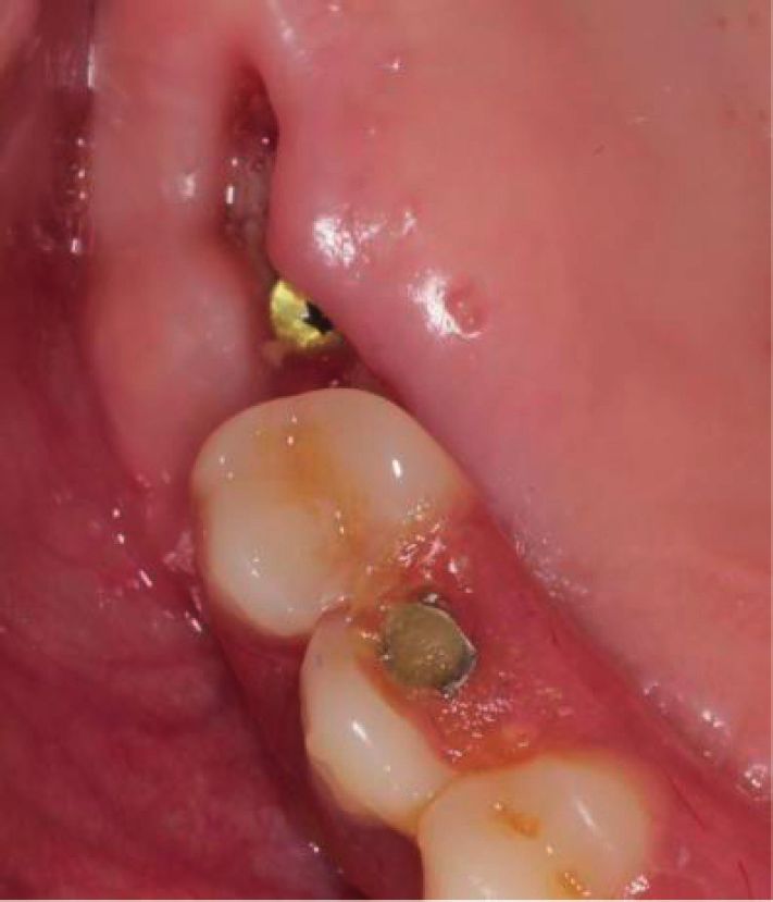
International Journal of Dentistry                                                                                                    3

                      Table 1: Quantitative characteristics of the patients according to age, gender, and nosology.
                                                                                                             Dental implants
                                                                 Primary implant         Age                 Gender
Diagnosis                                                                                                                       Total
                                                                 stability (N/cm2)     (years)     N (tooth        F (tooth    amount
                                                                                                   number)         number)
Partial edentulous maxilla delayed implantation (early                                                          5 (25, 26, 16,
                                                                      25–35            38–54     3 (26, 17, 17)                  8
loading with a prosthesis)                                                                                          16, 17)
Partial edentulous mandible delayed implantation (early                                                         5 (44, 47, 36,
                                                                      25–35            42–56       2 (36, 46)                    7
loading with a prosthesis)                                                                                          36, 37)
Partial edentulous maxilla delayed implantation (immediate
                                                                      35–45            41–46           1 (24)          1 (25)     2
loading with a prosthesis)
Partial edentulous maxilla (immediate postextraction
                                                                      30–35            34–40           1 (14)           —         1
implantation—early loading with a prosthesis)
Partial edentulous maxilla (immediate postextraction
                                                                      30–35            38–45            —         2 (46, 47)      2
implantation—early loading with a prosthesis)

                                     Table 2: Inclusion and exclusion criteria for the participants.
                                  Inclusion criteria                                              Exclusion criteria
            (i) Age > 21 years
            (ii) Absence of medical comorbidities
                                                                              (i) Inadequate oral hygiene
General     (iii) Absence of periodontal diseases
                                                                              (ii) Smoking
            (iv) Antagonist dentition
            (v) Availability for 20-week follow-up
            (i) Missing 1 or 2 teeth (included oral distal defects of the
                                                                              (i) Adjacent teeth with the presence of carious processes
            masticatory system—molars and premolars)
                                                                              (ii) Presence of periapical inflammation on adjacent teeth
            (ii) In the case of one-stage implantation, teeth with more
Local                                                                         (iii) Local inflammation of the periodontium
            than 80% decay or 3–4° mobility
                                                                              (iv) Mobility of adjacent teeth
            (iii) Plaque indicators throughout the oral cavity and bleeding
                                                                              (v) Mucosal disease
            indicators of less than 25%

          Figure 2: Prefabricated provisional abutment.                                   Figure 3: Cervico system.

    The customized provisional composite abutment is                        After the final setting in the Cervico system of the
fabricated using the Cervico system. The upper ring of the             necessary parameters for the fabrication of a customized
device is rotated until the desired size of the depression             provisional abutment, an analogue of the corresponding
matches the required size of the desired prosthetic con-               implantation system is fixed in the device. Its diameter is
nection at the base of the device. This information is                 completely identical to the dental implant, which will be
recorded in a special form, which in the future may be                 inserted into the bone tissue. The provisional abutment in
necessary for the orthopedic management of the patient. In             the analogue is fixed with an occlusal screw with a torque
addition to the selection of the shape for the future emer-            force of up to 15 N/cm2 (Figure 4).
gence profile, the Cervico device allows one to set the depth                A fluid light composite is introduced into the selected
of implant immersion in the bone of 0–4 mm (Figure 3).                 cell and illuminated with a polymerization lamp (Figure 5).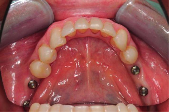
4                                                                                              International Journal of Dentistry

Figure 4: Placement of the prefabricated abutment into the
Cervico device.
                                                                       Figure 6: Customized provisional composite abutment.

                                                                   placement in an implant immediately, in case of achieving
                                                                   good primary implant stability, or after some time, with
                                                                   delayed implantation. In the latter case, an incision is made
                                                                   in the mucous membrane, an implant is found, the plug of
                                                                   the implant is unscrewed, the implant shaft is washed with
                                                                   an irrigation solution, and a gingiva former is placed, which
                                                                   is selected depending on the thickness of the mucosa that
                                                                   should rise no more than 1–2 mm above the gingiva. The
                                                                   gingiva former is necessary for the formation of soft tissues
                                                                   around the implant and for quick access to it during the
                                                                   prosthetics stage. The torque force when it is inserted into
                                                                   the implant is set to no more than 10 N/cm2.
                                                                       The standardized gingiva former does not completely
                                                                   recreate the contours of the future prosthesis and requires
                                                                   additional shaping of the gingiva with a provisional crown
                                                                   fabricated in a dental laboratory.
Figure 5: Stage of introducing the composite and polymerization.       In addition, there are laboratory methods for the fab-
                                                                   rication of customized titanium formers by the analogue
    After the composite has hardened, the customized               method, as well as current methods using CAD/CAM digital
provisional composite abutment is removed from the                 technologies.
Cervico device; the emergence profile is additionally finished
to ensure a smooth transition from the abutment neck to the        2.3. Statistical Analysis. The end point of the study was to
composite and then polished (Figure 6). After preliminary          determine the time of prosthetics from the digital impres-
assessment of the occlusal position, it is necessary to shorten    sion of the gingival profile to the final orthopedic rehabil-
the customized provisional composite abutment to the               itation for the developed technology and the conventional
antagonist teeth.                                                  approach using a prefabricated gingiva former. The implant
    A customized provisional composite abutment can be             was used as a statistical unit and analyzed. Statistical analysis
fabricated in advance before the surgery and then sterilized       was performed using NCSS 2020 software. Standard de-
in an autoclave at a pressure of 1.1 atm and a temperature of      scriptive methods such as median, frequency, minimum,
120°C for 45 minutes.                                              and maximum were used to determine sample character-
                                                                   istics. Quantitative data were compared between the groups
                                                                   using the Mann–Whitney U test and within groups using the
2.2. For Conventional Technology. The standardized gingiva         Wilcoxon test to assess the normality of the distribution. The
former is a titanium cylinder with a screw for insertion into      confidence interval was set at 95%.
the implant and is used for the formation of soft tissues
before the prosthetic stage. The gingiva former of many            3. Results
implant systems is available in diameters from 3 to 9 mm
(incisal, premolar, molar) and lengths from 1 to 7 mm. Most        All 20 patients underwent dental implant surgery with
are conical and cylindrical in shape. They are used for            primary stability of 25 to 45 N/cm2.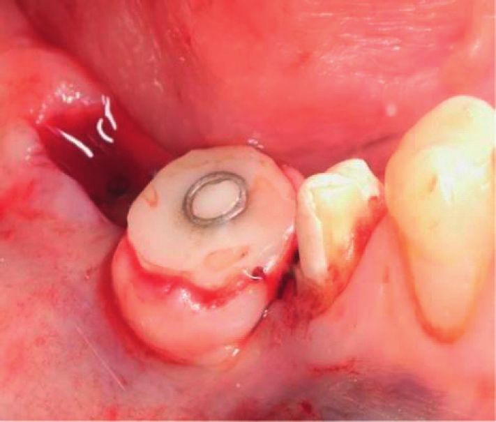
International Journal of Dentistry                                                                                           5

    Eleven patients received prefabricated gingiva formers,     emergence profile. The special program simulates a per-
ten of which had implants integrated into the mature bone.      manent implant-supported prosthesis. The prosthesis is
One patient received a prefabricated gingival former            fabricated of a biocompatible material—zirconium dioxide
inserted into the implant immediately after tooth extraction.   within three hours. A customized provisional composite
    Nine patients had a customized provisional composite        abutment is removed from the implant, the antiseptic ir-
abutment placed in the implant:                                 rigation of the internal shaft of the implant is performed, and
                                                                the permanent prosthesis is placed with a torque force of at
    (i) Immediately after tooth extraction—4 patients
                                                                least 30 N/cm2 (Figure 10).
   (ii) Delayed, in the mature bone—5 patients                      All this makes it possible in a few hours to provisionally
    The technique of fabrication and placement of the           restore a patient after surgery.
provisional composite abutment for one-stage implantation           The prefabricated gingiva former used in classical
immediately after tooth extraction included the following       delayed implantation in mature bone (Figure 11) has a small
stages (Figure 7):                                              diameter, which leads to the need for additional soft tissue
                                                                formation with a provisional crown within 7–14 days
     (i) After the placement of a customized provisional        (Figure 12).
         composite abutment into the implant area intra-            The time of placement of the dental prosthesis in the
         operatively, within 72 hours, its supragingival        implant ranged from 24 hours to 3 months. After 6–8 weeks,
         preparation using a turbine tip is performed           the X-ray control of the implant integration and assessment
    (ii) Scanning with a 3Shape intraoral scanner of a          of the emergence profile were performed.
         provisional composite abutment
   (iii) Modeling of the framework of a provisional pros-
         thesis and subsequent fabrication of a provisional     4. Discussion
         CAD/CAM crown from a PMMA (polymethyl                  The use of a standard gingiva former for immediate im-
         methacrylate) block                                    plantation leads to the need to suture the soft tissues with
   (iv) Treatment of a customized provisional composite         tension around the former, immobilizes the flap with ad-
         abutment with adhesive                                 ditional trauma, and, subsequently, results in soft tissue
    (v) Introducing a light composite into a provisional        deficiency.
         crown and gluing onto a customized composite               In particular, in immediate implantation in the masti-
         abutment                                               catory system, the space that is formed between the former
                                                                and the gingival flap negatively affects the tightness and,
   (vi) Final polymerization (occurs within 20 seconds)
                                                                subsequently, can cause delayed microbial contamination
    After the placement of the dental implant into the bone     around the implant, which is what happened to one patient
in the correct orthopedic position, including with respect to   (Figure 13).
the plane of the future suprastructure, a customized pro-           Lack of proper sealing can lead to loss of a clot or bone
visional composite abutment is placed on the implant in the     material placed in the space between the cortical plate and
oral cavity with a torque force on the screw of up to 15 N/     the implant. Tissue healing often occurs by secondary
cm2 (Figure 8).                                                 intention.
    The fit of the provisional abutment to the implant is            In both groups of patients, X-ray images were made
checked using an X-ray image (Figure 9). The shaft opening      directly during the surgery to check the accuracy of the
of the provisional abutment is closed with a Teflon tape and     placement of the superstructure with respect to the implant
sealed with a light composite. After the suprastructure has     platform.
been placed, nonresorbable sutures are placed to hold the           All participants were prosthetized with a provisional
flap around the customized provisional composite                 and, subsequently, with a permanent prosthesis, depending
abutment.                                                       on the primary bone stability of the implant.
    In the absence of primary implant stability (less than          Of the nine patients in the experimental group, 5 patients
25 N/cm2), the placement of a customized provisional            received early loading in the form of a provisional prosthesis
composite abutment is performed 2–6 months after the final       after the healing stage and final integration of the implant
implant integration is achieved.                                (6–8 weeks). The remaining four patients received early (2
    After the customized provisional composite abutment         patients) and immediate (2 patients) loading with a provi-
has formed the emergence profile and the implant has             sional crown on the day of surgery.
achieved the required final integration, as a rule, with early       At the stage of examination of the patients who un-
loading, there follows the transition to the stage of fabri-    derwent classical delayed implantation with a customized
cation of a permanent prosthesis, bypassing the provisional     provisional composite abutment (5 patients), on the 3rd–5th
crown, using digital technologies.                              day, it was determined that the wound healing occurred by
    Once the soft tissues have been formed, the provisional     primary intension and the sutures were good. They were
composite abutment is carefully removed with a torque key       removed on the 10–14th day. The epithelization of soft
and the emergence profile is assessed. A scan marker is          tissues was complete, and no inflammation was detected.
installed in the implant to scan the area of the formed         X-ray images taken to check the integration after 6 weeks
6                                                                                               International Journal of Dentistry

                  (a)                          (b)                          (c)                             (d)

Figure 7: Stages of application of a customized composite abutment with immediate loading: (a) preparation, (b) checking the fit, (c)
modeling of the provisional crown in the program, and (d) placement of the provisional crown.

                              Figure 8: Intraoperative placement of a provisional composite abutment.

                                                                    provisional composite abutment immediately after tooth
                                                                    extraction.
                                                                        X-ray images show the formation of the bone matrix
                                                                    after 6 weeks (Figure 15).
                                                                        Objectively, the soft tissues around the implant placed
                                                                    immediately are stable, tightly adhering to the customized
                                                                    provisional composite abutment. The sutures in 5–7 days
                                                                    after the implant placement are good; no tissue inflam-
                                                                    mation was detected.
                                                                        In the comparison group, consisting of 11 patients, a
                                                                    dental implantation surgery was performed and pre-
                                                                    fabricated gingiva formers were placed. Ten of them un-
                                                                    derwent delayed implantation in mature bone. One person
                                                                    in this group had an implant placed immediately after a
                                                                    tooth extraction. The primary stability of the implants
                                                                    ranged from 25 to 30 N/cm2.
Figure 9: Checking the fit of the provisional composite abutment.        X-rays after 6 weeks showed marginal resorption in one
                                                                    of 10 patients with an implant in the mature bone with a
                                                                    gingiva former (Figure 16).
showed no abnormalities in bone regeneration around the                 In 11 patients, the implants were placed in mature bone
implants (Figure 14).                                               and the delayed implantation with early loading after 6–8
   It is worth noting the absence of soft tissue inflammation        weeks was carried out. To shape the emergence profile, a
around the implants in four patients from the experimental          provisional milled crown was fabricated in the dental
group; the implants were placed together with a customized          laboratory.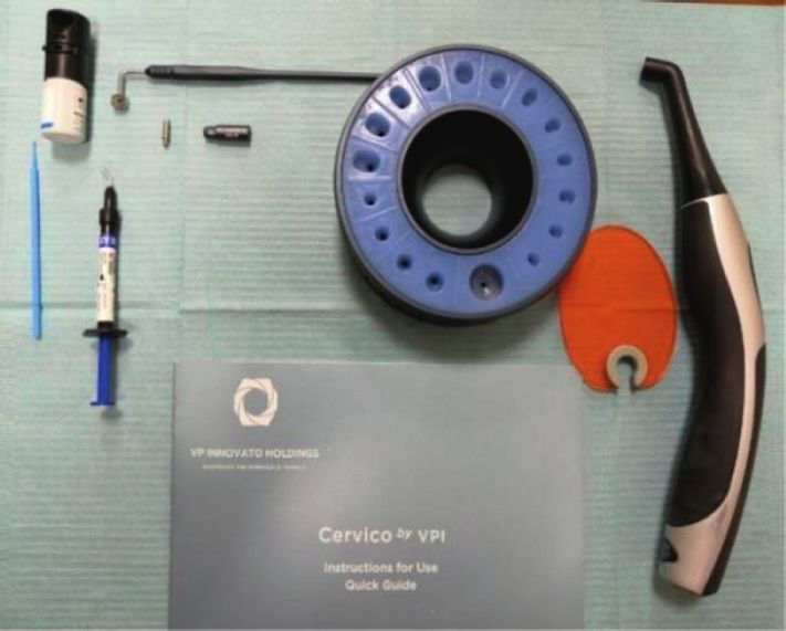
International Journal of Dentistry                                                                                                            7

          (a)                     (b)                       (c)                    (d)                     (e)                     (f )

                                                                      (g)

Figure 10: Stages of fabrication of a permanent prosthesis after the formation of the emergence profile with a customized composite
abutment: (a) planning, (b) a customized composite abutment, (c) the abutment placement (3 weeks after surgery), (d) formation of the
emergence profile (10 weeks), (e) intraoral scanning, (f ) planning of a permanent prosthesis in the program, and (g) placement of a
permanent prosthesis.

                                                 Figure 11: Placement of gingiva formers.

                  (a)                                 (b)                                 (c)                               (d)

Figure 12: Stages of soft tissue formation: (a) immediately after surgery, (b) after 6 to 8 weeks (with prefabricated gingiva former), (c) after
10 weeks (with provisional crowns), and (d) after 12 weeks of provisional crowns.

    The remaining four patients underwent immediate                         the following features on day 4–5 of the postoperative
postextraction implantation with the placement of pre-                      examination:
fabricated gingiva formers. Immediate loading with the
                                                                               (i) Soft tissues around the implant do not fit tightly in
prosthesis on the implants on the day of surgery was not
                                                                                   the zone of the gingiva former
performed.
    The only patient in the comparison group who under-                        (ii) The wound surface healing occurs by secondary
went immediate implantation with a gingiva former showed                            intention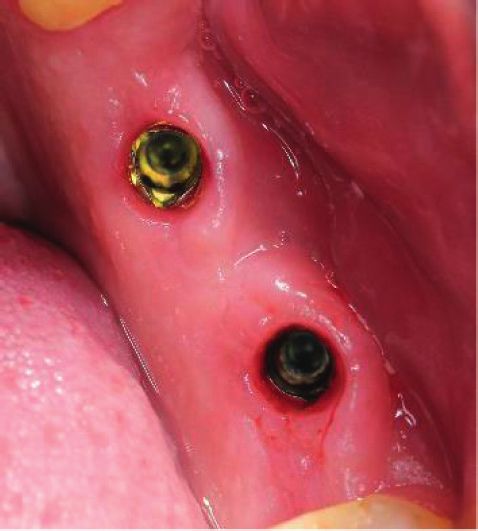
8                                                                                          International Journal of Dentistry

                                     Figure 13: Microbial contamination of soft tissues.

                                         Figure 14: Bone regeneration after 6 weeks.

                                          Figure 15: Formation of the bone matrix.

    Comparative characteristics of positive and negative        composite abutment. It is not always possible to achieve
aspects of the conventional and new methods are shown in        guaranteed success in soft tissue healing around gingiva
Table 3.                                                        formers.
    The risk of bacterial contamination of the implant area        The use of gingiva formers at the prosthetic stage is
after the placement of prefabricated gingiva formers is         accompanied by the formation of a small-diameter gingival
higher than that when using a customized provisional            profile. Subsequently, after the final implant integration, this
International Journal of Dentistry                                                                                                    9

                                             (a)                                           (b)

    Figure 16: Dynamics of bone resorption after the placement of a gingiva former: (a) the moment of surgery and (b) 6 weeks.

               Table 3: Comparative characteristics of positive and negative aspects of the conventional and new methods.
                                        Comparison group with gingiva former (11       Experimental group with provisional composite
                                                        patients)                                  abutment (9 patients)
Parameters                               Placement in     Immediate placement in         Placement in      Immediate placement in
                                        mature bone (10      implant after tooth        mature bone (5        implant after tooth
                                           patients)        extraction (1 patient)         patients)        extraction (4 patients)
Microbial contamination                  + (1 patient)                +                        –                       –
Bone augmentation loss                         –                      +                        –                       –
Narrow gingival profile requiring
                                                   +                   +                          –                     –
further shaping
Increased adhesion to plaque                       –                   –                          +                     +
The need for additional tissue
                                                   –                   +                          –                     –
immobilization
Tight sealing of the soft tissue-bone
                                                   +                   –                          +                     +
space
Quick formation of the necessary
gingival profile (according to the                  –                   –                          +                     +
shape of the tooth)
Increased requirements for the
                                                   –                   –                          +                     +
dentist’s manual skills
The possibility of obtaining
additional tissue volume through                   –                   –                          +                     +
grafting
Saving orthopedic rehabilitation
                                                   –                   –                          +                     +
time (7–21 days)
Note. +: yes; −: no.

                                         Table 4: Timing of the final orthopedic rehabilitation.
                                                        Number of implants and time of prosthetics in hours (from digital impression of
                                                                    the gingival profile to final orthopedic rehabilitation)
Patients                                                   Delayed implantation (early                    One-stage implantation
                                                                    loading)                           (immediate and early loading)
                                                            M                      F                      M                       F
                                  Mandible               1–84 ± 12            1–120 ± 12                  —                   2–72 ± 8
Experimental group
                                  Maxilla               1–104 ± 18             2–96 ± 12               1–60 ± 8              1–84 ± 10
                                  Mandible              1–432 ± 72            4–360 ± 60                  —                      —
Comparison group
                                  Maxilla               2–384 ± 68            3–360 ± 60              1–408 ± 72                 —

leads to additional formation of the gingival profile by a             the plaque due to the greater specific surface area of the
provisional crown, which increases the time of prosthetics.           porous, slightly roughened structure.
    The main minor disadvantage of using a customized                     This disadvantage is more than compensated by the
provisional composite abutment is the increased adhesion to           advantages of a customized provisional abutment: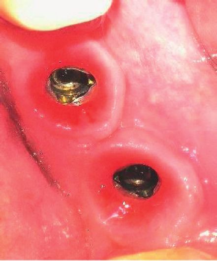
10                                                                                                  International Journal of Dentistry

      (i) The ability to seal the soft tissue-bone space               [2] A. A. Kulakov and A. Zh. Ashuev, “Immediate implantation
     (ii) No additional immobilization of tissues at the stage             and the role of early functional loading of the implant (ex-
                                                                           perimental study),” Stomatologiya, vol. 86, no. 1, pp. 23–27,
          of suturing a tooth socket in the case of immediate
                                                                           2007.
          implantation
                                                                       [3] T. G. Robustova and I. V. Fedorov, “Immediate implantation
     (iii) Quick formation of the necessary gingival profile,               technique for tooth extraction,” Problemy Stomatologii I
                                                                           Neirostomatologii, no. 1, pp. 34–38, 1998.
           taking into account the shape of the future
                                                                       [4] W. Becker, “Immediate implant placement: treatment plan-
           prosthesis
                                                                           ning and surgical steps for successful outcomes,” British
     (iv) No need for a provisional laboratory crown                       Dental Journal, vol. 201, no. 4, pp. 199–205, 2006.
      (v) Quick orthopedic rehabilitation of the patient               [5] E. G. Zuiderveld, H. J. A. Meijer, A. Vissink, and
                                                                           G. M. Raghoebar, “Immediate placement and provisionali-
    Table 4 shows comparative data on the timing of the final               zation of an implant after removal of an impacted maxillary
soft tissue formation for prosthetics in patients in the ex-               canine: two case reports,” International Journal of Implant
perimental group and the comparison group.                                 Dentistry, vol. 1, no. 1, p. 13, 2015.
    The above data show that the developed technology                  [6] L. Sheng, T. Silvestrin, J. Zhan et al., “Replacement of severely
makes it possible to spend much less time for orthopedic                   traumatized teeth with immediate implants and immediate
rehabilitation of patients. In the experimental group, there               loading: literature review and case reports,” Dental Trau-
was a statistically significant decrease in the time from                   matology, vol. 31, no. 6, pp. 493–503, 2015.
delayed implantation to one-stage implantation (Wil-                   [7] V. O. Samusenkov, Clinical and Microbiological Substantia-
coxon sign-rank test; p < 0.01). However, there was no                     tion of Provisional Prosthetics with Immediate Dental Im-
                                                                           plantation: Dissertation Abstract–Moscow, p. 24, Publishing
significant difference between timing depending on im-
                                                                           Moscow State University of Medicine and Dentistry of A.I.
plant placement—mandible or maxilla—in either group
                                                                           Evdokimov, Moscow, Russia, 2012.
(Wilcoxon sign-rank test, p > 0.05; Mann–Whitney U test;               [8] B. Tarazona, P. Tarazona-Álvarez, D. Peñarrocha-Oltra, and
p > 0.05).                                                                 M. Peñarrocha-Diago, “Relationship between indication for
    In the comparison group, the timing did not virtually                  tooth extraction and outcome of immediate implants: a
depend on the type of implantation—delayed or single—                      retrospective study with 5 years of follow-up,” Journal of
stage (Wilcoxon sign-rank test, p > 0.05) and was at least 360             Clinical and Experimental Dentistry, vol. 6, no. 4, pp. e384–8,
hours. This postpones the moment of orthopedic rehabili-                   2014.
tation of the patient, which can be avoided when using a               [9] A. Pozzi and P. Mura, “Immediate loading of conical con-
customized provisional composite abutment fabricated with                  nection implants: up-to-2-year retrospective clinical and ra-
the Cervico system.                                                        diologic study,” The International Journal of Oral &
                                                                           Maxillofacial Implants, vol. 31, no. 1, pp. 142–152, 2016.
                                                                      [10] J. Lee, D. Park, K.-T. Koo, Y.-J. Seol, and Y.-M. Lee,
5. Conclusions                                                             “Comparison of immediate implant placement in infected and
                                                                           non-infected extraction sockets: a systematic review and
The use of modern technologies for crown fabrication on an
                                                                           meta-analysis,” Acta Odontologica Scandinavica, vol. 76, no. 5,
implant by a direct digital method and the application of a
                                                                           pp. 338–345, 2018.
customized provisional composite abutment made it pos-                [11] S. Ramalingam, M. Al-Hindi, R. A. Al-Eid, and N. Nooh,
sible to significantly reduce the time of the patient’s pros-               “Clinical evaluation of implant survival based on size and site
thetic rehabilitation. Other advantages of the developed                   of placement: a retrospective study of immediate implants at
technology are the reduction in bacterial contamination in                 single rooted teeth sites,” The Saudi Dental Journal, vol. 27,
the bone formation zone, minimization of soft tissue is-                   no. 2, pp. 105–111, 2015.
chemia, acceleration of mucogingival and bone integration,            [12] Vorobiev A. A., Shemonaev V. I., Mikhalchenko D. V.,
and rapid formation of the desired emergence profile of the                 Velichko A. S. A view of the problem of dental implantation in
future prosthesis.                                                         the light of the current scientific concepts, bulletin of the
                                                                           volgograd scientific center of the russian academy of medical
                                                                           sciences 2, 2009. P. 19-24.
Data Availability                                                     [13] L. Schropp, L. Kostopoulos, and A. Wenzel, “Bone healing
                                                                           following immediate versus delayed placement of titanium
The data used to support the findings of this study are
                                                                           implants into extraction sockets: a prospective clinical study,”
available within the article.
                                                                           The International Journal of Oral & Maxillofacial Implants,
                                                                           vol. 18, pp. 189–199, 2003.
Conflicts of Interest                                                 [14] I. N. Dashevskii and P. S. Shushpannikov, “Dependence of the
                                                                           primary dental implant stability on the direction of the oc-
The authors declare that they have no conflicts of interest.                clusal loading,” Russian Journal of Biomechanics, vol. 23, no. 3,
                                                                           pp. 391–399, 2019.
References                                                            [15] H.-L. Huang, J.-T. Hsu, L.-J. Fuh, M.-G. Tu, C.-C. Ko, and
                                                                           Y.-W. Shen, “Bone stress and interfacial sliding analysis of
 [1] A. A. Kulakov and F. M. Abdullaev, “Immediate implantation            implant designs on an immediately loaded maxillary implant:
     in experiment and clinic,” Klinicheskaya Stomatologiya, no. 1,        a non-linear finite element study,” Journal of Dentistry,
     pp. 48–52, 2002.                                                      vol. 36, no. 6, pp. 409–417, 2008.
International Journal of Dentistry 11
[16] A. A. Lukyanenko and I. A. Kazantseva, “The experience of
using the resonant frequency method to assess the stability
and osseointegration of dental implants,” Sovremennye
problemy nauki i obrazovaniya, vol. 4, pp. 291–298, 2014.
[17] S. Yu. Ivanov, A. A. Muraev, E. A. Rukina, and A. A. Bunev,
“Method for immediate dental implantation,” Sovremennye
problemy nauki i obrazovaniya, vol. 5, pp. 230–245, 2015.
[18] N. A. Panakhov and T. G. Makhmudov, “The level of dental
implant stability at different periods of functioning//,”
Problemy stomatologii, vol. 14, no. 1, pp. 89–93, 2018.
[19] G. Mistry, O. Shetty, S. Shetty, and R. Singh, “Measuring
implant stability: a review of different methods,” Journal of
Dental Implants, vol. 4, no. 2, pp. 165–169, 2014.
[20] A. Marković, J. L. Calvo-Guirado, Z. Lazić et al., “Evaluation
of primary stability of self-tapping and non-self-tapping
dental implants,” A 12-Week Clinical Study Clinical Implant
Dentistry and Related Research, vol. 15, no. 3, pp. 341–349,
2013.
[21] H.-S. Ryu, C. Namgung, J.-H. Lee, and Y.-J. Lim, “The in-
fluence of thread geometry on implant osseointegration under
immediate loading: a literature review,” The Journal of Ad-
vanced Prosthodontics, vol. 6, no. 6, p. 547, 2014.
[22] G. Finelle and S. Lee, “Guided immediate implant placement
with wound closure by computer-aided design/computer-
assisted manufacture sealing socket abutment: case report,”
The International Journal of Oral & Maxillofacial Implants,
vol. 32, no. 2, pp. e63–e67, 2017.You can also read