Fact sheet Coronary heart disease - CPPE
←
→
Page content transcription
If your browser does not render page correctly, please read the page content below
Fact sheet
Coronary heart disease

Contents

Definition                               2

Prevalence and incidence                 4

Signs and symptoms                       4

Causes/risks factors                     5

Pathophysiology (mechanism of disease)   6

Prognosis and complications              8

Diagnosis/detection                      8

Pharmacological treatment                9

Non-pharmacological treatment            10

Patient support                          12

Further resources                        12

External websites                        13

References                               13

Page 1

Fact sheet
Coronary heart disease
Please note that links within this page may take you directly to the British Heart Foundation website where
you can find person-centred information on heart conditions and related terms.
Definition
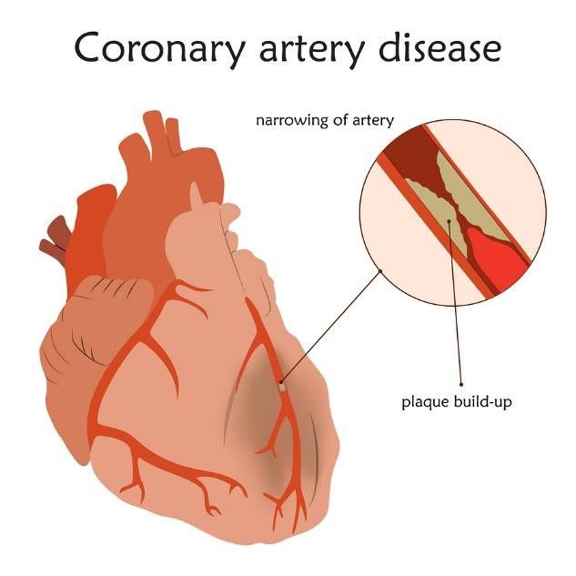
Coronary heart disease (CHD – also referred to as coronary artery
disease or ischaemic heart disease) is a type of cardiovascular
disease (CVD) where oxygenated blood is unable to perfuse the
myocardium (muscle tissue of the heart) due to narrowing of the
coronary arteries.
Most commonly this narrowing of the coronary arteries is a result of
atherosclerosis.1
Atherosclerosis is a condition where plaques form within the lining of the arteries. These plaques are
formed of white blood cells, lipids (cholesterol and fatty acids), calcium, and fibrous connective tissue. This
is collectively known as atheroma. Atheromatous plaques cause the lumen of the arteries to narrow and
blood flow to become restricted.
Sometime atheromatous plaques in the coronary arteries can rupture, causing a blood clot to form
(coronary thrombus). If this happens, the blood flow in that artery may be partially or completely blocked.2
Lumen
There are three main presentations of CHD: angina, myocardial infarction and heart failure. We will not be
covering heart failure on this page as it will be covered separately.
Angina
Angina is chest pain, caused by an insufficient blood supply to the myocardium. Pain may also be
experienced in the neck, shoulders, jaw, or arms.2,3
Page 2
Fact sheet
Coronary heart disease

Although commonly a symptom of CHD, angina is less commonly a result of a heart valve disease (for
example, aortic stenosis – narrowing of the aortic valve opening), hypertrophic cardiomyopathy, atrial
fibrillation, anaemia or hypertensive heart disease (raised pressure inside the heart).2 The British Heart
Foundation (BHF) also describes microvascular angina and coronary artery spasm.

Angina is classified as either the more common stable angina (angina pectoris) or more serious unstable
angina.4 The National Institute of Health and Care Excellence (NICE) clinical knowledge summary (CKS)
Angina states the following:
‘Stable angina usually occurs predictably with physical exertion or emotional stress, last for no more than
ten minutes (usually less) and is relieved within minutes of rest, as well as sublingual nitrates.
Unstable angina is new onset angina or abrupt deterioration in previously stable angina, often occurring at
rest. Unstable angina usually requires immediate admission or referral to hospital.’2

Myocardial infarction
Myocardial infarction (MI) is often referred to as a heart attack. It is defined as the death (necrosis) of an
area of myocardium due to an inadequate supply of oxygenated blood.

MI is usually caused by atheromatous plaque rupturing and forming a thrombus, as discussed under
atherosclerosis. Although it should be noted that a coronary thrombosis does not always cause an MI.

The size of thrombus and extent to which it affects blood flow affects whether it leads to no necrosis, a
relatively small amount of necrosis, or a more extensive area of necrosis. Collectively, this range of effects
is called acute coronary syndrome (ACS).5

Acute coronary syndrome (ACS)
NICE describes the way in which ACS is sub-divided as follows:

ST-elevation ACS (STE-ACS). This is typically caused by a complete blockage of an artery. ST relates to
the changes observed on an electrocardiogram (ECG); in this case, there is persistent elevation of the ST
wave. Most people with this type of ASC develop an ST-elevation MI (STEMI).

Non-ST-elevation ACS (NSTE-ACS). This typically reflects a partial or intermittent blockage of an artery.
Here the ECG does not show persistent ST-segment elevation, although there may be other ECG changes
or no changes.

NSTE-ACS is further divided into:
  • non-ST-elevation MI (NSTEMI): there is a rise in the blood troponin level
  • unstable angina: blood troponin level does not rise.5

ECGs show the electrical activity of the heart. In order to understand what this means, it is important to
understand the conduction pathways of the heart. If you would like to learn more about this, then watch the
following video by Handwritten Tutorials:

Page 3
Fact sheet
Coronary heart disease

Handwritten Tutorials – Conduction pathway of the heart

Return to contents

Prevalence and incidence
The BHF publishes data on mortality, morbidity, treatment and costs associated with cardiovascular
disease. They state that ‘CHD is the one of the UK’s leading causes of death’ and ‘it is also the leading
cause of death worldwide.’6

The BHF states that in England in 2015 there were 56,493 deaths due to CHD. Of these, 34,103 were men
and 22,390 were women. The total number of deaths is similar to the number attributable to dementia and
Alzheimer's (58,299), but less than those attributable to cancer (138,509) and respiratory disease
(70,111).7

For further information, visit the BHF’s Heart statistics publications page.

Return to contents

Signs and symptoms
Stable angina
The NHS outlines the following as symptoms of angina:
   • ‘feels tight, dull or heavy – it may spread to your left arm, neck, jaw or back
   • is triggered by physical exertion or stress
   • stops within a few minutes of resting’.4

ACS
The NHS outlines the following as symptoms for a heart attack, which differ considerably from the
symptoms listed for angina:
   • ‘chest pain – a sensation of pressure, tightness or squeezing in the centre of your chest
   • pain in other parts of the body – it can feel as if the pain is travelling from your chest to your arms
      (usually the left arm is affected, but it can affect both arms), jaw, neck, back and abdomen
   • feeling lightheaded or dizzy
   • sweating
   • shortness of breath

Page 4
Fact sheet
Coronary heart disease

   •     feeling sick (nausea) or being sick (vomiting)
   •     an overwhelming sense of anxiety (similar to having a panic attack)
   •     coughing or wheezing.’8

The NICE CKS Chest pain states to suspect ACS, if:
   • ‘Pain in the chest or other areas (for example the arms, back, or jaw) lasts longer than 15 minutes.
   • Chest pain is:
         o Associated with nausea and vomiting, sweating or breathlessness, or a combination of
             these.
         o Associated with haemodynamic instability (for example the person has a systolic blood
             pressure less than 90 mmHg).
         o Of a new-onset, or is the result of an abrupt deterioration of stable angina; with pain
             occurring frequently with little or no exertion, and often lasting longer than 15 minutes.
   • Do not use the person's response to glyceryl trinitrate to confirm or exclude a diagnosis of acute
      coronary syndrome.’

Note that generally the term haemodynamic instability is used to describe abnormal or unstable blood
pressure.

Return to contents

Causes/risk factors
Risk factors for CHD due to atheromatous plaques correlate with many of the risk factors for the overall
development of CVD. They can be classified as modifiable or non-modifiable.

Modifiable risk factors
  • Low blood level of high-density lipoprotein cholesterol (HDL-C)
  • High blood level of non-HDL-C
  • Hypertension
  • Diabetes
  • Smoking
  • Being overweight/obese
  • Inactivity
  • Unhealthy diet
  • Excessive alcohol consumption
  • Excessive stress9,10,11

For more information about modifiable risk factors, access Heart UK’s Risk factors for coronary heart
disease (CHD).

NHS Health Check is a scheme that ‘aims to promote and improve the early identification and
management of individual behavioural and physiological risk factors for vascular disease and the other
associated conditions.’12 For more information about NHS Health Checks, access CPPE’s Health Checks
gateway page.

Page 5
Fact sheet
Coronary heart disease
For more information on the prevention of CVD access, the NICE public health guideline Cardiovascular
disease prevention [PH25].
Non-modifiable risk factors
• Increasing age
• South Asian ethnic background and those with an African Caribbean background are at greater risk
of hypertension
• Family history of heart disease (family history of hypertension or hypercholesterolaemia increases
risk of developing these risk factors)9,10,11
Return to contents
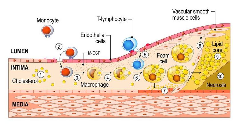
Pathophysiology (mechanism of disease)
As discussed in the above definition section, CHD is very often due to atherosclerosis and narrowing of the
coronary arteries.
Atherosclerosis is thought to be driven by a chronic inflammatory process within the arterial wall.13 The
diagram below shows a simplified outline of the way in which it is believed that atherosclerosis occurs.
Each step is numbered and the numbers correspond to the descriptions below.
Step 1 – it is thought that atherosclerosis starts when low-density lipoproteins (LDL), a type of cholesterol,
accumulate in the intima layer of the coronary artery wall, under the layer of endothelial cells
(endothelium) on its inner surface.14
Page 6
Fact sheet
Coronary heart disease

Step 2 – it is thought that an oxidised form of LDL (ox-LDL) plays an important role in the formation of
atheromatous plaques by increasing inflammation.15 The ox-LDL ‘activates’ the endothelial cells causing
monocytes (a type of white blood cell) to adhere (stick) to them.14,15

Step 3 – the monocytes (which can differentiate, or change into, other types of cells when responding to
stimuli) differentiate into macrophages (type of white blood cell).
Step 4 – the macrophages start to take up the cholesterol.16

Step 5 – T-lymphocytes (a type of white blood cell) enter the intima. Their role in atherosclerosis is not
fully understood, but it is thought that these cells can modulate the type and intensity of the inflammatory
response and may increase the activity of the macrophages.17

Step 6 – due to the uptake of cholesterol the macrophages differentiate into foam cells. These foam cells
are too large to pass back out of the intima through the endothelial cells. They stay in the intima and
stabilise the chronic inflammation.14 The accumulation of foam cells is known as a fatty streak.18

Steps 7 and 8 – the complex inflammatory process is thought to cause vascular smooth muscle cells to
migrate from the media layer towards the endothelial cells.19 When these smooth muscle cells reach the
endothelial cells, they produce fibrous tissue which forms a protective fibrous cap to try and contain the
plaque.20

Step 9 – a lipid core made from cholesterol develops. It is thought that this results from a combination of
lipids being deposited when foam cells undergo apoptosis (natural cell death) and also the aggregation of
free lipids within the media.21

Step 10 – when cells that have undergone apoptosis are not cleared away properly (due to defective
efferocytosis) this causes a necrosis to occur and a necrotic core forms.22

The lipid core and necrosis cause the plaque to swell, additionally the fibrous cap of the plaque can start to
degrade. This can result in the plaque rupturing, at which point platelets stick to the contents of the plaque
and form a blood clot.22 Atheromatous plaques that are at risk of rupture tend to have a thin fibrous cap, a
high lipid content, few smooth muscle cells, and a high proportion of macrophages and monocytes.23 This
blood clot can block the entire artery, meaning the oxygen-rich blood doesn’t reach the myocardium.

Stable angina
Atheromatous plaques affect the flow of blood through the coronary arteries; they also disrupt the
endothelium and affect its ability to fully dilate in response to increased oxygen demand.24 When the
demand for oxygenated blood is higher than the supply (without leading to necrosis), for example during
exercise, this can cause the pain experienced during stable angina.

ACS
If the atheromatous plaque changes acutely, this can trigger ACS. Changes can include endothelial
dysfunction, platelet aggregation, and spasm leading to plaque erosion, rupture, haemorrhage, and
thrombosis.25 STEMI is associated with major blockages to coronary arteries and a relatively large amount
of damage. NSTEMI is associated with less myocardial damage than STEMI, which may be due to there

Page 7
Fact sheet
Coronary heart disease

being a partial blockage or a smaller vessel being affected. Unstable angina is associated with a partial
blocking of an artery.26

When blood flow to the myocardium is reduced or blocked for around 30 to 60 minutes, this can lead to
cardiomyocyte (heart muscle cell) damage. This damage leads them to release a protein called troponin.

Return to contents

Prognosis and complications
NICE CKS offer excellent summaries of the prognosis for angina, Angina, prognosis, and the prognosis
and complications associated with an MI, MI – secondary prevention, Complications and prognosis.

A serious complication of CHD is cardiac arrest. For more information about cardiac arrest, visit the BHF’s
Cardiac arrest page.

Post MI, a process called cardiac (or ventricular) remodelling may occur. This can result in left ventricular
(LV) dysfunction and heart failure. For more information about remodelling and its prevention, access the
following articles:

The American College of Cardiology Foundation’s Causes and prevention of ventricular remodeling
after MI article.

Heart’s Left ventricular dysfunction: causes, natural history, and hopes for reversal.

Return to contents

Diagnosis/detection
Stable angina
Stable angina is diagnosed through clinical assessment, recognition of relevant risk factors and potentially
an ECG. Those who have experienced chest pain, but do not require a hospital admission, should be
referred to a specialist chest pain service to confirm or exclude angina.27 At this point a diagnostic test such
as a computerised tomography (CT) coronary angiogram, non-invasive functional testing or a percutaneous
coronary angiogram may be performed.28

NICE clinical guidance Recent-onset of chest pain of suspected cardiac origin: assessment and
diagnosis [CG95] recommends that the typicality of chest pain is assessed as follows:
   • ‘Presence of three of the features below is defined as typical angina.
   • Presence of two of the three features below is defined as atypical angina.
   • Presence of one or none of the features below is defined as non-anginal chest pain.
Anginal pain is:
   • constricting discomfort in the front of the chest, or in the neck, shoulders, jaw or arms
   • precipitated by physical exertion
   • relieved by rest or GTN within about five minutes.’

The reason for this classification is to help clinicians determine the likelihood of pain being related to CHD;
it can also be used to help determine which tests should be undertaken to aid a diagnosis.

Page 8
Fact sheet
    Coronary heart disease

    ACS
    NICE quality standard Acute coronary syndromes in adults [QS68] states that ‘Acute myocardial
    infarction can have a poor prognosis so prompt and accurate diagnosis is important to ensure that
    appropriate treatment and care is offered as soon as possible.’

    ACSs are medical emergencies.29 The BHF and NHS both advise people who are experiencing the
    symptoms of a heart attack to immediately dial 999.30,31

    Diagnosis of ACS is made by taking a clinical history, conducting an assessment, taking a 12-lead ECG
    and can be aided by measuring blood troponin level.28

    More information about diagnosis and assessment of those who present with chest pain can be found in
    NICE guideline Recent-onset chest pain of suspected cardiac origin: assessment and diagnosis
    [CG95].

    Return to contents

    Pharmacological treatment
    Stable angina
    Read Sections 1.3 General principles for treating people with stable angina and 1.4 Anti-anginal drug
    treatment of NICE clinical guidance Stable angina: management [CG126] that outline which antianginal
    therapies should be offered, in which order they should be offered, and the additional add-on therapies
    which are recommended.

    Additionally the Scottish Intercollegiate Guidelines Network (SIGN) national clinical guideline Management
    of stable angina [SIGN 151] provides evidence-based recommendations and best practice guidance on the
    management of patients with stable angina.

    ACS
    Initial management
    The following NICE guidance outlines recommendations for early treatment options in those presenting with
    ACS:
•
    NICE clinical guideline Acute coronary syndromes [NG185]

    Ongoing management
    Those who experience ACS are usually offered the following therapies:32,33,34
       • angiotensin-converting enzyme inhibitor (ACE inhibitor) to reduce the cardiac remodelling (changes
          in ventricular size and shape)
       • dual antiplatelet therapy (aspirin plus a second antiplatelet agent) to reduce the risk of coronary
          thrombosis
       • beta-blocker to reduce heart rate, limit myocardial oxygen demand and reduce the incidence of
          arrhythmias and cardiac death35
       • high-potency statin to reduce LDL levels.

    Page 9
Fact sheet Coronary heart disease Read Section 1.4 Drug therapy for secondary prevention which outlines the standard pharmacological therapies and refers to further guidance as necessary. Age and Ageing’s article, Acute coronary syndrome management in older adults: guidelines, temporal changes and challenges, explores the management of older people who have experienced ACS and the available guidance. British National Formulary (BNF) The BNF offers a treatment summary for the initial and long-term management of Acute coronary syndromes which also links to the individual monographs for each recommended medicine. The BHF’s Tests information booklet offers simple explanations about which tests are commonly offered to help diagnose heart diseases. Return to contents Non-pharmacological treatment There are several non-pharmacological treatment options which can be offered to those with both stable angina and ACS. Information and support As part of long-term management, information and support can be provided. This should include information about a person’s condition and the way it may affect their lifestyle. The person should also be encouraged to ask questions about their condition and be offered support to manage it. Information about how to reduce cardiovascular risk should be offered.36,37 Detailed information about the recommended lifestyle advice can be found on the NICE CKS MI - secondary prevention, Lifestyle advice page. This information also applies to those who have stable angina.36,37 Cardiac rehabilitation Cardiac rehabilitation involves exercise and information, and should be offered to all patients who experience ACS. For more information, you can watch the following BHF video, which can be found on their Cardiac rehabilitation page: Page 10
Fact sheet
Coronary heart disease

British Heart Foundation - Joining a cardiac rehabilitation programme

For a person’s perspective of cardiac rehabilitation, access Moving forward after a heart attack - Mark's
story.
Coronary angioplasty and stents
Coronary angioplasty, which is also known as percutaneous coronary intervention (PCI) or percutaneous
transluminal coronary angioplasty (PTCA), can be used to improve symptoms of angina or in an emergency
situation to open a blocked coronary artery and hence re-perfuse the heart.

Watch the BHF video Your guide to angioplasty and stents, which can be found on their Coronary
angioplasty and stents page, to learn about what happens during an angioplasty and the use of stents.

The location of the stents may be described in terms of which coronary artery they have been placed in.
For more information about the anatomy of the coronary arteries, visit the Heart & Vascular Institute’s
Anatomy and Function of the Coronary Arteries page. In most circumstances a drug-eluting stent is
used.

Page 11
Fact sheet
Coronary heart disease

The technology for stents is still evolving and knowing the type of stent used is important for deciding on
the duration of dual antiplatelet therapy. Dual antiplatelet therapy should be offered in accordance with the
stent’s instructions for use (IFU), which are device-specific.38

Coronary bypass surgery
Coronary bypass surgery involves grafting a blood vessel to bypass a blockage in a coronary artery. The
graft is referred to as a coronary artery bypass graft (CABG).

Watch the BHF’s video Your guide to Coronary Bypass Surgery, heart disease treatment which can be
found on their Coronary bypass surgery page to learn about what happens during coronary bypass
surgery.

Return to contents

Patient support
The BHF offers information about specific conditions and support for those with heart conditions. More
information can be found on their Information & support page.

The NHS has a dedicated Coronary heart disease page with information on symptoms, causes,
diagnosis, treatment, recovery and prevention.

Return to contents

Further resources
CPPE’s Ischaemic heart disease gateway page contains further learning on stable angina, smoking
cessation and links to NICE guidance.

The following Geriatric medicine article looks at the management of angina in the elderly population
specifically, Angina: management in the elderly.

The European Society of Cardiology’s Antiplatelets therapy, Antiplatelet therapy in ischemic heart
disease explores the differences between antiplatelet therapies.

Return to contents

Page 12
Fact sheet
Coronary heart disease

External websites
CPPE is not responsible for the content of any non-CPPE websites mentioned on this page or for the
accuracy of any information to be found there.

All web links were accessed on 30 January 2020.

Return to contents

References
   1. Waller B. Chapter 35. Nonobstructive atherosclerotic and nonatherosclerotic coronary heart
       disease. In: Hurst’s The Heart. McGraw-Hill Education. 2011.
   2. National Institute for Health and Care Excellence. Clinical knowledge summary. Angina. Definition.
       Revised November 2020.
   3. British Heart Foundation. Angina – Causes, symptoms & treatments.
   4. NHS. Angina. Overview. March 2018.
   5. National Institute for Health and Care Excellence. Clinical knowledge summary. MI – secondary
       prevention. Definition. March 2019.
   6. British Heart Foundation. Heart statistics.
   7. British Heart Foundation. Cardiovascular disease statistics 2017.
   8. NHS. Heart attack. Symptoms. November 2019.
   9. British Heart Foundation. Healthy hearts – Risk factors for coronary heart disease.
   10. Heart UK. Risk factors for coronary heart disease (CHD).
   11. National Institute for Health and Care Excellence. Clinical knowledge summary. CVD risk
       assessment and management. Risk factors for CVD. March 2019.
   12. Public Health England. NHS Health Checks: applying All Our Health. December 2019.
   13. Mallat Z, Taleb S, Ait-Oufella H, Tedgui A. The role of adaptive T cell immunity in
       atherosclerosis. The Journal of Lipid Research. 2009; 50: S364-S369.
   14. Čejkova S, Králová-Lesná I, Poledne R. Monocyte adhesion to the endothelium is an initial
       stage of atherosclerosis development. Cor et Vasa. 2016; 58(4): e419-e425.
   15. Li D, Mehta J L. Oxidized LDL, a critical factor in atherogenesis. Cardiovascular Research.
       2005; 68(3): 353-354.
   16. Remmerie A, Scott C L. Macrophages and lipid metabolism. Cellular Immunology. 2018; 330: 27-
       42.
   17. de Boer O J, Becker A E, van der Wal A C. T lymphocytes in atherogenesis – functional
       aspects and antigenic repertoire. Cardiovascular Research. 2003; 60(1): 78-86.
   18. Crowther, M A. Pathogenesis of atherosclerosis. Hematology. 2005; 2005(1): 436-441.
   19. Rudijanto A. The Role of Vascular Smooth Muscle Cells on The Pathogenesis of
       Atherosclerosis. Indonesian Journal of Internal Medicine. 2007; 39(2): 86-93.
   20. Frink R J. Inflammatory Atherosclerosis: Characteristics of the Injurious Agent. Sacramento
       (CA): Heart Research Foundation; 2002. Chapter 2, The Smooth Muscle Cell. The Pivot in
       Atherosclerosis.
   21. Guyton J R and Klemp K F. Development of the Lipid-Rich Core in Human Atherosclerosis.
       Arteriosclerosis, Thrombosis, and Vascular Biology. 2018; 16(1): 4-11.
   22. Moore K J and Tabas I. The Cellular Biology of Macrophages in Atherosclerosis. Cell. 2011;
       145(3): 341-355.
   23. BMJ Best Practice. Unstable angina. Aetiology. September 2018.

Page 13
Fact sheet
Coronary heart disease
24. Albrecht S. The Pathophysiology and Treatment of Stable Angina Pectoris. US Pharmacist.
August 2018.
25. Buja L M. The Pathobiology of Acute Coronary Syndromes: Clinical Implications and Central
Role of the Mitochondria. Texas Heart Institute Journal. 2013; 40(3): 221-228.
26. National Institute for Health and Care Excellence. Diagnostics guidance. Myocardial infarction
(acute): Early rule out using high-sensitivity troponin tests (Elecsys Troponin T high-
sensitive, ARCHITECT STAT High Sensitive Troponin-I and AccuTnI+3 assays. October 2014.
27. National Institute for Health and Care Excellence. Clinical knowledge summary. Angina.
Confirming the diagnosis. Revised November 2020.
28. National Institute for Health and Care Excellence. Clinical guideline [CG95]. Chest pain of recent
onset: assessment and diagnosis. November 2016.
29. National Institute for Health and Care Excellence. Quality standard [QS68]. Acute coronary
syndromes in adults. September 2014.
30. British Heart Foundation. Heart attack. October 2019.
31. NHS. Heart attack. Overview. November 2019.
32. BMJ Best Practice. Unstable angina. Treatment algorithm. September 2018.
33. BMJ Best Practice. ST-elevation myocardial infarction. Treatment algorithm.
34. BMJ Best Practice. Non-ST-elevation myocardial infarction. Treatment algorithm. December
2018.
35. National Institute for Health and Care Excellence. NICE guideline [NG185]. Acute
coronary syndromes. November 2020.
36. National Institute for Health and Care Excellence. Clinical knowledge summary. Angina. Scenario:
New diagnosis. Revised November 2020.
37. National Institute for Health and Care Excellence. Clinical knowledge summary. MI – secondary
prevention. Scenario: Secondary prevention. March 2019.
38. National Institute for Health and Care Excellence. Technology appraisal [TA71]. Guidance on the
use of coronary artery stents. July 2008.
Return to contents
Last review: January 2021
Next review due: December 2021
Page 14You can also read