Fungus in a Viral Land - OSF
←
→
Page content transcription
If your browser does not render page correctly, please read the page content below
Fungus in a Viral Land
             Orbital Mucormycosis in Patients With COVID-19 Infection.

Sufia Imam1, Dattatreya Mukherjee1, Suriya N Harikrishnan1, A.R Sinha2

        1. Dept of Ophthalmology, First Affiliated Hospital of Jinan University,
           P.R China and International School, Jinan University, P.R China
        2. Geomedi Teaching University, Georgia

                             This is a Preprint, Not Peer Reviewed

The coronavirus disease 2019(COVID-19) infection caused by the novel severe acute
respiratory syndrome coronavirus 2 (SARS-CoV-2) may be associated with a wide range of
disease patterns, ranging from mild to life-threatening pneumonia[1].
During the current pandemic of COVID-19, a myriad of manifestations and complications has
emerged and are being reported frequently. We are discovering patients with COVID-19 are at
increased risk of acute cardiac injury, arrythmias, thromboembolic complications (pulmonary
embolism and acute stroke), and secondary infections[2]. Recently, we have noticed that there
is an increase in the incidence of invasive fungal infections in COVID-19 disease manifesting
as orbital compartment syndrome,orbital cellulitis and rhino-orbital-cerberal mucormycosis.
Several such cases have been reported during the course of COVID-19 disease or as a sequelae.
In general mucormycosis infections are rare and opportunistic and are associated with high
mortality ranging from 25-62%[3].

Mucormycosis is an invasive fungal infection caused by mold fungi of the genus Mucor
Rhizopus, Rhizomucor and Absidia, which are in the Mucorales order of the Zygomycetes
class [4]. The most common type is Rhizopus Oryzae and approximately 60% of
mucormycosis cases in humans; it is responsible for 90% of the rhino-orbital-cerebral form
[5]. The pathogenesis of mucormycosis include killing of mucorales by mononuclear and
polymorphonuclear phagocytes of normal hosts through generation of oxidative metabolites
and defensins, hence neutropenic patients and those with dysfunctional phagocytes are
susceptible to develop invasive mucormycosis[6,7]. A hallmark of mucormycosis infection is
the presence of extensive angioinvasion with resultant vessel thrombosis, infarction and tissue
necrosis[8].
In COVID-19 there is endothelial barrier disruption, dysfunctional alveolar-capillary oxygen
transmission and impaired oxygen diffusion capacity[9]. Profound lymphopenia with reduced
numbers of T lymphocytes, CD4+T, and CD8+T cells, may alter the immune response of
COVID-19 patients enhancing the risk of invasive fungal infections[10].
We conducted a literature search online and read several published papers on ‘mucormycosis
post COVID-19 infection. Here we present to you the risk factors, clinical profile,
investigations, treatment and prevention of this rare disease which is causing havoc during the
COVID-19 pandemic.

Risk Factors: Preexisting morbidities like uncontrolled diabetes mellitus and lung disease,
ventilator-associated pneumonia, neutropenia, elevated free iron levels, deferoxamine,
hematological malignancies, stem cell transplants, and organ transplant patients on
immunosuppressants, use of corticosteroids and tocilizumab, iatrogenic immunosuppressions,
COVID-19 infection & its treatment causing immunosuppression.

1 |P a g e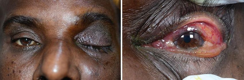
Clinical Profile: fever, pain, redness, and periocular swelling, acute and progressive,
drooping of eyelids, limitation of ocular movements, and painful loss of vision. The
progression is usually rapid, an average of two days from the onset. Eyelid and periocular
edema, complete ptosis, total ophthalmoplegia, proptosis and relative afferent pupillary
defect, unilateral facial or orbital pain, headache, periocular swelling, double vision or
diminution of vision,congestion with conjunctival edema and signs of exposure keratitis,
chemosis, sinusitis, nasal discharge and neurological signs and symptoms if intracranial
extension is present. The patients may exhibit one or more of the above mentioned signs and
symptoms depending on the severity of disease.

Figure 1: Clinical picture of Orbital Mucormycosis with (a) left periocular edema, complete ptosis and (b)
proptosis, conjunctival congestion, and severe chemosis.( Sen M, Lahane S, Lahane TP, Parekh R, Honavar
SG. Mucor in a Viral Land: A Tale of Two Pathogens. Indian J Ophthalmol 2021;69:244-52.)

Figure 2: Axial scan of MRI orbit and brain of the same patient showing mild proptosis of the left eye,
diffusion involvement of the ethmoid and sphenoid sinus, orbital apex (white arrow) and extension into the
cavernous sinus.( Sen M, Lahane S, Lahane TP, Parekh R, Honavar SG. Mucor in a Viral Land: A Tale of Two
Pathogens. Indian J Ophthalmol 2021;69:244-52.)

Investigations:Proven mucormycosis can be defined as histopathologic, cytopathologic or
direct microscopic examination showing fungal hyphae in biopsy specimen with associated
tissue damage, or a positive culture result. Serology- antigen and b-antibody, (1,3) D-Glucan
and Galactomannan detection by serum, molecular identification (PCR). Magnetic resonance

2 |P a g e
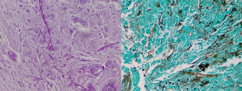
imaging (MRI) of orbit, brain, and paranasal sinuses with or without computed tomography
(CT) could be performed for assessing extent of the disease.

Figure 3(a–f ) Axial, magnetic resonance imaging scans showing intra-orbital extension of mucormycosis.
(Sharma S, Grover M, Bhargava S, Samdani S, Kataria T. Post coronavirus disease mucormycosis: a deadly
addition to the pandemic spectrum. J Laryngol Otol 2021;1–6. https://doi.org/10.1017/ S0022215121000992).

Figure 4: HPE of a patient showing aseptate hyphae with angioinvasion and Giemsa
stain of the same showing black fungal elements in green background.(Mishra N et al. Int J
Otorhinolaryngol Head Neck Surg. 2021 May;7(5):867-870).

Prevention   1.Avoid extensive use of steroids/monoclonal antibodies/broad-spectrum
antibiotics during the treatment course of COVID-19 infection (may lead to the
development/exacerbation of a preexisting fungal disease).
 2. Early identification of fungal co-infections may significantly reduce morbidity and
 mortality.
 3. Use of therapeutic agents should be monitored to achieve a therapeutic effect at the lowest
 dose for the shortest durations.
 4. Avoid Nosocomial Infections.

3 |P a g e
Treatment

Figure 4: Proposed guideline for surgical debridement and orbital exenteration for
rhino-orbito-cerebral mucormycosis based on disease extent.(Sen M, Lahane S, Lahane TP,
Parekh R, Honavar SG. Mucor in a Viral Land: A Tale of Two Pathogens. Indian J Ophthalmol 2021;69:244-52).

Conclusion: With the advent of COVID-19 Pandemic there has been turns and twists in terms
of its pathophysiology, diagnosis, management, sequelae and complications[11].
As we learn more about COVID-19 and continue seeing more patients with this illness, we
likely will continue seeing other unique presentations of other disease entities. Its association
with invasive mucormycosis is dangerous and must be given serious consideration
immediately. We also have to confirm whether this ‘Black Fungus’ is a previously
undiagnosed mucor infection which may have been aggravated or it may have subsequently
developed during the course of COVID-19 infections.
Clinical suspicion and early treatment with surgical debridement are key to preventing
mortality and morbidity.

REFERENCES

1. Mehta S, Pandey A (September 30, 2020) Rhino-Orbital Mucormycosis Associated With COVID-
 19. Cureus 12(9): e10726. DOI 10.7759/cureus.10726.

4 |P a g e
2. Amanda Werthman-Ehrenreich (September 4, 2020)Mucormycosis with orbital compartment
  syndrome in a patient with COVID-19.https://doi.org/10.1016/j.ajem.2020.09.032

 3. Mishra N et al. Int J Otorhinolaryngol Head Neck Surg. 2021 May;7(5):867-870.

 4. Eucker J, Sezer O, Graf B, Possinger K. Mucormycoses. Mycoses. 2001;44(7):253-260.

 5. Sugar AM. In: Mandell GL, Bennett JE, Dolin R(eds) Mandell, Douglas, and Bennett’s principles
  and practice of infectious diseases (5th edn), Churchill Livingstone, New York, USA, 2000.

 6. Waldorf AR. Pulmonary defense mechanisms against opportunistic fungal pathogens. Immunol
    Ser. 1989;47:243-71.

 7. Waldorf AR, Ruderman N, Diamond RD. Specific susceptibity to mucormycosis in murine
  diabetes and bronchoalveolar macrophage defense mechanisms against Rhizopus. J Clin Invest.
  1984;74(1):150-60.

 8. Bouchara JP, Oumeziane NA, Lissitzky JC, Larcher G, Tronchin G, Chabasse D. Attachment of
  spores of the human pathogenic fungus Rhizopus oryzae to extracellular matrix components. Eur J
  Cell Biol. 1996;70(1):76-83.

 9. Wiersinga WJ, Rhodes A, Cheng AC, Peacock SJ, Prescott HC. Pathophysiology, transmission,
   diagnosis, and treatment of coronavirus disease 2019 (COVID-19): a review. JAMA.
   2020;324(4):782-93.

 10. Gangneux JP, Bougnoux ME, Dannaoui E, Cornet M, Zahar JR: Invasive fungal diseases during
COVID-19: we should be prepared. J Mycol Med. 2020, 30:100971. 10.1016/j.mycmed.2020.100971.

 11. Wuhan City Health Committee. Wuhan Municipal Health and Health Commission’s briefing on the
  current pneumonia epidemic situation in our city 2019.
  In:http://wjw.wuhan.gov.cn/front/web/showDetail/2019123108989 [14 January 2020].

 5 |P a g e
You can also read