Future Unruptured Intracranial Aneurysm Growth Prediction using Mesh Convolutional Neural Networks
←
→
Page content transcription
If your browser does not render page correctly, please read the page content below
Future Unruptured Intracranial Aneurysm
                                               Growth Prediction using Mesh Convolutional
                                                           Neural Networks

                                               Kimberley M. Timmins1 , Maarten J. Kamphuis2 , Iris N. Vos1 , Birgitta K.
                                                      Velthuis2 , Irene C. van der Schaaf*2 , and Hugo J. Kuijf*1
arXiv:2207.13518v3 [eess.IV] 25 Aug 2022

                                                1
                                                    Image Sciences Institute, University Medical Center Utrecht, Utrecht, The
                                                                  Netherlands. k.m.timmins@umcutrecht.nl
                                                2
                                                    Department of Radiology, University Medical Center Utrecht, Utrecht, The
                                                                                  Netherlands.
                                                                              *Joint last author

                                                     Abstract. The growth of unruptured intracranial aneurysms (UIAs) is
                                                     a predictor of rupture. Therefore, for further imaging surveillance and
                                                     treatment planning, it is important to be able to predict if an UIA is
                                                     likely to grow based on an initial baseline Time-of-Flight MRA (TOF-
                                                     MRA). It is known that the size and shape of UIAs are predictors of
                                                     aneurysm growth and/or rupture. We perform a feasibility study of us-
                                                     ing a mesh convolutional neural network for future UIA growth predic-
                                                     tion from baseline TOF-MRAs. We include 151 TOF-MRAs, with 169
                                                     UIAs where 49 UIAs were classified as growing and 120 as stable, based
                                                     on the clinical definition of growth (>1 mm increase in size in follow-up
                                                     scan). UIAs were segmented from TOF-MRAs and meshes were automat-
                                                     ically generated. We investigate the input of both UIA mesh only and
                                                     region-of-interest (ROI) meshes including UIA and surrounding parent
                                                     vessels. We develop a classification model to predict UIAs that will grow
                                                     or remain stable. The model consisted of a mesh convolutional neural
                                                     network including additional novel input edge features of shape index
                                                     and curvedness which describe the surface topology. It was investigated
                                                     if input edge mid-point co-ordinates influenced the model performance.
                                                     The model with highest AUC (63.8%) for growth prediction was using
                                                     UIA meshes with input edge mid-point co-ordinate features (average F1
                                                     score = 62.3%, accuracy = 66.9%, sensitivity = 57.3%, specificity =
                                                     70.8%). We present a future UIA growth prediction model based on a
                                                     mesh convolutional neural network with promising results.

                                                     Keywords: meshes · aneurysms · growth prediction · geometric deep
                                                     learning · topology.

                                           1        Introduction

                                           Approximately 3% of the general population has a unruptured intracranial aneurysm
                                           (UIAs) [5]. If an UIA ruptures, it leads to subarachnoid haemorrhage with a
2 K.M.Timmins et al.
high mortality and morbidity rate. Neurosurgical or endovascular treatment can
prevent UIAs from rupture, but carry a considerable risk. Therefore a balanced
decision based on the rupture and treatment complication risk must be made [1].
UIA growth is an important rupture risk factor [9], and if detected, preventative
treatment should be considered. Most UIAs are monitored, using Time-of-Flight
Magnetic Resonance Angiographs (TOF-MRAs) or Computed Tomography An-
giographs (CTAs). Currently, 2D size measurements of the UIAs are made and
an aneurysm will be considered to be growing if there is a change in size (>1
mm) [6]. UIA shape is also known to be different in aneurysms that grow [2].
The ELAPSS score [2] is a clinical score for UIA growth prediction based on
patient and aneurysm characteristics. The predictors are: Earlier subarachnoid
hemorrhage, aneurysm Location, Age, Population, aneurysm Size and Shape.
Shape is assessed visually as ’Regular’ or ’Irregular’.
As computer-aided radiology tools continue to be developed, 3D volume and
morphology measurements of UIAs can be made [19], including to distinguish
between growing and stable aneurysms [13,12,22]. UIA rupture risk prediction
models have been developed based on morphological parameters, as well as clas-
sical parameters [10,14]. More recently, some prediction models for aneurysmal
stability and growth have been proposed [15,3].
Liu et al.[15] investigated predicting aneurysm stability using machine learn-
ing regression models and 12 morphology radiomics features. The dataset in-
cluded 420 aneurysms (4 - 8 mm). Instability was defined as ruptured within a
month, growth or adjacent structure compressive symptoms. They determined
flatness to be the most important morphological predictor of aneurysm stabil-
ity. Bizjak et al.[3] found using point clouds with PointNet++ for future UIA
growth prediction had a higher accuracy than other machine learning models
based on morphological parameters. The method was performed using only 44
UIAs, where 25 growing and 19 stable. UIAs were visually inspected in 3D to be
classified as growing or stable.
Various different morphology measurements and definitions of growth or sta-
bility have been used in these studies, making it difficult to make direct compar-
isons. However, it is clear that UIA shape and surface topology is an important
predictor of future UIA growth and that deep learning methods may have an ad-
vantage over using predefined morphology parameters. Geometric deep learning
methods are well suited to this problem, as they accurately describe the shape
and topology of a surface by using point clouds or meshes [4]. Meshes may have a
preference over point clouds as they include connectivity information, providing
more information about the surface topology. Segmented UIA meshes could be
used as we already know UIA shape is a growth predictor growth. Alternatively,
parent vessels in a Region-of-Interest (ROI) around the UIA could be used which
includes UIA-vessel configuration and exact UIA segmentation is not required.
MeshCNN[7] is a convolutional neural network (CNN) developed for classifi-
cation and segmentation problems using 3D triangular meshes. Convolutions and
pooling are performed on edges of the meshes, based on an edge neighbourhood.
Five relative scale, translation and rotation invariant geometric edge are deter-Future UIA Growth Prediction with MeshCNNs 3
mined for each edge as input features for the model. These five geometric features
are: the dihedral angle, two inner angles and two edge-length ratios. MeshCNN
has only been used for a few medical imaging classification and segmentation
problems, including age prediction based on the neonatal white matter cortical
surface[24] and UIA segmentation from a parent vessel[18]. In our previous work,
we proposed a modified version of MeshCNN for UIA detection based on brain
vessel surface meshes[20].
In this paper, we propose a prediction model for future UIA growth from
baseline TOF-MRAs using a mesh convolutional neural network. We investigate
the use of meshes of UIAs alone, and region-of-interest (ROI) meshes including
the UIA with parent vessels as input for these models and their performance
for future UIA growth prediction. We also investigate the addition of edge mid-
point co-ordinate input features of the meshes and the impact on the model
performance.
2 Materials and Methods
2.1 Dataset
The dataset consisted of 151 baseline Time-of-Flight MRAs (TOF-MRAs) taken
from routine clinical scans. We included patients with UIAs who met the fol-
lowing inclusion criteria: 1) A TOF-MRA or CTA was available at baseline
and follow-up, 2) the follow-up scan was performed at least 6 months after the
baseline scan, and 3) the patient had at least 1 untreated UIA present on both
baseline and follow-up imaging. The most recent follow-up scan in which the UIA
remained untreated and unruptured was used for growth assessment. Fusiform
and ruptured aneurysms were excluded. All scans were made from the Univer-
sity Medical Center Utrecht between 2006 and 2020. The average time between
baseline and follow-up scans was 5.2 ± 3.3 years (range: 1 - 16 years) The mean
baseline aneurysm size was 5.0 ± 2.2 mm with a range of 1.3 – 14.7 mm. Manual
2D length and width UIA measurements were performed in IntelliSpace Por-
tal (Philips Healthcare) by an experienced neuro-radiologist (I.C.v.d.S.) and a
trained PhD-student (M.J.K.) according to standard clinical protocol. Growth
was defined as a ≥ 1.0 mm increase in any direction between the baseline and
follow-up scan[6]. Based on this definition, UIAs were categorised as either ‘grow-
ing’ (30%, n= 49) or ‘stable’ (70%, n = 120).
2.2 Methods
Input Mesh Generation All baseline TOF-MRAs were pre-processed using
an N4 bias field correction algorithm and z-score normalised before being re-
sampled to have voxel size 0.357 mm x 0.357 mm x 0.500 mm (median of the
dataset). All UIA and ROI selection, mesh generation and processing was per-
formed completely automatically based on UIA annotations.4 K.M.Timmins et al.
UIA mesh generation UIA meshes were generated and pre-processed automati-
cally based on the TOF-MRAs and UIA annotations. UIAs were manually seg-
mented from the TOF-MRAs using annotations drawn on axial slices in in-
house-developed software implemented in MeVisLab (MeVis Medical Solutions)
(performed by I.C.v.d.S. and M.J.K). A triangular mesh was automatically fit-
ted to the outside of the UIA surface using a Marching Cubes algorithm [17].
All UIA meshes were down-sampled to 1000 edges and included just the UIA
and no other vessels.
Region-of-Interest (ROI) mesh generation ROI meshes were automatically gen-
erated from the TOF-MRAs using the UIA segmentations. An existing 3D U-net
was used to automatically perform full vessel segmentation from the scans[23].
Based on the UIA segmentation, a region-of-interest (ROI) including only the
UIA and parent vessels was made. The centre-of-mass of the UIA segmentation
was determined and the ROI included all connected vessels (and UIA) within a
20 mm cube around the centre-of-mass. A mesh was automatically fitted to the
outside of the UIA and parent vessel surface using a Marching Cubes algorithm
[17]. All ROI meshes were down-sampled to 2000 edges.
Input edge features Based on the generated UIA and ROI meshes, new input
edge features were automatically determined per edge. These were shape index,
curvedness and edge mid-point co-ordinates. These further edge features (shape
index, curvedness and mid-point co-ordinates) could then be included as input
to the network, in addition to the original five geometric edge features.
Shape index and curvedness are rotation and translation invariant measures
which describe the topology of the UIA surface. The invariant nature of these
novel input edge features ideal for use in MeshCNN. It is known from our previ-
ous work in UIA detection that the addition of both shape index and curvedness
as input edge features improve the performance of the original MeshCNN [21].
Shape descriptor values; shape index and curvedness, were calculated for each
vertex on the mesh surface using the standard formulae [11]. An edge was then
given a shape descriptor value (shape index or curvedness), as being the average
of the values at the corresponding end vertices of the edge.
The addition of edge mid-point co-ordinate values was suggested in the orig-
inal MeshCNN paper [7]. We experiment with including these co-ordinates in
our models as we know location is important as an aneurysm growth predictor
[2]. Edge mid-point co-ordinates (x,y,z) were determined as the average of the
world co-ordinates of the corresponding end vertices of the edge.
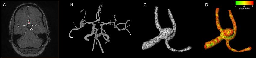
Figure 1 shows an example generation of a ROI mesh including shape index
values determined for each edge.
Model Implementation A ConvNet style network was set up based on our
modified MeshCNN framework [20] including four convolutional layers and four
pooling layers. Four different model configurations were investigated. The first
model (uia model) had UIA meshes only as input, with 1000 edges. Pooling layerFuture UIA Growth Prediction with MeshCNNs 5
Fig. 1. Example generation of input region-of-interest (ROI) mesh including parent
vessels and UIA. A: TOF-MRA with annotated UIA shown overlaid in red. B: Vessel
segmentation performed using 3D U-net [23]. C: ROI selection including UIA and par-
ent vessels, followed by mesh generation. D: Shape index determination for each edge,
to be used as an additional input feature alongside curvedness and edge coordinates.
configuration for the UIA model was: 750, 600, 500, 400. The second model
(roi model) had ROI meshes including UIA and parent vessels as input. The
pooling layer configuration for the ROI model was: 1500, 1200, 1000, 800. All
models were made to include shape index and curvedness as additional input fea-
tures to the original five edge geometric features of MeshCNN. This meant that
there were seven input edge features as standard. For each different input, two
models were trained. The first with the seven input edge features (uia model 1,
roi model 1), and the second including edge mid-point co-ordinates (x,y,z) as
further additional input features (uia model 2, roi model 2), meaning there were
ten input edge features. No augmentation was used.
For all models, all other hyperparameters were kept the same, and as similar
to the original paper as possible[7]. Both a weighted data sampler and weighted
cross-entropy loss function were used, based on the class distribution of growing
and stable UIAs (0.7 to growing, 0.3 to stable). Batch normalisation was used
with a batch size of 50 meshes and a learning rate of 0.0002. The classification
model was trained to predict future growth of the UIA as defined by the clinical
definition, whereby output was one of the two classes: growing or stable. All
experiments were performed using five-fold cross-validation where the validation
splits were made randomly and kept the same for each experiment. The models
were trained for a maximum of 200 epochs with validation every 5 epochs and
the model with the highest average F1 score for each split was selected. The
model was implemented in Python 3.8.5 with Pytorch version 1.8.0 on a NVIDIA
TITAN X Pascal (12GB) GPU with CUDA version 11.2.
For final model assessment, we determined the classification accuracy, growth
prediction sensitivity and specificity, where the metrics were averaged across all
validation splits. A true positive was considered a correctly identified growing
UIA, a true negative was a correctly identified stable UIA. Sensitivity and Speci-
ficity were determined using these definitions, therefore high sensitivity suggests
the model is good at detecting growing UIAs and high specificity suggests the
model is good at detecting stable UIAs. We plotted the mean ROC curve and6 K.M.Timmins et al.
calculated the mean area under the curve (AUC) for each model, as the average
over all validation splits for each model.
3 Results
Results of the growth prediction models averaged across all validation splits
are summarised in Table 1. Figure 2 shows ROC curves for all of the mod-
els. Roi model 1, using ROI meshes and no edge mid-point co-ordinates, had
the highest accuracy (0.761), F1 score (0.681) and specificity (0.883) suggesting
it performs optimally for stable aneurysm detection. Uia model 2, using UIA
meshes and including edge mid-point co-ordinates, had the highest sensitivity
for growth detection. Overall, both the second models including edge mid-point
co-ordinates had higher AUC and sensitivity values but slightly lower accuracy
and F1 scores.
Table 1. Classification metrics for each model. F1 score is the average of F1 score for
each class (growing and stable). A true positive was considered a correctly identified
growing UIA, a true negative was a correctly identified stable UIA. Sensitivity and
Specificity were determined using these definitions. AUC is the area under the mean
ROC curve in Figure 2. Values are provided as mean (standard deviation) across all
validation splits (standard deviation)
Model Accuracy F1 score Sensitivity Specificity AUC
uia model1 0.704 (0.077) 0.617 (0.062) 0.389 (0.048) 0.833 (0.121) 0.620 (0.119)
uia model2 0.669 (0.061) 0.623 (0.073) 0.573 (0.155) 0.708 (0.075) 0.638 (0.116)
roi model1 0.761 (0.017) 0.681 (0.021) 0.458 (0.038) 0.883 (0.030) 0.606 (0.056)
roi model2 0.713 (0.075) 0.650 (0.077) 0.498 (0.090) 0.781 (0.110) 0.622 (0.064)
4 Discussion
In this paper, we demonstrate that a future UIA growth prediction model could
be developed using a mesh convolutional neural network, which considers the
topology of UIAs and their parent vasculature. We found that adding edge
mid-point co-ordinates as input features to the network increases the AUC and
sensitivity of growth prediction but reduces the overall accuracy of the model
(uia model 2, roi model 2). Using ROI meshes as opposed to UIA meshes alone,
improved the accuracy and F1 score of the model but has a decreased AUC
for growth prediction. A sensitive growth prediction model should consider us-
ing UIA meshes as input and including edge mid-point co-ordinates as input
features (uia model 2).
We found using UIA meshes alone (uia model 2) improved the AUC relative
to using the ROI including parent vessels (roi model 2). This suggests that it is
the topology of the aneurysm surface itself which is high indicative of growth orFuture UIA Growth Prediction with MeshCNNs 7
Mean ROC curves for all m odels
1.0
0.8
True Posit ive Rat e
0.6
0.4
uia_m odel_1
0.2 uia_m odel_2
roi_m odel_1
roi_m odel_2
0.0
0.0 0.2 0.4 0.6 0.8 1.0
False Posit ive Rat e
Fig. 2. ROC curves of all trained models for growth prediction classification. Each line
is the mean of the performance across all cross validation splits for each model. The
black dotted line indicates a classifier which would give random choice.
stability as opposed to UIA configuration relative to parent vessels. This result
is similar to previous studies, where measurements of just the UIA distinguish
growing and stable UIAs [13,12,22]. However, it is worth noting that using a
ROI as opposed to the UIA mesh does not greatly reduce the performance of
the method. A ROI mesh, is easier to achieve in the clinic as it requires only
a click of a centre point to select the ROI. Whereas, currently the UIA meshes
require accurate manual UIA segmentation. Therefore, a ROI model have more
clinic applicability and has adequate performance for UIA growth prediction.
The inclusion of input edge mid-point co-ordinate features increased the AUC
and growth prediction sensitivity. This is likely because the co-ordinate provides
aneurysm location information to the network. Location is a known predictor of
growth [2]. In the original MeshCNN paper [7] it was commented that adding
in edge co-ordinates reduced the model performance, possibly due to removing
the rotation, translation and uniform scaling in-variance of the usual relative
geometric input edge features. However, in real-life applications, such as in med-
ical images, the co-ordinates give important information about the location of
lesions. Therefore, the addition of these features only appears to improve the
performance in this scenario. Further studies could investigate the use of rel-
ative position input features, to ensure the in-variance to rotation, translation
and scaling is kept. Another possibility could be to include position/location,8 K.M.Timmins et al.
and potentially other known growth predictors, as global features in the final
layers of the network.
The models all had a relatively high specificity, suggesting they perform
well for detecting stable UIAs. This may be useful in clinic to identify those
UIAs which are stable and do not need further investigation. In our study, we
had a relatively large class imbalance of only 30% growing UIAs to 70% stable
UIAs. Although weighted loss functions and samplers were used, this does not
eliminate the class imbalance. In the future, a more balanced dataset, including
more growing UIAs could be used. The validation results displayed a large range
in sensitivity, and it was also clear, that the model tended to over-fit relatively
quickly to the training set. This is due, in part, to the heterogeneous nature of the
UIAs and configurations leading to the validation sets being quite different to the
training data. This could be improved by including more training and validation
data. Furthermore, a larger dataset would allow for independent evaluation on
a separate test set.
The ELAPSS growth prediction score was determined to have a c-statistic
(AUC) of 0.69 in an external validation study [8]. Our model performed only
slightly inferior to this (AUC = 0.64), suggesting that our model has compa-
rable performance to current clinical prediction models. Future studies should
consider combining the patient characteristics used in the ELAPSS score, with
the aneurysm characteristics used in our model.
Our proposed method did not perform as well as the method using Point-
Net++ put forward by Bizjak [3] (accuracy = 82%). This may be for a vari-
ety of reasons. Firstly, our dataset was imbalanced (30% growing, 70% stable)
compared to the dataset they used which included more growing than stable
aneurysms. Secondly, in Bizjak et al. they assess growth visually on the pre-
processed 3D meshes. Instead, our model can provide a prediction for growth as
is currently clinically assessed and accepted in the clinic. Future studies should
investigate different definitions of growth, and as computer aided tools for UIA
diagnosis and assessment continue to be developed and improve, a definition for
volumetric growth should be considered [19,16]. It is difficult to make a compar-
ison of our model to the study by Liu et al. [15] as they are predicting aneurysm
stability, which included rupture and not just growth. Furthermore, they have
a larger dataset of all aneurysms larger than 4 mm. However, future studies
could investigate if our mesh based model could also predict rupture/aneurysm
instability as well as growth.
In our previous paper [20], we demonstrated the mesh convolutional neural
networks could be used for a modality independent UIA detection method. Based
on these results, we believe that our growth prediction method could also be
modality independent. This would be helpful in the clinic, where UIAs are often
assessed or followed-up with different modalities such as CTA or DSA.Future UIA Growth Prediction with MeshCNNs 9
5 Conclusion
We present a future UIA growth prediction model using a mesh convolutional
neural network. We demonstrate that both UIA and ROI meshes can be used as
input for such a prediction model, and that edge mid-point co-ordinates improve
the growth prediction sensitivity. This model may have potential clinical use as
an aid for radiologists assessing potential future UIA growth.
6 Acknowledgements
We acknowledge the support from the Netherlands Cardiovascular Research Ini-
tiative: An initiative with support of the Dutch Heart Foundation, CVON2015-08
ERASE and CVON2018-02 ANEURYSM@RISK.
References
1. Algra, A.M., Lindgren, A., Vergouwen, M.D., Greving, J.P., Van Der Schaaf,
I.C., Van Doormaal, T.P., Rinkel, G.J.: Procedural Clinical Complica-
tions, Case-Fatality Risks, and Risk Factors in Endovascular and Neu-
rosurgical Treatment of Unruptured Intracranial Aneurysms: A System-
atic Review and Meta-analysis. JAMA Neurology 76(3), 282–293 (2019).
https://doi.org/10.1001/jamaneurol.2018.4165
2. Backes, D., Rinkel, G., Greving, J., Velthuis, B.K., Murayama, Y., Takao,
H., Ishibashi, T., Igase, M., TerBrugge, K., Agid, R., Jaaskelainen, J., Lind-
gren, A., Koivisto, T., Von Und Zu Fraunberg, M., Matsubara, S., Mo-
roi, J., Wong, G., Abrigo, J., Igase, K., Matsumoto, K., Wermer, M.,
Van Walderveen, M.A., Algra, A.: ELAPSS score for prediction of risk of
growth of unruptured intracranial aneurysms. Neurology 88(17), 1600–1606 (2017).
https://doi.org/http://dx.doi.org/10.1212/WNL.0000000000003865
3. Bizjak, Z., Pernus, F., Spiclin, Z.: Deep Shape Features for Predicting Future
Intracranial Aneurysm Growth. Frontiers in Physiology 12(July), 1–10 (2021).
https://doi.org/10.3389/fphys.2021.644349
4. Cao, W., Yan, Z., He, Z., He, Z.: A Comprehensive Survey on
Geometric Deep Learning. IEEE Access 8, 35929–35949 (2020).
https://doi.org/10.1109/ACCESS.2020.2975067, https://ieeexplore.ieee.
org/document/9003285/
5. Greving, J.P., Wermer, M.J., Brown, R.D., Morita, A., Juvela, S., Yonekura, M.,
Ishibashi, T., Torner, J.C., Nakayama, T., Rinkel, G.J., Algra, A.: Development
of the PHASES score for prediction of risk of rupture of intracranial aneurysms:
A pooled analysis of six prospective cohort studies. The Lancet Neurology 13(1),
59–66 (2014). https://doi.org/10.1016/S1474-4422(13)70263-1
6. Hackenberg, K.A., Algra, A., Salman, R.A.S., Frösen, J., Hasan, D., Juvela, S.,
Langer, D., Meyers, P., Morita, A., Rinkel, G., Etminan, N., Suarez, J.I., Mac-
donald, R.L., Amin-Hanjani, S., Brown, R.D., de Oliveira Manoel, A.L., Derdeyn,
C.P., Keller, E., LeRoux, P.D., Mayer, S., Rufennacht, D., Stienen, M.N., Torner,
J., Vergouwen, M.D., Wong, G.K., Bijlenga, P., Ko, N., McDougall, C.G., Mocco,
J., Murayama, Y., Werner, M.J., Damani, R., Broderick, J., Dhar, R., Jauch, E.C.,10 K.M.Timmins et al.
Kirkpatrick, P.J., Martin, R.H., Muehlschlegel, S., Mutoh, T., Nyquist, P., Olson,
D., Mejia-Mantilla, J.H., van der Jagt, M., Bambakidis, N., Brophy, G., Bulsara,
K., Claassen, J., Connolly, E.S., Hoffer, S.A., Hoh, B.L., Holloway, R.G., Kelly,
A., Nakaji, P., Rabinstein, A., Vajkoczy, P., Woo, H., Zipfel, G.J., Chou, S., Doré,
S., Dumont, A.S., Gunel, M., Kasuya, H., Roederer, A., Ruigrok, Y., Vespa, P.M.,
Sarrafzadeh-Khorrasani, A.S., Hackenberg, K.A., Huston, J., Krings, T., Lanzino,
G., Meyers, P.M., Wintermark, M., Daly, J., Ogilvy, C., Rhoney, D.H., Roos, Y.B.,
Siddiqui, A., Frösen, J., Langer, D.J., Hanggi, D., Schweizer, T., Visser-Meily, J.,
Amos, L., Ludet, C., Moy, C., Odenkirchen, J., Ala’i, S., Esterlitz, J., Joseph,
K., Sheikh, M.: Definition and Prioritization of Data Elements for Cohort Stud-
ies and Clinical Trials on Patients with Unruptured Intracranial Aneurysms: Pro-
posal of a Multidisciplinary Research Group. Neurocritical Care 30, 87–101 (2019).
https://doi.org/10.1007/s12028-019-00729-0
7. Hanocka, R.: MeshCNN: A network with an edge. ACM Transactions on Graphics
38(4) (2019). https://doi.org/10.1145/3306346.3322959
8. Sánchez van Kammen, M., Greving, J.P., Kuroda, S., Kashiwazaki, D., Morita,
A., Shiokawa, Y., Kimura, T., Cognard, C., Januel, A.C., Lindgren, A., Koivisto,
T., Jääskeläinen, J.E., Ronkainen, A., Pyysalo, L., Öhman, J., Rahi, M., Kuhmo-
nen, J., Rinne, J., Leemans, E.L., Majoie, C.B., Vandertop, W.P., Verbaan, D.,
Roos, Y.B., Berg, R.v.d., Boogaarts, H.D., Moudrous, W., Wijngaard, I.R.v.d.,
Hove, L.t., Teo, M., George, E.J.S., Hackenberg, K.A., Abdulazim, A., Etmi-
nan, N., Rinkel, G.J., Vergouwen, M.D.: External Validation of the ELAPSS
Score for Prediction of Unruptured Intracranial Aneurysm Growth Risk. Jour-
nal of Stroke 21(3), 340–346 (9 2019). https://doi.org/10.5853/jos.2019.01277,
http://j-stroke.org/journal/view.php?doi=10.5853/jos.2019.01277
9. van der Kamp, L.T., Rinkel, G.J.E., Verbaan, D., van den Berg, R., Van-
dertop, W.P., Murayama, Y., Ishibashi, T., Lindgren, A., Koivisto, T.,
Teo, M., St George, J., Agid, R., Radovanovic, I., Moroi, J., Igase, K.,
van den Wijngaard, I.R., Rahi, M., Rinne, J., Kuhmonen, J., Boogaarts,
H.D., Wong, G.K.C., Abrigo, J.M., Morita, A., Shiokawa, Y., Hackenberg,
K.A.M., Etminan, N., van der Schaaf, I.C., Zuithoff, N.P.A., Vergouwen,
M.D.I.: Risk of Rupture After Intracranial Aneurysm Growth. JAMA Neurology
(8 2021). https://doi.org/10.1001/jamaneurol.2021.2915, https://jamanetwork.
com/journals/jamaneurology/fullarticle/2783663
10. Kim, H.C., Rhim, J.K., Ahn, J.H., Park, J.J., Moon, J.U., Hong, E.P., Kim, M.R.,
Kim, S.G., Lee, S.H., Jeong, J.H., Choi, S.W., Jeon, J.P.: Machine Learning Appli-
cation for Rupture Risk Assessment in Small-Sized Intracranial Aneurysm. Journal
of Clinical Medicine 8(5), 683 (2019). https://doi.org/10.3390/jcm8050683
11. Koenderink, J., Doorn, A.: Surface shape and curvature scales. Image and Vision
Computing 10(8), 557–564 (1992)
12. Leemans, E.L., Cornelissen, B.M., Slump, C.H., Majoie, C.B., Cebral, J.R.,
Marquering, H.A.: Comparing Morphology and Hemodynamics of Stable-versus-
Growing and Grown Intracranial Aneurysms. American Journal of Neuroradiology
40(12), 2102–2110 (2019). https://doi.org/10.3174/ajnr.A6307
13. Leemans, E.L., Cornelissen, B.M.W., Said, M., van den Berg, R., Slump,
C.H., Marquering, H.A., Majoie, C.B.L.M.: Intracranial aneurysm growth:
consistency of morphological changes. Neurosurgical Focus 47(1), E5 (7
2019). https://doi.org/10.3171/2019.4.FOCUS1987, https://thejns.org/view/
journals/neurosurg-focus/47/1/article-pE5.xmlFuture UIA Growth Prediction with MeshCNNs 11
14. Liu, J., Chen, Y., Lan, L., Lin, B., Chen, W., Wang, M., Li, R., Yang, Y.,
Zhao, B., Hu, Z., Duan, Y.: Prediction of rupture risk in anterior communicating
artery aneurysms with a feed-forward artificial neural network. European Radiol-
ogy 28(8), 3268–3275 (2018). https://doi.org/10.1007/s00330-017-5300-3
15. Liu, Q., Jiang, P., Jiang, Y., Ge, H., Li, S., Jin, H., Li, Y.: Predic-
tion of Aneurysm Stability Using a Machine Learning Model Based on
PyRadiomics-Derived Morphological Features. Stroke 50(9), 2314–2321 (2019).
https://doi.org/10.1161/STROKEAHA.119.025777
16. Liu, X., Haraldsson, H., Wang, Y., Kao, E., Ballweber, M., Martin, A.,
McCulloch, C., Faraji, F., Saloner, D.: A Volumetric Metric for Mon-
itoring Intracranial Aneurysms: Repeatability and Growth Criteria in a
Longitudinal MR Imaging Study. American Journal of Neuroradiology (6
2021). https://doi.org/10.3174/ajnr.A7190, http://www.ajnr.org/lookup/doi/
10.3174/ajnr.A7190
17. Lorensen, W.E., Cline, H.E.: Marching Cubes: A High Resolution 3D Surface Con-
struction Algorithm. ACM siggraph computer graphics 21(4), 163–169 (1987)
18. Schneider, L., Niemann, A., Beuing, O., Preim, B., Saalfeld, S.: MedmeshCNN -
Enabling meshcnn for medical surface models. Computer Methods and Programs
in Biomedicine 210, 106372 (2021). https://doi.org/10.1016/j.cmpb.2021.106372,
http://arxiv.org/abs/2009.04893
19. Timmins, K.M., Kuijf, H.J., Vergouwen, M.D., Otten, M.J., Ruigrok, Y.M.,
Velthuis, B.K., van der Schaaf, I.C.: Reliability and agreement of 2d and
3d measurements on mras for growth assessment of unruptured intracra-
nial aneurysms. American Journal of Neuroradiology 42(9), 1598–1603 (7
2021). https://doi.org/10.3174/ajnr.A7186, http://www.ajnr.org/lookup/doi/
10.3174/ajnr.A7186
20. Timmins, K.M., Schaaf, I.C., Vos, I.N., Ruigrok, Y.M., Velthuis, B.K., Kuijf, H.J.:
Deep learning with vessel surface meshes for intracranial aneurysm detection.
In: Iftekharuddin, K.M., Drukker, K., Mazurowski, M.A., Lu, H., Muramatsu,
C., Samala, R.K. (eds.) Medical Imaging 2022: Computer-Aided Diagnosis.
p. 110. SPIE (4 2022). https://doi.org/10.1117/12.2610745, https://www.
spiedigitallibrary.org/conference-proceedings-of-spie/12033/2610745/
Deep-learning-with-vessel-surface-meshes-for-intracranial-aneurysm-detection/
10.1117/12.2610745.full
21. Timmins, K.M., van der Schaaf, I.C., Vos, I.N., Ruigrok, Y.M., Velthuis, B.K.,
Kuijf, H.J.: Geometric Deep Learning using Vascular Surface Meshes for Modality-
Independent Unruptured Intracranial Aneurysm Detection. Under Review (2022)
22. Timmins, K., Kuijf, H., Vergouwen, M., Ruigrok, Y., Velthuis, B., van der
Schaaf, I.: Relationship between 3D Morphologic Change and 2D and 3D Growth
of Unruptured Intracranial Aneurysms. American Journal of Neuroradiology (2
2022). https://doi.org/10.3174/ajnr.A7418, http://www.ajnr.org/lookup/doi/
10.3174/ajnr.A7418
23. de Vos, V., Timmins, K., van der Schaaf, I., Ruigrok, Y., Velthuis, B., Kuijf, H.J.:
Automatic cerebral vessel extraction in TOF-MRA using deep learning (February
2021), 83 (2021). https://doi.org/10.1117/12.2581226
24. Vosylius, V., Wang, A., Waters, C., Zakharov, A., Ward, F., Le Folgoc, L., Cu-
pitt, J., Makropoulos, A., Schuh, A., Rueckert, D., Alansary, A.: Geometric Deep
Learning for Post-Menstrual Age Prediction Based on the Neonatal White Matter
Cortical Surface. Lecture Notes in Computer Science (including subseries Lecture12 K.M.Timmins et al.
Notes in Artificial Intelligence and Lecture Notes in Bioinformatics) 12443 LNCS,
174–186 (2020). https://doi.org/10.1007/978-3-030-60365-6 17You can also read