Guidance on using shielding on
patients for diagnostic radiology
applications
←
→
Page content transcription
If your browser does not render page correctly, please read the page content below

Guidance on using shielding on patients for diagnostic radiology applications
Guidance on using shielding on patients for diagnostic radiology applications
Guidance on using shielding on patients for diagnostic radiology
applications
A joint report of the British Institute of Radiology (BIR), Institute of Physics and Engineering
in Medicine (IPEM), Public Health England (PHE), Royal College of Radiologists (RCR), Society
and College of Radiographers (SCoR) and the Society for Radiological Protection (SRP).
Working Party Members
Peter Hiles, BIR (Chair) Yvonne Sullivan, PHE
Elizabeth Benson, BIR Guy Hickson, RCR
Helen Hughes, BIR Phil Cosson, SCoR
Rob Loader, IPEM Lynda Johnson, SCoR
Dan Shaw, IPEM David Dommett, SRP
Sue Edyvean, PHE
For queries relating to this document, please contact communications@bir.org.uk.
About The British Institute of Radiology
The British Institute of Radiology is an international membership organisation for everyone
working in imaging, radiation oncology and the underlying sciences.
Our aims are to:
support the work of our members and their colleagues to achieve professional
excellence
provide continuing professional development for our multidisciplinary community
publish cutting-edge research for our authors and readers across the world
influence and connect with the wider professional sector.
Published by the British Institute of Radiology. This is an open access publication distributed
under the terms of the Creative Commons Attribution 4.0 International Licence, which
permits unrestricted use, distribution and reproduction in any medium, provided the
original author and source are credited.
British Institute of Radiology
48–50 St John Street
London
EC1M 4DG
Tel: 020 3668 2220
www.bir.org.uk
Published March 2020
BIR Registered charity number 215869
2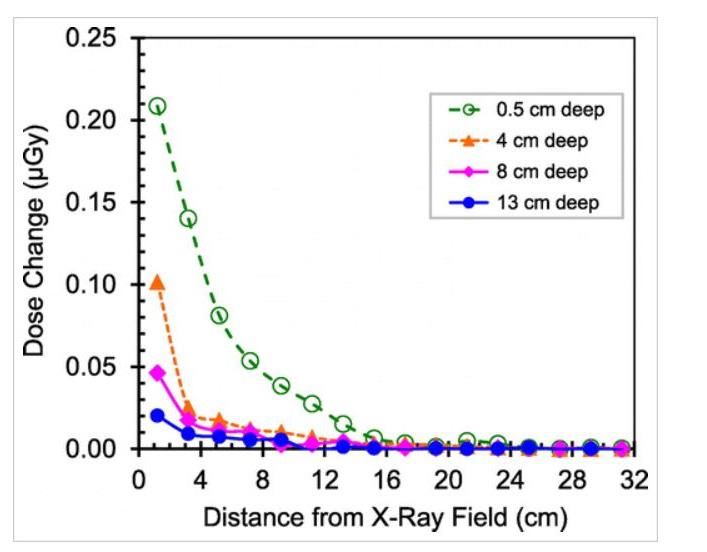
Guidance on using shielding on patients for diagnostic radiology applications

Contents

Executive summary                                                               5
1.   Introduction                                                               6
         1.1 Background
         1.2 Scope
         1.3 Aim
2.   General requirements for patient contact shielding                         8
         2.1 Radiation safety culture
         2.2 Context for applying shielding
         2.3 Medical device and product marking
         2.4 Recommendations for local practice
3.   Radiation dose and risk                                                    12
         3.1 Historical perspective
         3.2 Stochastic and hereditary risk
         3.3 Introduction to levels of risk
         3.4 Optimisation and applying the ALARP principle
4.   Sources of radiation exposure                                              21
         4.1 Primary radiation
         4.2 Secondary radiation
         4.3 Summary
5.   Operator responsibilities                                                  31
         5.1 Overview
         5.2 Communication
         5.3 Consent
         5.4 Patient complaints and duty of candour
         5.5 Summary
6.   Clinical service requirements for patient shielding                        38
         6.1 Overview
         6.2 Priorities in imaging
         6.3 Training
         6.4 Continuing Professional Development (CPD)
         6.5 Procurement, storage and maintenance of patient contact shields
         6.6 Applying patient contact shielding
         6.7 Leadership
         6.8 Continuous Quality Improvement (CQI)
         6.9 Repeat exposures
         6.10 Summary
7.   Shielding in general radiography                                           45
         7.1 Organs at risk
         7.2 The anatomy and concept behind shield application

                                                       3
Guidance on using shielding on patients for diagnostic radiology applications

           7.3 In-beam protection (Primary beam)
           7.4 Outside beam protection
           7.5 Influence of shielding on equipment function and image quality
           7.6 Special patient groups
           7.7 Recommendations for local practice
8.     Shielding in diagnostic and interventional fluoroscopy                    50
           8.1 Organs at risk
           8.2 The anatomy and concept behind shield application
           8.3 Influence of shielding on equipment function
           8.4 Special patient groups
           8.5 Recommendations for local practice
9.     Shielding in CT                                                           54
           9.1 Organs at risk
           9.2 The anatomy and concept behind shield application
           9.3 In-beam protection (Primary beam)
           9.4 Outside beam protection
           9.5 Influence of shielding on equipment function and image quality
           9.6 Special patient groups
           9.7 Recommendations for local practice
10.    Shielding in mammography                                                  78
           10.1 The anatomy and concept behind shield application
           10.2 Influence of shielding on equipment function and image quality
           10.3 Special patient groups
           10.4 Recommendations for local practice
11.    Shielding in dental radiography                                           80
           11.1 Organs at risk
           11.2 The anatomy and ideas behind shield application
           11.3 In-beam protection (primary beam)
           11.4 Outside beam protection
           11.5 Influence of shielding on equipment function and image quality
           11.6 Special patient groups
           11.7 Recommendations for local practice
12.    Glossary                                                                  86

                                                       4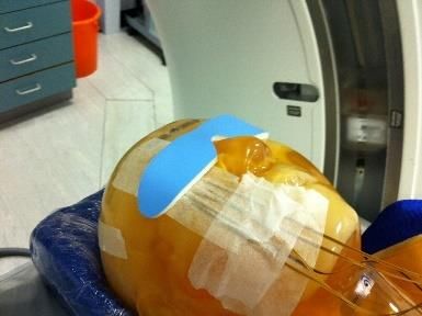
Guidance on using shielding on patients for diagnostic radiology applications

Executive summary

Over the last 70 years or so, it has been a common practice amongst radiological
professionals to place radiation protective material directly on the surface of a patient
during radiodiagnostic procedures to help reduce the dose to critical organs. This has led to
the expectation amongst patients and professionals alike that this would continue.
However, an increasing number of studies have raised concerns regarding the efficacy and
effectiveness of such ‘contact shielding’. This has led to an inconsistency in application and,
in some cases, friction between patients demanding shielding and professionals judging it is
unnecessary or even potentially harmful.

Therefore a working party consisting of representatives from various UK radiological
professional bodies was established to consider the evidence-base for patient contact
shielding and produce a consensus of opinion as to what constitutes best and agreed
practice, with the aim of improving consistency in application of such shielding.

This work challenges the historical perspective that using contact shielding only provides a
benefit for the patient. Rather it suggests that contact shielding can adversely interfere with
the imaging (leading to a repeat test) and, if misplaced or allowed to move during an
examination, can actually lead to increased patient radiation exposure, rather than the
reverse. Overall, the findings suggest that contact shielding provides minimal or no benefit
and professionals should concentrate on other areas of radiation protection which are more
effective in optimising the patient radiation exposure.

The recommended cessation of the widespread practice of applying patient contact
shielding requires a major cultural change in outlook regarding radiation safety and practice
amongst medical professionals, educators, regulators and the public alike. The adoption of
these guidelines into clinical practice will therefore also require a suitable education
programme which could incorporate some of the material provided here.

                                                       5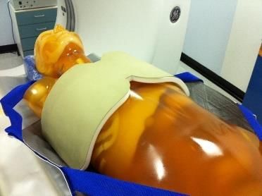
Guidance on using shielding on patients for diagnostic radiology applications

Chapter 1 Introduction

1.1 Background

The use of shielding, generally in the form of lead rubber, applied directly to patients has
been practised for many years to reduce the dose to critical organs, notably the gonads.
However, some studies have questioned the efficacy of using such shielding,1,2,3 while
others have highlighted the inconsistencies in application.4 These self-same studies have
called for national guidance to help reduce variations in approach. In addition, new designs,
applications and materials for patient contact protection have appeared on the market.5
Therefore a working party consisting of representatives from various UK radiological
professional bodies was established to consider the evidence-base for patient shielding and
produce a consensus of opinion as to what constitutes best and agreed practice, with the
aim of improving consistency in application of such shielding.

1.2 Scope

This guidance is intended to cover radiation protection applied directly to patients
undergoing diagnostic and interventional X-ray procedures within the healthcare sector,
hereafter referred to as patient contact shielding. It does not include shielding built into the
imaging equipment or in the room design and excludes ad hoc protection not actually
placed on the patient (e.g. the use of shielding on incubators in neonatal intensive care
units, as it does not touch the baby).

1.3 Aim

The aim of this document is to provide general guidance on why patient contact shielding
may be required and when and where it might be used, with the intention of reducing
confusion and improving consistency in practice across the UK. The number of X-ray
procedures is vast, thus providing advice for each examination and individual projection
would be problematic. Therefore the approach taken is to provide generalised reasons and
evidence (where available) for why protection may or may not be applicable.

The overriding consideration throughout this guidance is the patient’s needs, both in terms
of risk reduction and reassurance. The guidance therefore starts by addressing what levels
of risk might be involved, the place of shielding within an imaging task, and what sources of
radiation require attenuating. The area of staff/patient interaction, including the patient’s
expectations and staff concerns regarding physically applying shielding to a person, is also
addressed (see chapters 5 and 6). This is followed by several chapters describing how these
issues relate to specific imaging modalities. These have been designed to be read by those

                                                       6
Guidance on using shielding on patients for diagnostic radiology applications

who may only be interested in an individual modality and so they intentionally include some
repetition of text.

In order to provide a clear way forward, recommendations for local practice are provided at
the end of each chapter, along with the relevant evidence (references).

References

    1. Fawcett SL, Gomez AC, Barter SJ, Ditchfield M and Set P. More harm than good? The
       anatomy of misguided shielding of the ovaries. Br. J. Radiol. 2012; 85: e442–e447.
    2. Marsh RM and Silosky M. Patient shielding in diagnostic imaging: discontinuing a
       legacy practice. Am. J. Roentgenol. 2019; 212:1–3.
    3. McKenney S, Gingold E and Zaidi H. Point/Counterpoint: Gonad shielding should be
       discontinued for most diagnostic imaging exams. Med. Phys. 2019; 46(3), 1111–
       1114.
    4. Hayre CM, Blackman S, Carlton K and Eyden A. Attitudes and perceptions of
       radiographers applying lead (Pb) protection in general radiography: An ethnographic
       study. Radiography 2018; 24: e13–e18.
    5. AAPM, 2017. Position statement on the use of Bismuth shielding for the purpose of
       dose reduction in CT scanning. American Association of Physicists in Medicine, Policy
       Number PP 26-B. https://www.aapm.org/publicgeneral/BismuthShielding.pdf
       [Accessed 29.10.2019].

                                                       7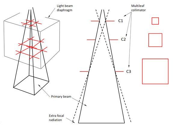
Guidance on using shielding on patients for diagnostic radiology applications
Chapter 2 General requirements for patient contact shielding
2.1 Radiation safety culture
In the UK, the exposure of patients is governed by the Ionising Radiation (Medical Exposure)
Regulations,1,2 IR(ME)R. These regulations encapsulate the fundamental radiation safety
principle of keeping patients doses ‘as low as reasonably practicable’, or ALARP.3
In general terms this is achieved by:
First, ensuring practitioners and operators are suitably trained, entitled and
competent.
Second, requiring each individual X-ray examination to be ‘justified’, meaning the
benefits outweigh the risks of the exposure. Justification involves making sure the
most appropriate examination takes place, including considering other imaging
modalities which do not involve ionising radiation.
Third, requiring the exposure to be ‘optimised’, to ensure that patient doses arising
from the exposure are kept as low as reasonably practicable (ALARP) consistent with
the intended purpose. This includes both exposure parameters and equipment
maintenance.
In applying these principles to the area of patient protection it is important to recognise that
optimisation of protection is not about minimising radiation dose, but rather balancing
detriments and benefits.4 It therefore involves managing the patient dose in line with the
intended medical purpose. For example, applying protection to reduce the dose while
increasing the risk of obscuring important diagnostic information is contrary to good
medical practice and is not sound radiological protection.
2.2 Context for applying shielding
It is important that the application of patient contact shielding, if required, should only take
place once all other dose reduction techniques (e.g. selection of exposure factors,
collimation) have been applied.
It is often assumed that shielding always improves patient safety, but this is not necessarily
the case. The use of shielding in diagnostic imaging should be guided by the supporting
evidence and the focus should be on what is safest for the patient. This guidance aims to
illustrate best practice and provides the scientific evidence to enable IR(ME)R1,2 operators
(e.g. radiographers, radiologists, dentists, radiology assistant practitioners) to communicate
with patients and those who care for them to provide adequate information in order to
reach agreement on the appropriate use of shielding. Every individual has a right to request
or refuse shielding and should be supported to make their own decision.5
8
Guidance on using shielding on patients for diagnostic radiology applications

While any dose reduction is desirable, in the context of the low levels of dose from
diagnostic radiology, other factors must be considered as strong influences on the decision
to use or withhold shielding. These are considered throughout this document. For example:
psychological factors such as whether it is a reassuring or alarming process to use a shield;
accuracy of imaging relating to the proximity of the shield to the imaged area; and practical
and comfort issues such as the position and weight of the shielding.

Appropriately trained staff with the knowledge and skills to listen as well as provide
adequate information should facilitate all discussions around the use of patient contact
shielding. Where appropriate, for example if the individual is particularly anxious or requires
additional reassurance, operators should take time to explain the function of shielding as
part of a multifactorial and overarching dose reduction strategy. The priority remains to
achieve a suitable diagnostic image, where benefit outweighs risk. If the patient/individual
chooses a course of action that might increase their risk in terms of radiation dose, it is the
operator’s responsibility to take action to prevent harm. Each decision to use shielding
should be relevant to the individual circumstances.

2.3 Medical Device and product marking

Patient contact shielding devices should be available, where appropriate, and may include
proprietary gonad shields and aprons and various in-house modified lead-rubber shapes. If
they are to be procured they require suitable marking (such as CE6) to indicate that the
manufacturer has checked that these products conform to the relevant safety, health or
environmental requirements. By this means the manufacturer is confirming that the product
is suitable to be sold throughout the specified market. At the time of writing (February
2020), the UK and EU are negotiating product safety marking.7

Users should be aware that if an item is purchased with the intention of placing it on a
patient to protect them from radiation then it is a medical device 8 and should be marked as
such. This includes gonad, eye, thyroid and breast protective products. The manufacturer of
such products should provide appropriate operating instructions, including when it can and
cannot be used9.

If, however, an item is purchased with the intention of using it for occupational protection
and as a patient shield, then it is both a medical device and personal protective equipment
(PPE).8 This could include “half-aprons” and dental aprons. In such cases the product should
be marked as a medical device. The manufacturer must also fulfil the relevant requirements
for the production of personal protective equipment.

                                                       9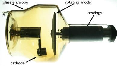
Guidance on using shielding on patients for diagnostic radiology applications

In the case of shielding designed in-house for a particular application then, provided it is not
then transferred or sold as a product, it is not classed as a medical device with regard to the
legislation and controls8.

2.4 Recommendations for local practice

The overall conclusion from the available evidence is that patient contact shielding is not
generally required in diagnostic and interventional radiology. The fundamental reason
being to protect the patient, given that the use of patient contact shielding can often
actually lead to an increase in patient dose (due to the need to repeat an examination or
interference with automatic dose control systems). Even outside the primary radiation
beam, efforts spent on correct positioning and optimising protocol parameters can lead to
dose savings which are more significant than applying patient contact shielding. This overall
conclusion is in line with the recent American Association of Physicists in Medicine position
statement regarding gonad and fetal shielding.10

Few exceptions have been identified, but these may occur where a particular patient care
pathway requires a number of repeat examinations where patient contact shielding may be
applied, particularly in the case of paediatric patients.

The recommended cessation of the widespread practice of applying patient contact
shielding requires a major cultural change in outlook regarding radiation safety and practice
amongst medical professionals, educators, regulators and the public alike. The adoption of
these guidelines into clinical practice will therefore also require a suitable education
programme which could incorporate some of the material provided here.

  Scenario                            Recommendation                        Comments

  Patient contact shielding in        Not recommended                       Anticipate very few specific
  diagnostic and                                                            situations where this does
  interventional radiology                                                  not apply.

References

    1. UK Government, 2017. The Ionising Radiation (Medical Exposure) Regulations 2017
       (No. 1322).
       http://www.legislation.gov.uk/uksi/2017/1322/pdfs/uksi_20171322_en.pdf
       [Accessed 29.10.2019].
    2. UK Government, 2018. The Ionising Radiation (Medical Exposure) Regulations
       (Northern Ireland) 2018. Statutory Instrument 2018 No. 17.
       http://www.legislation.gov.uk/nisr/2018/17/contents/made [Accessed 29.10.2019].

                                                      10
Guidance on using shielding on patients for diagnostic radiology applications
3. European ALARA Network. Optimization of Radiation Protection. ALARA: A Practical
Guidebook 2019. https://www.eu-alara.net/index.php/activities/documents-related-
to-alara/330-optimization-of-radiological-protection-alara-a-practical-
guidebook.html [Accessed 29.10.2019].
4. ICRP, 2013. Radiological protection in paediatric diagnostic and interventional
radiology. International Commission on Radiological Protection, Publication 121.
Ann. ICRP 42(2) (2013).
5. The Society and College of Radiographers, 2018. Obtaining consent: a clinical
guideline for the diagnostic imaging and radiotherapy workforce.
https://www.sor.org/sites/default/files/document-
versions/obtaining_consent_170118.pdf [Accessed 05.10.2018].
6. UK Government Guidance 2019a. CE marking. https://www.gov.uk/guidance/ce-
marking [Accessed 07.01.2020].
7. UK Government Guidance 2019b. Prepare to use the UKCA mark after Brexit.
https://www.gov.uk/guidance/prepare-to-use-the-ukca-mark-after-brexit [Accessed
07.02.2020].
8. MHRA, 2014. Borderlines with Medical Devices. Medicine and Healthcare Products
Regulator Agency, Guidance on legislation.
https://www.gov.uk/government/publications/borderlines-with-medical-devices
[Accessed 29.10.2019].
9. HSE, 2014. Safe Use of Work Equipment. Provision and Use of Work Equipment
Regulations 1998. Health and Safety Executive, Approved Code of Practice and
Guidance L22, 4th Edition (2014). http://www.hse.gov.uk/pubns/books/l22.htm
[Accessed 29.10.2019].
10. AAPM 2019. Position statement on the use of patient gonadal and fetal shielding.
American Association of Physicists in Medicine. Policy number PP 32-A.
https://www.aapm.org/org/policies/details.asp?id=468&type=PP¤t=true
[Accessed 29.10.2019].
11Guidance on using shielding on patients for diagnostic radiology applications
Chapter 3 Radiation dose and risk
3.1 Historical perspective
Past practice in radiation protection has been based on the dose range and associated risk
estimates prevalent at the time. However, the levels of dose and estimates of risk have
changed over the years (e.g. since some operators qualified), requiring continuous revision
of local practice in-line with current knowledge and advice. For example, in the UK the mean
entrance surface dose for an AP pelvis radiograph (where gonad protection may be
considered) dropped by a factor of 10 between 1900 and 1958 1,2 and then by a further
factor of 6 by 20103, see Figure 3.1.
Figure 3.1 Example change in mean entrance surface dose values with time for an AP Pelvis
radiograph. Based on doses reported in the literature.1 ,2, 3
3.1.1 Organs at risk (OAR)
Knowledge of the radiosensitivity of various tissues and organs has also changed with time
as new information and evidence became available. For example, the tissue weighting factor
(WT), is a relative measure of the risk of stochastic effects (see section 3.2) that might result
from irradiation of that specific tissue. Table 3.1 summarises the tissue weighting factors
recommended by the International Commission on Radiological Protection (ICRP) over a
thirty year period.4,5,6
12Guidance on using shielding on patients for diagnostic radiology applications
Table 3.1 Changes in the International Commission on Radiological Protection (ICRP)
recommended tissue weighting factors with tissue/organ and time
Tissue or ICRP recommended tissue weighting factor (wT)
organ
(in order of ICRP 26 ICRP 60 ICRP 103
wT) (1977) 4 (1990) 5 (2007)6
Bone marrow 0.12 0.12 0.12
(red)
Breast 0.15 0.05 0.12
Colon 0.12 0.12
Lung 0.12 0.12 0.12
Stomach 0.12 0.12
Gonads 0.25 0.20 0.08
Bladder 0.05 0.04
Liver 0.05 0.04
Oesophagus 0.05 0.04
Thyroid 0.03 0.05 0.04
Bone surface 0.03 0.01 0.01
Brain 0.01
Salivary glands 0.01
Skin 0.01 0.01
Sub Total 0.70 0.95 0.88
Remainder 0.30 0.05 0.12
tissues*
Total 1.00 1.00 1.00
* From ICRP 1036, remainder tissues are mean doses to adrenals, extrathoracic (ET) region,
gall bladder, heart, kidneys, lymphatic nodes, muscle, oral mucosa, pancreas, prostate
(male), small intestine, spleen, thymus, uterus/cervix (female)
13Guidance on using shielding on patients for diagnostic radiology applications
Organs such as colon and stomach have been given specific weighting factors in recent
years, but of particular importance for these guidelines are the significant changes in w T for
the gonads and breast tissue (see Figure 3.2). These figures would suggest that dose
optimisation processes should concentrate on those organs with the highest wT and much
less on those with a low wT, such as the gonads. The latest wT figures in Table 3.1 suggest
contributions from just five organs (bone marrow, breast, colon, lung and stomach) make up
60% of the total risk.
Figure 3.2 Tissue weighting factor versus year of recommendation by the ICRP for two
particular tissue types.4, 5, 6
3.1.2 Heritable effects
Information on hereditary (or genetic) effects of radiation was developed almost entirely
from animal experiments in the 1950s. This gave rise to considerable interest in measuring
gonad doses (e.g. the Committee on Radiological Hazards to Patients set up in 1956 under
the chairmanship of Lord Adrian2) and the introduction of gonad shielding methods during
the next few decades. However, more recently genetic risk estimations in human
populations have concluded that there is no direct evidence of a radiation associated excess
of heritable disease.6 This change in emphasis is illustrated in Figure 3.3 where the decrease
in estimated genetic risk has led directly to the significant reduction in the tissue weighting
factor for the gonads from 0.2 to 0.08 (see also Table 3.1).
14Guidance on using shielding on patients for diagnostic radiology applications
Figure 3.3. Illustrating how, over the past half century, the concern regarding exposure to
ionising radiation has changed from heritable (genetic) effects to carcinogenesis. [Used with
permission from Hall 2009.7]
The effects and risks from exposure to ionising radiation depend upon many factors, such as
the absorbed dose, the dose rate, quality of radiation, specifics of the tissue irradiated and
other factors such as the age and sex of the individual. The hereditary risks from irradiation
that might result in effects to offspring of humans appear to be much lower than the
stochastic effect of cancer induction and are now so low they are rarely considered.
Therefore carcinogenesis is currently considered the most important stochastic effect at
absorbed doses of less than 1 Gy. The risk of cancer induction varies widely across different
tissues; however, the risk of fatal radiation-induced cancer for a general population
following chronic exposure6 is about 5% Sv−1. Due to difficulties in obtaining accurate
evidence, quantification of cancer risk at doses of less than 0.1 Gy remains problematic.8
3.1.3 Eye lens risk
For a long time the lens of the eye has been regarded as radiosensitive in a deterministic
manner and therefore requires a threshold radiation dose to be exceeded before lens
opacities will develop. In recent years, a number of new studies have suggested an elevated
risk for cataract development in populations exposed to doses of ionising radiation below
the previously assumed thresholds.9 Deterministic thresholds for the lens of the eye
(radiation induced cataract) are now considered to be 0.5 Gy,6,9,10 with some authors raising
the possibility of no threshold at all.11 The particular form of cataract associated with
15Guidance on using shielding on patients for diagnostic radiology applications
ionising radiation is Posterior Subcapsular Cataract (PSC). Therefore minimising the eye lens
dose remains an important consideration in radiation protection.
3.2 Stochastic and hereditary risk
Radiation effects are divided into the categories of stochastic effects, tissue effects
(deterministic) and genetic effects. At dose levels commonly found in diagnostic radiology,
the overwhelming effect in both adult and paediatric patients is believed to be an increased
incidence and associated mortality from stochastic effects.12 Heritable effects to offspring
can be considered a negligible risk for the expected gonad doses associated with diagnostic
radiology (including CT).6
The significance of these radiation effects is dependent on the biological sex and age of the
patient at the time of the exposure and generally the younger the patient the more
important the effect is. The increased risk for a given dose for younger age groups reflects
the increased radiation organ sensitivity during development and the longer life expectancy
of the child, during which time a cancer can become established and develop.
Cancer incidence also varies considerably between the different organs, with the female
breast being the most radiosensitive at birth and the thyroid and female breast showing the
greatest decrease in radiosensitivity with age (see Figure 3.4).
Figure 3.4 Lifetime risk of cancer incidence by organ and age for a composite Euro-American
female population (% per Gy) (data from the Health Protection Agency 2011.13)
16Guidance on using shielding on patients for diagnostic radiology applications 3.3 Introduction to levels of risk In order to help prioritise dose reduction techniques, such as the use of patient contact shielding, it may be useful to consider a general scale of risk related to radiation dose, such as that provided in Table 3.2. Although the tabulated radiation dose is related to effective (whole body) dose, whereas patient shielding is used to minimise individual organ doses, the risk levels and descriptions are nevertheless a useful general guide. For instance, where the estimated examination dose is already in the ‘negligible’ risk category, then to provide shielding which halves the dose will have little, if any, effect on the overall risk to the patient and would suggest optimisation efforts would be better employed elsewhere. Table 3.2 Lifetime cancer induction risk categorisation for medical exposures, based on Martin et al.14 Effective dose Lifetime cancer Risk description Example radiological (mSv) induction risk procedure
Guidance on using shielding on patients for diagnostic radiology applications

3.4 Optimisation and applying the ALARP principle

How are we to balance risk and benefit and how far do we go to achieve ALARP? It is not
just about aiming for radiation doses ‘as low as’ conceivably possible, but keeping in mind
the idea of ‘reasonably practicable’.16

Some useful advice regarding ALARP has been provided by the Health and Safety
Executive,17 who suggest that ALARP does not mean that every measure that could possibly
be taken (however theoretical) to reduce risk must be taken and it does not represent zero
risk. The risk from an activity can never be entirely eliminated unless the activity is stopped.
Applying the ALARP principle is dependent upon the exercising of professional judgement
and experience, as well as following any consensus on ‘good practice’ established by
stakeholders. In addition, decisions about what is ALARP should be affected by changes in
knowledge about the size or nature of the risk presented by a hazard. If the evidence shows
the hazard presents a significantly lower risk than previously thought, then a relaxation in
controls may be accepted provided the new arrangements ensure the risks are ALARP.

Therefore, the process of applying optimisation and ALARP in diagnostic radiology should
concentrate on minimising the primary beam dose and prioritising protection for the ‘high
risk’ organs mentioned in sections 3.1 and 3.2. In the context of applying surface shielding
this includes the eye lens and breast tissue (and thyroid in paediatric patients). However,
primary beam shielding has the potential to increase dose when using automatic exposure
controls and risks obscuring pathology and adversely affecting clinical findings, potentially
leading to repeat exposures.

These considerations should guide clinical practice. For example, in comparing AP and PA
projections for abdominal radiography,18 the anatomically anterior positioning of the
stomach, colon and liver mean they would receive higher organ doses in an AP projection
than a PA projection and thus make a greater contribution to effective dose. Similarly, in the
selection of tube potential (kV) or filtration for a radiographic examination, increasing the
kV will give more penetrating radiation, lowering the dose to more superficial tissues, while
the effect on doses to tissues deep within the body near to the image receptor will be
minor.19

The decision regarding whether any further dose reduction (e.g. from secondary radiation)
is necessary, should take into account the levels of risk highlighted in section 3.3. It is
suggested that if the dose has been reduced to the ‘negligible’ risk level, no further action
need be taken to satisfy ALARP.

Other considerations which might influence the optimisation process could include cases
where a patient is likely to undergo multiple or sequential examinations.

                                                      18
Guidance on using shielding on patients for diagnostic radiology applications
References
1. Kotre CJ and Little BG. Patient and staff radiation doses from early radiological
examinations (1899-1902). Br. J. Radiol. 2006; 70: 837–842.
2. Adrian, 1966. Final report of the Adrian Committee on Radiological hazards to
patients. London, HMSO. https://archive.org/details/op1269041-1001/page/n1
[Accessed 29.10.2019].
3. HPA, 2012. Doses to patients from radiographic and fluoroscopic X-ray imaging
procedures in the UK – 2010 review. Health Protection Agency Report HPA-CRCE-
034. https://www.gov.uk/government/publications/radiographic-and-fluoroscopic-
X-rays-patient-doses [Accessed 29.10.2019].
4. ICRP, 1977. Recommendations of the International Commission on Radiological
Protection. ICRP Publication 26. Ann. ICRP 1 (3).
5. ICRP, 1991. 1990 Recommendations of the International Commission on Radiological
Protection. ICRP Publication 60. Ann. ICRP 21 (1–3).
6. ICRP, 2007. The 2007 Recommendations of the International Commission on
Radiological Protection. ICRP Publication 103. Ann. ICRP 37 (2–4).
7. Hall EJ. Is there a place for quantitative risk assessment? J. Radiol. Prot. 2009; 29:
A171–A184.
8. Ross JC, Vilić D and Fongenie B. Reforming the debate around radiation risk. J. Radiol.
Prot. 2019; 39: 635–640.
9. ICRP, 2012. ICRP statement on tissue reactions and early and late effects of radiation
in normal tissue and organs – threshold doses for tissue reactions in radiation
protection context. International Commission on Radiological Protection, Publication
118. Ann. ICRP 41 (1–2).
10. ICRP Statement on Tissue Reactions. ICRP reference 4825-3093-1464. Commission
Approved Statement 21.4.2011. http://www.icrp.org/docs/2011%20Seoul.pdf
[Accessed 22.07.2019].
11. Michel M, Jacob S, Roger G, Pelosse B, Laurier D, Le Pointe HD and Bernier MO. Eye
lens radiation exposure and repeated head CT scans: A problem to keep in mind. Eur.
J. Radiol. 2012; 81(8): 1896–900.
12. IAEA, 2013. Dosimetry in diagnostic radiology for paediatric patients. International
Atomic Energy Agency, Human Health Series No. 24. https://www-
pub.iaea.org/books/iaeabooks/8965/dosimetry-in-diagnostic-radiology-for-
paediatric-patients [Accessed 29.10.2019].
13. HPA, 2011. Radiation risks from medical X-ray examinations as a function of age and
sex of the patient. Health Protection Agency Report HPA-CRCE-028.
https://www.gov.uk/government/publications/medical-x-rays-radiation-risks-by-
age-and-sex-of-patient [Accessed 29.10.2019].
14. Martin CJ, Vassileva J, Vano E, Mahesh M, Ebdon-Jackson SE, Ng KH, Frush DP, Loose
R and Damilakis J. Unintended and accidental medical radiation exposures in
radiology: guidelines on investigation and prevention. J. Radiol. Prot. 2017; 37: 883–
906.
14. Health Risks from Exposure to Low Levels of Ionizing Radiation: BEIR VII Phase 2.
Washington, DC: National Research Council; 2006. Report No.: 978-0-309-09156-5
19Guidance on using shielding on patients for diagnostic radiology applications
15. Coates R. International Radiation Protection Association, President’s Blog. IRPA,
Bulletin 20 (December 2018). http://www.irpa.net/page.asp?id=54592 [Accessed
29.10.2019].
16. HSE, 2018. ALARP “at a glance”. Health and Safety Executive web-based guidance.
http://www.hse.gov.uk/risk/theory/alarpglance.htm [Accessed 18.11.2018]
17. Martin CJ, Sutton DG and Sharp PF. Balancing patient dose and image quality. Appl.
Radiat. Isot. 1999; 50: 1–19.
18. Martin CJ and Sutton DG. Practical Radiation Protection in Healthcare. 2 nd edition
(2014). Oxford University Press: Oxford.
20Guidance on using shielding on patients for diagnostic radiology applications
Chapter 4 Sources of radiation exposure
Effective use of protective equipment requires a clear understanding of the sources of
ionising radiation that a patient is exposed to while undergoing a radiological investigation.
These sources include the primary beam and secondary radiation from several sources.
Knowledge of the relative intensity of each of these sources should guide radiation
protection practice. There has been confusion evident in the literature concerning the
terminology, generation process, and relative intensity of some of these sources of
secondary radiation. This chapter aims to provide the current knowledge available to help
guide protection practice for all professional groups engaged in the examination of patients.
4.1 Primary radiation
This is the radiation emitted from the X-ray tube in the intended field of irradiation. Dose
rates within the primary beam can be relatively high. There are broadly three categories of
dose rate commonly delivered to the surface of the patient as primary radiation in
radiological examinations. One is between 1 and 10 mGy s-1 and would include fluoroscopic
exposures. The second category typically ranges between 15 and 25 mGy s-1 and would
include projection radiographs, dental examinations, angiographic acquisitions,
fluorography acquisitions and mammograms. The third can deliver 50 to 100 mGy s -1 and is
exclusively for computed tomography (CT) examinations. These dose rates are at least fifty
times the dose rate from even the most significant source of secondary radiation, it is
therefore extremely important to limit the area of the primary beam.
4.1.1 Collimation
The size of the primary beam is controlled by means of a collimation system. An example of
the effectiveness of the collimators in general radiography is given in Figure 4.1, which
demonstrates the steep decline in radiation output close to the collimator edge. The insert
in this figure illustrates the low output levels measured outside of the collimated region, in
this case dropping to less than 1% of the primary beam output within approximately 25 mm.
Therefore efficient use of collimators is a significant contributor to optimisation of patient
dose.
Operators must include all anatomy and pathology indicated by the examination protocol.
Ensuring this is achieved in just a single exposure is a skilled task. Inadequate collimation
(use of large field sizes, or too small a field size requiring a repeated exposure) has been
shown to be a major cause of increased risk to patients, especially children and
neonates.1,2,3 There is a fine balance between adequate visualisation of anatomy and
pathology on the one hand and beam size limitation for radiation protection purposes on
the other.
21Guidance on using shielding on patients for diagnostic radiology applications
Figure 4.1 An example of the change in radiation output with distance from the centre of the
X-ray beam for a conventional radiographic X-ray tube, in the directions parallel to and
perpendicular to the X-ray tube Anode Cathode (A/C) axis. The collimated X-ray beam edge
in this example is within 3 mm of the light beam edge, as shown. The insert highlights the
response close to the field edge, where the radiation output falls to less than 1% within 25
mm.
4.2 Secondary radiation
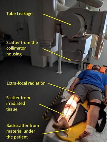
All other sources of radiation within the X-ray room are termed secondary radiation. Figure
4.2 highlights secondary sources of radiation for a projection radiography situation, namely
tube leakage, extra focal radiation and several sources of scattered radiation. These
secondary sources are also present in other modalities, such as CT and fluoroscopy.
Patients may be completely unaware of these sources. Conversely, they may be
unnecessarily anxious about the risks posed by them. Therefore, the absence of measures to
protect against them may need to be explained.
22Guidance on using shielding on patients for diagnostic radiology applications
Figure 4.2 Secondary sources of radiation present during projection radiography.
4.2.1 Tube housing radiation leakage
Leakage radiation is the term given to radiation escaping the X-ray tube housing other than
through the tube port. This must be limited to less than 1 mGy hr-1 averaged over an area of
1 m2 at a distance of 1 metre from the focal spot.4 In practice, the dose rate from leakage
radiation in a properly designed and maintained system5 will be less than 0.3 mGy hr-1.
4.2.2 Scatter from tube, filtration and housing
Scatter in the tube and housing is a well-known source of secondary radiation; it is
generated as the primary beam passes through the construction elements of the tube,
coolant, tube housing and the collimator. This scatter will give rise to very low levels of
additional dose for the patient. It is common to have a transmission ionisation chamber
attached to the front of the collimator. This can be a source of additional scattered
radiation.
4.2.3 Extra-focal radiation
This occurs adjacent to the collimated X-ray field and is generated by energised electrons in
the tube that interact with parts of the anode other than the focal spot. This should not be
confused with the penumbra of the primary beam; it is of lower intensity but affects a much
larger area.
23Guidance on using shielding on patients for diagnostic radiology applications
Figure 4.3. A typical rotating anode X-ray tube. The cathode is the source of energised
electrons, which travel to the anode on initiation of exposure
(https://upload.wikimedia.org/wikipedia/commons/d/d5/Rotating_anode_X-
ray_tube_%28labeled%29.jpg).
Figure 4.3 identifies the components of a typical rotating anode X-ray tube. Electrons
released from the heated cathode filament are accelerated across the near total vacuum in
the tube by electrostatic forces. Various design elements help to focus the majority of
accelerating electrons to interact with the anode in a very small area, known as the focal
spot. However, this focusing is not perfect. Electrons can diverge from the accelerated
electron beam. Because they are generated away from the focus, any X-ray photons
generated from these electron interactions are termed extra-focal, or off-focus radiation.
Their exit trajectory from the tube housing and collimator are different from that expected
and illuminated by the light field. They can emerge from the collimator more divergent from
the central ray than the primary field. These photons are therefore present across the
illuminated field and also beyond it. Modern multi-leaf collimators are designed with extra
collimation leaves close to the tube port (Figure 4.4, C1) to reduce the area irradiated by
extra-focal radiation as much as possible.
24Guidance on using shielding on patients for diagnostic radiology applications
Figure 4.4 Extra-focal radiation and its trajectory from the X-ray tube and collimator. This
diagram shows a multi-leaf collimator designed to reduce off-focal radiation to the
minimum.
The effects of extra-focal radiation can clearly be seen using modern digital detectors with a
wide dynamic range (Figure 4.5). The use of extra-focal shielding immediately adjacent to
the collimated primary beam has been advocated by several authors.6,7 However, risks to
the patient from the focal/extra-focal radiation is generally agreed to be small, due to the
primary to extra-focal radiation ratio being of the order of 500:1.8,9,10
25Guidance on using shielding on patients for diagnostic radiology applications Figure 4.5 The window width of this image has been decreased. The image of soft tissue and bone from extra-focal radiation outside of the primary field of irradiation is now evident. 4.2.4 Scatter from irradiated objects The patient themselves and the patient support are a source of secondary radiation during exposure. Internal scatter within the patient is difficult to quantify but can be the major source of secondary radiation to an organ outside the primary beam. It is very difficult to shield one part of the patient from another internally. Work by Iball and Brettle (2010)11 provides evidence to suggest that it is the predominant component of any radiation dose measured within the patient close to the primary beam (
Guidance on using shielding on patients for diagnostic radiology applications

theoretical possibility that scatter leaving the patient might be backscattered towards the
patient from any shielding applied, such as a drape or pad for the protection of cardiologists
fingers. Their modelling suggests there may be an effect, but it is small, superficial, and falls
off rapidly away from the primary X-ray field (Figure 4.6).

Figure 4.6 Reduction in radiation dose with distance from the primary X-ray field (from
reference 13, used with permission).

Iball et al (2008)14 modelled patient dose from secondary sources of radiation in Computed
Tomography, where the dose rates and duration of the primary beam are high. Figure 4.7
shows the relative contribution of the three types of scatter (internal, external, and
backscatter from an applied shield) to the fetus. This shows that the backscatter from the
applied shielding towards the patient is the smallest and its insignificance as a contributor to
patient dose is apparent when it is remembered that all these contributions are many
magnitudes smaller than the incident primary beam dose rates.

                                                      27
Guidance on using shielding on patients for diagnostic radiology applications
Figure 4.7 The relative contributions of the three sources of secondary radiation to the total
fetal dose at 140 kVp. Used with permission from Iball et al (2008).14
4.3 Summary
The primary beam provides significantly higher dose rates than all sources of secondary
radiation, as illustrated in Table 4.1. Therefore optimisation techniques which limit the
primary beam size and position will have far greater impact upon patient dose than any
efforts spent reducing the exposure from secondary radiation sources. After that, if
additional shielding is deemed necessary to reduce secondary radiation, then it will be most
effective close to the beam edge.
Table 4.1 provides a comparison of the three categories of primary beam dose rates
typically encountered and the likely dose rates from secondary sources thus generated. It is
useful for comparison However, it is important to remember that patient dose will depend
on the duration of the exposure as well as the dose rate. This can be as short as a few
milliseconds in projection radiography, compared with CT sequences of several seconds and
fluoroscopy exposure times with a potential duration of many minutes. Any risk benefit
calculation regarding the potential application of any contact shielding must take into
account the likely dose rate (and duration of exposure) of the sources the shielding might
attenuate.
28Guidance on using shielding on patients for diagnostic radiology applications
Table 4.1 Example dose rates (in mGy s-1) at 75 cm from the tube focus, due to various
radiation sources, for three X-ray imaging modalities.
Source of radiation exposure Dose Rate (mGy s-1) at 75 cm
Fluoroscopy Projection CT
Radiography
Primary beam 5 25 50
Extra-focal (0.2% of primary) 0.01 0.05 0.10
Scatter from irradiated objects 0.001 0.005 0.010
Tube housing leakage 0.0001 0.0001 0.0001
References
1. Datz H, Bader D, Sadetzki S, Juster-Reicher A, Marks K, Smolkin T, Zangen S and
Margaliot M. The additional dose to radiosensitive organs caused by using under-
collimated X-ray beams in neonatal intensive care radiography. Rad. Prot. Dosim.
2008; 130(4): 518–524.
2. Fauber TL and Dempsey MC. X-ray Field Size and Patient Dosimetry. Radiol. Technol.
2013; 85(2): 155–161.
3. Stollfuss J, Schneide, K and Krüger-Stollfuss I. A comparative study of collimation in
bedside chest radiography for preterm infants in two teaching hospitals. Eur. J.
Radiol. Open. 2015; 2: 118–122.
4. BSI, 2010. Medical electrical equipment. Particular requirements for the basic safety
and essential performance of X-ray tube assemblies for medical diagnosis. BS EN
60601-2-28, London, BSI (2010).
5. Tsalafoutas IA. Excessive leakage radiation measured on two mobile X-ray units due
to the methodology used by the manufacturer to calculate and specify the required
tube shielding. Br. J. Radiol. 2006; 79(938): 162–164.
6. Hawking N and Sharp T. Decreasing Radiation Exposure on Pediatric Portable Chest
Radiographs. Radiol. Technol. 2013; 85(1): 9–16.
7. Mekis N, Zontar D and Skrk D. The effect of breast shielding during lumbar spine
radiography. Radiol. Oncol. 2013; 47(1): 26–31.
8. Miettunen R. Measurement of extra-focal radiation by computed radiography. Br. J.
Radiol. 1992; 65: 238–241.
9. Birch R. The spectrum and intensity of extra-focal (off-focus) radiation. Br. J. Radiol.
1976; 49:951–955.
10. Thomas SR, Freshcorn JE, Krugh KB, Henry GC, Kereiakes JG and Kaufman RA.
Characteristics of extra-focal radiation and its potential significance in pediatric
radiology. Radiology 1983; 146: 793–799.
29Guidance on using shielding on patients for diagnostic radiology applications
11. Iball GR and Brettle DS. Organ and effective dose reduction in adult chest CT using
abdominal lead shielding. Br. J. Radiol. 2011; 84: 1020–1026.
12. Culp M and Barbara J. Shield placement: Effect on exposure. Radiol. Technol. 2014;
85(4): 369–376.
13. Matyagin YP and Collins PJ. Effectiveness of abdominal shields in chest radiography:
a Monte Carlo evaluation. Br. J. Radiol. 2016; 89(1066): 20160465.
14. Iball G, Kennedy EV and Brettle DS. Modelling the effect of lead and other materials
for shielding of the fetus in CT pulmonary angiography, Br. J. Radiol. 2008; 81(966):
499–503.
30Guidance on using shielding on patients for diagnostic radiology applications
Chapter 5 Operator responsibilities
5.1 Overview
The rare application of patient contact shielding should be justified and employers should
develop clear criteria for its use. The operator must be adequately trained (see chapter 6)
and aware of their responsibilities when using patient contact shielding. In general this
should be covered by education at pre-registration level, local training and Continuous
Professional Development (CPD) programmes.
Patients should experience the same high standards of care regardless of where their
medical exposure takes place. Guidance provides scientific evidence to support assistant
practitioners (AP), radiographers, radiologists and others acting as IR(ME)R1,2 operators to
offer safe, high quality and consistent radiographic care to patients.
The operator should be familiar with the guidance and employers should support the
operator by providing written procedures, time and suitable equipment for staff to carry out
their duties as IR(ME)R operators.
The operator is an IR(ME)R-entitled duty holder responsible for practical aspects of the
exposure and for complying with the employer’s procedures.1,2 The use of patient contact
shielding in diagnostic imaging is a practical aspect. This guidance recommends its use only
in specific circumstances informed by recent and relevant evidence. Operators should be
further guided by what matters to the patient,3 taking care to ensure operator actions result
in an overall net benefit to the patient.
Operators should take care to ensure the patient understands the function of shielding as
the final element in a comprehensive and individualised dose reduction strategy. Where
indicated, it should be integral to the benefit risk conversation with the patient. Operators
should be respectful of individual choice and non-judgmental; the operator has a
responsibility to keep the patient safe and to take action to prevent harm. Shielding devices
should be appropriately used and accurately positioned to provide efficient protection to
the relevant body part.4
It is considered good practice to have a written procedure for the use of patient shielding
which should contain inclusion criteria. It may be helpful to incorporate scenarios to
illustrate how and when patient shielding should be used. It is important to note that local
procedures should allow for the professional judgement of the operator in individual
circumstances. The operator should document reasoned decisions that do not comply with
the procedure. Procedures should include a process to manage the need for repeat
exposures and how this is recorded (see chapter 6).
31Guidance on using shielding on patients for diagnostic radiology applications
5.2 Communication
Historical practice means that for some time there is likely to be a natural expectation that
patient contact shielding is used. Operators may need to take time to explain to the patient
the rationale for not using it until this becomes normalised practice. In the rare
circumstances where its use is advocated, operators should be adequately trained to do
so.1,2 The application of shielding directly onto the skin or clothing of a patient can be a
sensitive task. The patient should be provided with adequate information, prior to
placement, which explains the associated benefits and risks of using the shielding. Good
communication, where the conversation is supported by knowledge and evidence, helps
nurture trust between the patient and the operator and is likely to result in a higher rate of
acceptable diagnostic images. The priority should always be achieving a high quality
diagnostic image where benefit outweighs risk.5
When communicating the benefit and risk of using patient contact shielding the following
points should be considered:
Is the patient/their representative/the referrer asking for patient contact shielding
contrary to recommended guidelines? In these circumstances, is the operator
confident to respond to challenges regarding the absence of shielding and if not why
not?
Does the evidence support the use of patient contact shielding for this examination?
(See chapter 3.)
Is there a local procedure for this examination? (See chapter 6.)
Does the patient meet the inclusion criteria? (See chapter 6.)
Is the operator/trainee adequately trained/supervised to use the shielding? (See
chapter 6.)
Has the application of local procedures for transgender or gender non-conforming
individuals been considered?
Is there anything in the clinical information for this patient that precludes the use of
patient contact shielding?
Is its use justified? (Consider the risk of the patient being unable to comply and the
effect on image quality.)
Is the patient contact shielding fit for purpose? (Approved for use, free from defect,
clean and the correct size – include special considerations in neonatal care.)
Will it do any harm to the patient or adversely affect image quality if it is used
contrary to local procedures or professional guidance? Decisions made in these
circumstances should be documented along with the rationale for doing so.
Is it safe to delay the examination if the patient is still insisting on the use of patient
contact shielding contrary to advice? Is the patient likely to be significantly reassured
if patient contact shielding is used, even if it is unlikely to afford them any radiation
protection? (N.B. it is not recommended that patient contact shielding is used as a
32Guidance on using shielding on patients for diagnostic radiology applications
means of reassurance. This should be addressed through appropriate one to one
communication.)
The specific needs of paediatric patients should be taken into consideration and techniques
used to aid communication and nurture confidence (for example play specialists and
distraction techniques). The use of patient contact shielding, where indicated, must be the
final step in an overarching optimisation strategy.
The following scenarios are provided to illustrate some of the challenges and suggested
outcomes operators may experience in practice.
Scenario 1
A two year old child arrives for a chest X-ray. They are upset and distracted by the
unfamiliar environment. The operator, who is a radiographer in this case, explains the
benefits and risks of the exposure to the child’s parent. In accordance with the locally
agreed procedure, patient contact shielding is not required for this examination. The
radiographer provides assurance that the potential harm from a repeat exposure is
considered a greater risk than the exposure from scattered radiation. They further explain
that the priority for optimising the child’s exposure is close collimation of the primary
beam in order to avoid irradiating organs unnecessarily. The parent is reassured and
agrees to the examination proceeding without the use of patient contact shielding. A play
specialist works with the operator and parent and helps calm the child who manages to sit
still in the required position for the chest X-ray.
The risk of patient contact shielding moving and obscuring the lung bases should be
balanced against the risk of a repeat exposure and the anticipated benefit from reducing
dose from scattered radiation.
Scenario 2
A pregnant 25 year old female attends from the emergency department for a CT
pulmonary angiogram. There is a high clinical suspicion of pulmonary embolism. The
patient is very unwell and is also distressed about the safety of her unborn child during the
scan. She is insisting on the use of patient contact shielding for her abdomen and pelvis.
There is a local procedure for pregnant patients undergoing CT which necessitates a
consultant referrer to consultant practitioner referral pathway and recommends patient
shielding is not used. The examination is justified with instructions for additional
optimisation by using a reduced scan length. The radiographer explains the benefits and
risks of the scan to the patient including the local policy not to use patient shielding. The
patient is not convinced as she is aware that radiation can cause cancer. Consequently the
patient becomes more distressed despite the efforts of the radiographer and the
emergency department staff to reassure her. The radiographer discusses the benefits and
risks with the referrer and the practitioner.
33You can also read