Gut microbiota transplantation for colonization of germ-free mice
←
→
Page content transcription
If your browser does not render page correctly, please read the page content below
ll
                                                                                                               OPEN ACCESS

Protocol
Gut microbiota transplantation for
colonization of germ-free mice

                                                                                                      Jocelyn M. Choo,
                                                                                                      Geraint B. Rogers
                                                                                                      jocelyn.choo@sahmri.com
                                                                                                      (J.M.C.)
                                                                                                      geraint.rogers@sahmri.
                                                                                                      com (G.B.R.)

                                                                                                      Highlights
                                                                                                      Describes
                                                                                                      preparation and
                                                                                                      transplantation of gut
                                                                                                      microbiota into germ-
                                                                                                      free mice

                                                                                                      Strict anaerobic
                                                                                                      processing of donor
                                                                                                      cecal microbiota is
                                                                                                      essential

                                                                                                      Faithful microbiome
                                                                                                      replication requires
                                                                                                      multiple instillations
                                                                                                      by gavage

                                                                                                      Adaptable to
                                                                                                      different gut
                                                                                                      materials and
The use of germ-free mice is integral to the understanding of host-gut microbiome relationships.      highlights
Such models rely on faithful replication of the donor microbiome to establish causal effects of the   recommended
gut microbiota on host pathophysiology. This protocol describes the preparation and transfer of       validation steps
donor microbiota, focusing on strict anaerobic processing methods and multiple instillations by
gavage for optimal gut microbiota recovery.

                                                                                                      Choo & Rogers, STAR
                                                                                                      Protocols 2, 100610
                                                                                                      September 17, 2021 ª 2021
                                                                                                      The Authors.
                                                                                                      https://doi.org/10.1016/
                                                                                                      j.xpro.2021.100610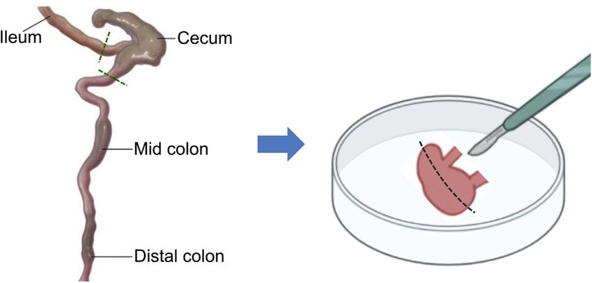
ll
                                                                                                                                         OPEN ACCESS

Protocol
Gut microbiota transplantation for colonization of germ-
free mice
Jocelyn M. Choo1,2,3,4,* and Geraint B. Rogers1,2,*
1Microbiome    and Host Health, South Australia Health and Medical Research Institute, Adelaide 5000, Australia
2Microbiome    Research Laboratory, College of Medicine and Public Health, Flinders University, Bedford Park 5042, Australia
3Technical   contact
4Lead   contact
*Correspondence: jocelyn.choo@sahmri.com (J.M.C.), geraint.rogers@sahmri.com (G.B.R.)
https://doi.org/10.1016/j.xpro.2021.100610

SUMMARY
The use of germ-free mice is integral to the understanding of host-gut micro-
biome relationships. Such models rely on faithful replication of the donor micro-
biome to establish causal effects of the gut microbiota on host pathophysiology.
This protocol describes the preparation and transfer of donor microbiota,
focusing on strict anaerobic processing methods and multiple instillations by
gavage for optimal gut microbiota recovery.
For complete details on the generation and use of this protocol, please refer to
Choo and Rogers (2021).

BEFORE YOU BEGIN
Reagent preparation

        Timing: 1 h preparation time, 10 days wait time

1. Prepare sterile 30% glycerol in phosphate-buffered saline (PBS) solution (pH 7.4).
2. Prepare sterile 20% glycerol in PBS solution (pH 7.4).
3. Prepare sterile PBS solution (pH 7.4), stored in a Falcon tube.
4. Place all reagents in anaerobic chamber for at least 10 days prior to use.
5. Prepare sterile pre-reduced PBS solution (pH 7.4), stored in an anaerobic culture tube (for
   example, Hungate tube).
   a. Prepare sterilized Hungate tube as detailed in Figure 1.
   b. In an anaerobic chamber, use a sterile syringe and 23G needle to transfer sterile pre-reduced
      PBS solution into Hungate tube.
6. Pre-weigh a sterile, capped, 50 mL Falcon tube to at least two decimal places (for use in step-by-
   step 5d).

     Alternatives: An anaerobic bag (for example, the Spilfyter Hands-in-Bag Atmospheric Cham-
     bers) may be considered as a cost-effective alternative to the anaerobic chamber to maintain
     an anaerobic environment. Equipment details are available from the supplier (https://us.vwr.
     com/store/product/4775838/spilfyter-hands-in-bag-atmospheric-chambers) and usage
     method is demonstrated in Weldy et al., 2020; Methods Video S1.

Suitability of donor and recipient mice

        Timing: >10 days

                                                                                   STAR Protocols 2, 100610, September 17, 2021 ª 2021 The Authors.       1
                                                  This is an open access article under the CC BY license (http://creativecommons.org/licenses/by/4.0/).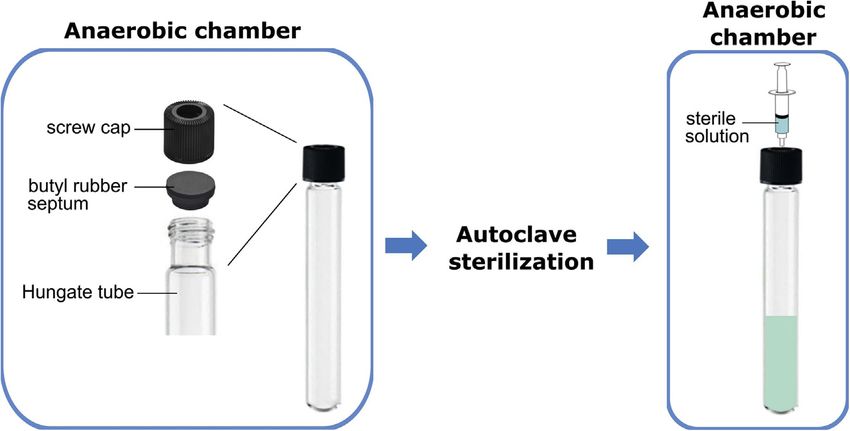
ll
     OPEN ACCESS
                                                                                                                       Protocol

Figure 1. Preparation of Hungate tube
Seal Hungate tube using a butyl rubber septum, followed by a screw cap in an anaerobic chamber. Sterilize the sealed
Hungate tube by autoclaving. Following transport of the Hungate tube into an anaerobic chamber, transfer the
appropriate solution into the Hungate tube using a sterile syringe and needle. Further information on the Hungate
tube technique is accessible from the Leibniz Institute DSMZ (https://www.dsmz.de/fileadmin/Bereiche/
Microbiology/Dateien/Kultivierungshinweise/Kultivierungshinweise_neu_CD/englAnaerob.pdf).

7. All animal procedures need to be approved by an Institutional Animal Care and Use Committee.
   All experiments in this protocol were approved by the South Australia Health and Medical
   Research Institute Animal Ethics Committee (SAM# 378).
8. Germ-free mice should be housed 2 to 5 animals of the same sex per cage, ensuring a random
   distribution of variables such as age and litter.
   a. House mice in a temperature-controlled room (22 G 2 C) on a 12-h light/dark cycle.
9. Acclimatize mice to facility conditions for at least 10 days prior to inoculation.

    Note: Adult male mice should not be cohoused unless they were housed together before sex-
    ual maturity, which is reached between 40 to 60 days of age.

    Note: Factors that are known to contribute to variation in the gut microbiota include the sex
    (Elderman et al., 2018), genotype (Ericsson et al., 2017), diet (Rausch et al., 2016), and
    bedding type (Rausch et al., 2016). These variables should be kept consistent across donor
    mice, wherever possible.

    Note: This protocol was used for gut microbiota colonization of 6 to 7-week old female
    C57BL/6 germ-free mice using material from C57BL/6 donor mice. The protocol can be
    used for other adult donor or germ-free mouse sex, genotype and ages 6 weeks or older.

KEY RESOURCES TABLE

REAGENT or RESOURCE                              SOURCE                                 IDENTIFIER
Chemicals, peptides, and recombinant proteins
13 Phosphate-buffered saline (pH 7.4)            Thermo Fisher Scientific               10010
Glycerol                                         Thermo Fisher Scientific               17904
Experimental models: Organisms/strains
Conventional mice                                The Jackson Laboratory                 C57BL/6
Germ-free mice                                   Translational Research                 C57BL/6
                                                 Institute, Queensland, Australia
                                                                                           (Continued on next page)

2     STAR Protocols 2, 100610, September 17, 2021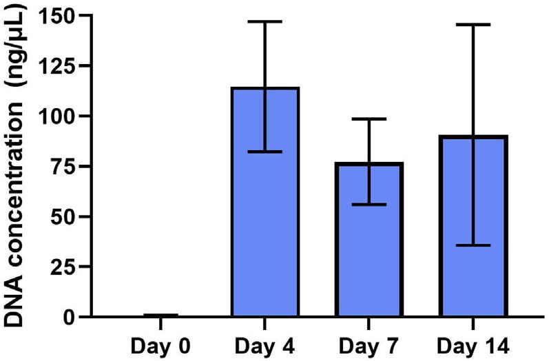
ll
Protocol                                                                                                                OPEN ACCESS

Continued
REAGENT or RESOURCE                           SOURCE                           IDENTIFIER
Other
Anaerobic chamber                             COY                              7150220
Hungate Tube (16 3 125 mm)                    Bellco Glass                     2047-16125
Corning 100 mm Cell Strainer                  Corning                          431752
Pestle for cell strainer                      Sigma-Aldrich                    Z742105
Petri dish 90 mm                              Thermo Fisher Scientific         LBS60014X
Sterile spatula                               Sigma-Aldrich                    CLS3005-100EA
Disposable scalpel                            Adelab Scientific                LV-SCP21
Syringe (1 mL, 10 mL, 30 mL)                  BD                               309659, 303134, 309651
Needle (23G)                                  Terumo                           219912
Gavage needle                                 Instech                          N/A
Minisart syringe filter (0.22 mm)             Sartorius                        16532
Vortex mixer                                  Ratek                            VM1-FT
QIAGEN DNeasy PowerLyzer PowerSoil kit        QIAGEN                           12855-100
Qubit dsDNA HS Assay Kit                      Thermo Fisher Scientific         Q32851
Qubit Flex Fluorometer                        Thermo Fisher Scientific         Q33327
Tubes with screw cap (10 mL, 50 mL)           Thermo Fisher Scientific         348224, 339653
Teklad Global 18% Rodent Diet                 Envigo                           2018S

MATERIALS AND EQUIPMENT

Pre-reduced 15% glycerol-phosphate buffered saline solution
Reagent                                       Final concentration                               Amount
Glycerol                                      15%                                               7.5 mL
PBS (pH 7.4)                                  n/a                                               42.5 mL
Total                                         Not applicable                                    50 mL

Pre-reduced 30% glycerol-phosphate buffered saline (PBS) solution
Reagent                                      Final Concentration                                Amount
Glycerol                                     30%                                                15 mL
PBS (pH 7.4)                                 n/a                                                35 mL
Total                                        Not applicable                                     50 mL

    Note: All solution can be stored at room temperature (22 G 2 C) for up to a year.

        CRITICAL: The solution should be filter-sterilized into a sterile 100 mL bottle by passing
        the solution through a 0.22 mm pore size syringe filter using a syringe with luer lock, or
        an equivalent device with a similar pore size, such as a 0.2 mm sterile bottle top filter. A
        bottle with a large aperture is recommended to enable rapid reduction of the solution.
        The bottle containing the solution should be placed with the seal cap loosened in the
        anaerobic chamber at room temperature (22 G 2 C) for at least 10 days prior to use.
        The mixture should be swirled at least once a day to aid complete reduction.

    Alternatives: Sterile PBS solution (pH7.4) can also be prepared in-house using the published
    protocol (Cold Spring Harbor Laboratory Press, 2006, https://doi.org/10.1101/pdb.rec8247).

STEP-BY-STEP METHOD DETAILS
Harvest and preparation of pooled donor cecal material

        Timing: 2.5 h

                                                                                         STAR Protocols 2, 100610, September 17, 2021   3
ll
OPEN ACCESS
Protocol
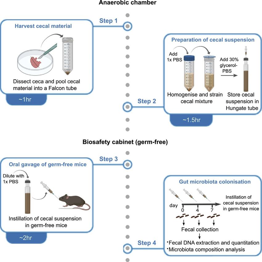
Preparation of cecal suspension under anaerobic conditions for inoculation into germ-free mice. The
commensal gut microbiota is dominated by anaerobic bacteria. Processing of donor material under
strict anaerobic conditions is therefore essential in maintaining viability across constituent taxa. The
time duration indicated for this section is an approximate for processing the ceca of three to five
mice of the same group.
1. Perform humane killing of mice according to ethics approval.
2. Position mouse in a supine position and perform a vertical midline incision to expose the gastro-
intestinal organs.
3. Dissect cecum from the gastrointestinal tract using a sterile scalpel (Figure 2).
a. Perform a horizontal dissection at the junction between the cecum and ileum (0.5 cm from cecum).
b. Perform a horizontal dissection at the junction between the cecum and colon (0.5 cm from cecum).
4. Transfer cecum into a 10 mL Falcon tube containing 5 mL of pre-reduced 15% glycerol.
5. In an anaerobic chamber, place the cecum onto a sterile petri dish to harvest cecal material from
the tissue encasing.
a. Perform a longitudinal incision on the cecum using a sterile scalpel to expose cecal material.
b. Use a sterile spatula to scrape and transfer cecal material into a pre-weighed empty 50 mL
Falcon tube.
c. Repeat this process for the remaining ceca from mice belonging to the same donor group.
d. Pool all cecal material into the pre-weighed 50 mL Falcon tube.
6. Record weight of the 50 mL Falcon tube containing the cecal material and determine net weight.
7. Dilute cecal sample in 43 volume of pre-reduced PBS.
a. For example, use 400 mL of PBS for every 100 mg cecum material.
8. Vortex cecal mixture for 1 min to obtain a homogeneous suspension.
a. Use a pipette tip to disperse large pieces of material where necessary.
9. Place a 100 mm cell strainer onto a fresh sterile 50 mL Falcon tube (‘‘suspension tube’’).
10. Pipette the cecal material onto the cell strainer.
11. Allow the cecal suspension (supernatant) to flow into the 50 mL Falcon tube for 5 min.
a. Use a sterile pestle to extract remaining supernatant in the cell strainer.
b. Discard the cell strainer containing the remaining cecal material.
12. Dilute the cecal suspension with 13 volume of pre-reduced 30% glycerol.
13. Using a sterile syringe and 23G needle, draw up the cecal suspension from the ‘‘suspension
tube’’ and transfer cecal suspension into a sealed Hungate tube.
a. Aliquot the appropriate volume of cecal suspension into separate Hungate tubes.
Figure 2. Dissection of cecum in donor mice
Cecum is isolated from the gastrointestinal tract by performing a horizontal dissection at the junction between the cecum and
the ileum, and between the cecum and colon, as indicated by the green dotted lines. The dissection section of the
gastrointestinal tract should be within 0.5 cm of the cecum. A longitudinal incision is performed on the cecum (black dotted
lines) for cecal material collection. Image of the mouse gastrointestinal tract is adapted from (Treuting et al., 2018).
4 STAR Protocols 2, 100610, September 17, 2021ll
Protocol OPEN ACCESS
b. Allocate one Hungate tube per cage for each gavage day.
14. Store cecal suspension at 80 C until use.
Note: Ideally, the dissected ceca in 15% glycerol should be processed immediately after har-
vest to obtain cecal suspension, in order to reduce freeze-thaw of cecal material. Freeze-thaw
may affect the viability of bacteria as detailed in the limitations section. Where not possible,
the dissected ceca can be stored in its entirety in 15% glycerol at 80 C, and preparation of
cecal suspension should be performed as soon as possible.
Note: Use a wide-bore tip when pipetting cecal material (steps 8 and 9) to prevent blockage.
All material and reagents used should be sterile and aseptic technique practiced throughout.
Anaerobic conditions should be maintained throughout the processing of donor material.
Note: The total volume of cecal suspension to aliquot into separate Hungate tubes (step 13a)
is calculated based on (n 3 75 mL) + 200 mL. ‘n’ denotes the number of mice per cage. The
addition of 200 mL to the total cecal suspension volume is to ensure sufficient volume of ma-
terial for oral gavage per cage.
Note: Pooled material from three ceca per group should provide sufficient material to perform
the gut microbiota colonization step on 15 to 20 mice per group.
Pause point: Cecal suspension in Hungate tube can be kept frozen at 80 C until required
for instillation into germ-free mice.
CRITICAL: Avoid repeated freeze-thaw of cecal suspensions to prevent loss of bacterial
viability.
Gut microbiota colonization of germ-free mice using cecal suspension
Timing: 7 days
Oral gavage of germ-free mice with cecal suspension is performed to achieve gut microbiota
establishment.
15. Thaw Hungate tubes containing aliquots of the donor cecal suspension on ice on bench top
(approximately 30 mins).
16. Transport germ-free mice, sterile pre-reduced PBS in Hungate tube, and cecal suspension in
Hungate tube, into a sterilized biosafety cabinet in the germ-free animal facility.
17. Dilute cecal suspension in Hungate tube using 23 volume of sterile pre-reduced PBS.
a. Add the required volume of PBS (in Hungate tube) into the Hungate tube containing cecal
suspension using a sterile syringe and 23G needle.
b. Vortex the diluted cecal suspension for 1 min.
18. Perform oral gavage using 150 mL of the cecal suspension according to institutional animal
research facility safety operating practices (SOPs).
a. Draw up cecal suspension using a sterile syringe and 23G needle.
b. Replace 23G needle with a gavage needle for oral gavage.
19. Repeat steps 16–18 for oral gavage at day 4 and day 7.
Optional: Collect fecal sample from individual mice prior to oral gavage at day 0, day 4, day 7,
and appropriate time points thereafter for fecal DNA quantitation. Fresh fecal samples are
collected using a sterile toothpick into a sterile 1.5-mL microcentrifuge tube. DNA extraction
from fecal samples can be performed using the QIAGEN DNeasy PowerLyzer PowerSoil kit, or
STAR Protocols 2, 100610, September 17, 2021 5ll
OPEN ACCESS
Protocol
an equivalent DNA extraction kit. Quantitation of the extracted DNA can be performed using
the Qubit dsDNA HS assay kit, or an equivalent fluorometric quantification assay. For high
stringency bacterial DNA determination, quantitative PCR of the bacterial 16S rRNA gene
can be performed on the extracted DNA samples, as described in Choo and Rogers, 2021.
Optional: Gut microbiota composition of colonized germ-free mice can be determined using
16S rRNA amplicon sequencing to determine similarity to the donor gut microbiota, as
described in Choo and Rogers, 2021.
Note: Place thawed Hungate tube containing cecal suspension on ice on bench top until trans-
ported into the biosafety cabinet. Thawed cecal suspension can be kept at room temperature
in the biosafety cabinet and should be used within 2.5h for oral gavage.
Note: All procedures should be performed in a cleaned biosafety cabinet according to germ-
free animal techniques. Prior to transfer of equipment into the biosafety cabinet, the external
surfaces of all equipment should be sterilized using a suitable disinfectant solution according
to animal facility SOPs. It is recommended to use one Hungate tube per cage to minimize the
risk of contamination.
Note: Where animal research facility SOP for oral gavage is not available, a reference for the
procedure is available at https://ouv.vt.edu/content/dam/ouv_vt_edu/sops/small-animal-
biomedical/sop-mouse-oral-gavage.pdf
EXPECTED OUTCOMES
The cecal suspension preparation is expected to yield approximately 105 – 106 16S rRNA copies/mL
based on quantitative PCR of the 16S rRNA gene (Choo and Rogers, 2021).
Fecal bacterial load level of colonized germ-free mice is expected to reach the maximal average G
standard deviation of 96.0 G 32.9 ng/mL by day 4 or day 7 (Figure 3). Detailed fecal bacterial load of
colonized germ-free mice from day 0 to day 70 and those of the donor mice is indicated in Choo and
Rogers, 2021; Figure 1. The dissimilarity/similarity of donor-recipient microbiota composition, and
bacterial relative abundance in the recipient from day 4 to day 70 compared to the donor, is indi-
cated in Figures 4 and 6, respectively, of Choo and Rogers, 2021.
LIMITATIONS
Several factors may influence gut microbiota colonization of germ-free mice, including the age of germ-
free mice, the gut material used for oral gavage, and the microbial community of donor mice. Previous
microbiota colonization studies in conventional mice suggests that microbiota engraftment may be more
effective when performed on younger mice (Le Roy et al., 2019). Multiple gavages into germ-free mice is
expected to increase donor-recipient compositional similarity compared to those receiving only a single
Figure 3. Concentration of DNA extracted from
fecal samples of colonized germ-free mice
DNA was extracted from fecal samples collected from
colonized germ-free mice at baseline (day 0), day 4,
day 7 and day 14 following the first gavage. The bar
and error bars represent the mean and standard
deviation, respectively.
6 STAR Protocols 2, 100610, September 17, 2021ll
Protocol OPEN ACCESS
gavage (Eberl et al., 2020; Choo and Rogers, 2021), although this effect is dependent on the microbial
community (Choo and Rogers, 2021). Given that engraftment of the transplanted microbiota is less effi-
cient in a stable microbial community compared to a depleted intestinal microbiota environment (Le Roy
et al., 2019), the inoculation of microbiota into germ-free mice during the early time points is expected to
be more effective for microbiota colonization.
The method can be adapted for preparing gavage material from fecal samples or other sections of
the gastrointestinal tract. However, optimization of the dilution method will need to be performed
for these samples. It should be noted that fecal samples have larger exposure to atmospheric oxy-
gen, which can reduce viability of bacteria (Papanicolas et al., 2019). These microbiota changes can
reduce the efficiency of colonization and/or skew the recapitulation of the gut microbiota. In addi-
tion, the bacterial load and composition of the donor microbial community may be altered by inter-
ventions such as antibiotic treatment. Compositional differences of the gut microbiota can influence
the efficiency of gut microbiota colonization in germ-free mice (Choo and Rogers, 2021). Where sig-
nificant reduction in bacterial load is anticipated, the dilution factor should be optimized. The in vitro
viability of murine microbiota were not affected in fecal samples that were frozen at 20 C for up to
one month (Takahashi et al., 2018), although bacterial viability have been reported to reduce by up
to half in freeze-thawed human fecal samples (Papanicolas et al., 2019). Therefore, cecal suspension
should be frozen at 80 C and the number of freeze-thaw cycles minimized where possible.
TROUBLESHOOTING
Problem 1
Difficulty in pipetting cecal material due to blockage in pipette tip (step 10).
Potential solution
Use a wide bore tip when pipetting cecal material. Disperse large pieces of material prior to pipet-
ting suspension.
Problem 2
Low volume recovery of cecal suspension (step 11).
Potential solution
Use a pestle to dislodge cecal material blocking the cell strainer and to extract the remaining fluid.
Use a new cell strainer if processing large volumes of cecal material.
Problem 3
Low bacterial load levels in cecal suspension (step 13 and Expected Outcomes section).
Potential solution
Where an intervention, such as antibiotic treatment, is expected to reduce the total gut bacterial load in
donor mice, fecal samples should be collected from donor mice for DNA extraction and quantification
prior to processing cecal material. The dilution factor and glycerol concentrations of cecal suspensions
should be adjusted accordingly to achieve sufficient bacterial cells for oral gavage.
Problem 4
Cecal suspension too viscous to pass through a plastic oral gavage needle (step 18).
Potential solution
Reusable metal oral gavage needle that has been sterilized by autoclaving can be used to perform oral
gavage of the cecal suspension. Personnel should be adequately trained and adhere to the SOP at all times.
Problem 5
Cecum is not available for harvesting of inoculum material (step 3).
STAR Protocols 2, 100610, September 17, 2021 7ll
OPEN ACCESS
Protocol
Potential solution
Cecal material is used for the preparation of inoculum material as it yields a larger amount of material
and has minimal exposure to oxygen. Where the cecum is unavailable for harvest, material from
other regions of the lower gastrointestinal tract, such as the distal colon or feces, may be considered
as a replacement to cecal material. Higher compositional similarity and microbiota stability is re-
ported between the cecum, distal colon and feces, which microbiota composition differed to those
of the small intestinal tract (Gu et al., 2013). However, the dilution method should be optimized and
the constraints of using these material are indicated in the limitations section.
RESOURCE AVAILABILITY
Lead contact
Further information and requests for resources and reagents should be directed to and will be ful-
filled by the lead contact, Jocelyn Choo (jocelyn.choo@sahmri.com).
Material availability
This study did not generate new unique reagents.
Data and code availability
Original data for fecal bacterial load in colonized germ-free mice based on quantitative PCR of the
16S rRNA gene is available. [https://doi.org/10.1016/j.isci.2021.102049]
ACKNOWLEDGMENTS
We thank Samay D. Trec and Mariah Turelli from the SAHMRI-PIRL Germ-Free animal facility for
contributing to the work. G.B.R. is supported by a Matthew Flinders Research Fellowship and a Na-
tional Health and Medical Research Council Senior Research Fellowship (GNT1155179). Images
were created using GraphPad Prism 9 and Biorender.com.
AUTHOR CONTRIBUTIONS
Conceptualization, J.M.C. and G.B.R.; methodology, J.M.C.; formal analysis, J.M.C.; investigation,
J.M.C.; writing – original draft, J.M.C. and G.B.R.; writing – review & editing, J.M.C. and G.B.R.
DECLARATION OF INTERESTS
The authors declare no competing interests.
REFERENCES
Choo, J.M., and Rogers, G.B. (2021). Establishment Gu, S., Chen, D., Zhang, J., Lv, X., Wang, K., Duan, variation in the C57BL/6J fecal microbiota
of murine gut microbiota in gnotobiotic mice. L., Nie, Y., and Wu, X. (2013). Bacterial community across German animal facilities. Int. J. Med.
iScience 24, 102049. mapping of the mouse gastrointestinal tract. PLoS Microbiol. 306, 343–355.
One 8, e74957.
Eberl, C., Ring, D., Münch, P.C., Beutler, M., Basic, Takahashi, M., Ishikawa, D., Sasaki, T., Lu, Y.J.,
M., Slack, E.C., Schwarzer, M., Srutkova, D., Lange, Le Roy, T., Debédat, J., Marquet, F., Da-Cunha, C., Kuwahra-Arai, K., Kamei, M., Shibuya, T.,
A., Frick, J.S., et al. (2020). Reproducible Ichou, D., Guerre-Millo, M., Kapel, N., Aron- Osada, T., Hiramatsu, K., and Nagahara, A.
colonization of germ-free mice with the oligo- Wisnewsky, J., and Clément, K. (2019). (2018). Faecal freezing preservation period
mouse-microbiota in different animal facilities. Comparative evaluation of microbiota engraftment influences colonization ability for faecal
Front. Microbiol. 10, 2999. following fecal microbiota transfer in mice models: microbiota transplantation. J. Appl. Microbiol.
age, kinetic and microbial status matter. Front. 126, 973–984.
Elderman, M., Hugenholtz, F., Belzer, C., Microbiol. 9, 3289.
Boekschoten, M., van Beek, A., de Haan, B.,
Savelkoul, H., de Vos, P., and Faas, M. (2018). Sex Papanicolas, L.E., Choo, J.M., Wang, Y., Leong, Treuting, P., Dintzis, S., and Montine, K.S. (2018).
and strain dependent differences in mucosal L.E.X., Costello, S.P., Gordon, D.L., Wesselingh, Lower gastrointestinal tract. In Comparative
immunology and microbiota composition in mice. S.L., and Rogers, G.B. (2019). Bacterial viability in Anatomy and Histology, Second edition (Academic
Biol. Sex Differ. 9, 26. faecal transplants: which bacteria survive? Press), pp. 213–228.
EBioMedicine 41, 509–516.
Ericsson, A.C., Personett, A.R., Turner, G., Weldy, M., Evert, C., Dosa, P.I., Khoruts, A., and
Dorfmeyer, R.A., and Franklin, C.L. (2017). Variable Rausch, P., Basic, M., Batra, A., Bischoff, S.C., Sadowsky, M.J. (2020). Convenient Protocol for
colonization after reciprocal fecal microbiota Blaut, M., Clavel, T., Glasner, J., Production and Purification of Clostridioides
transfer between mice with low and high richness Gopalakrishnan, S., Grassl, G.A., Gunther, C., difficile Spores for Germination Studies. STAR
microbiota. Front. Microbiol. 8, 196. et al. (2016). Analysis of factors contributing to Protocols 1, 100071.
8 STAR Protocols 2, 100610, September 17, 2021You can also read