Hands Free Technique: New Tool Possibility for Image Capture of Sublingual Microcirculation with Handheld Vital ...
←
→
Page content transcription
If your browser does not render page correctly, please read the page content below
Int. J. Odontostomat.,
15(1):181-188, 2021.

    Hands Free Technique: New Tool Possibility for Image
     Capture of Sublingual Microcirculation with Handheld
                   Vital Microscopy – HVM

             Técnica de Manos Libres: Nueva Posibilidad de Herramienta para Capturar
            imágenes de Microcirculación Sublingual con Microscopía Vital Portátil – HVM

                                                     José Custódio Feres Vieira

VIEIRA, J. C. F. Hands free technique: new tool possibility for image capture of sublingual microcirculation with handheld
vital microspopy – HVM. Int. J. Odontostomat. 15(1):181-188, 2021.

        ABSTRACT: New microcirculatory imaging techniques allowed direct observation of microcirculation at the bedside.
This study presents a new device that assists the operator with the unprecedented Hands Free technique. To this end, a
replica of Handheld Vital Microscopy was developed to simulate the method of capturing the image in the sublingual area,
the most used site to assess microcirculation in critically ill patients. We achieved a reduction in the displacement of microscope
replica with a Hands Free method . The immediate consequence is an increase in the stability of HVM replica by 75 times,
or more, over the current 4 seconds, during its contact with the sublingual tissue. The device also offers better control of the
pressure of the tip of the HVM replica over the sublingual area. The results demonstrated that the Hands Free technique,
operating in the same sublingual area for 900 seconds, should allow for future research aimed at therapeutic maneuvers in
patients with serious illnesses.

      KEY WORDS: hands free technique, stabilizer, HVM imaging, artefacts, sublingual, microcirculation, SDF
imaging, IDF imaging.

INTRODUCTION

       The central function of microcirculation is the                portable, allows direct view of microcirculation (Groner
exchange of oxygen and nutrients between the                          et al., 1999). After OPS came other generations of
intravascular and adjacent cells (Tsai et al., 2003).                 HVM, such as: SDF, Sidestream Dark Field (Goedhart
Microcirculation is the set of capillary vessels,                     et al., 2007) and, the most recent, CytoCam (Aykut
arterioles and venules with a diameter of less than                   et al., 2015), an IDF-based device.
100 micrometers (Ince, 2005). As the change in
microcirculation precedes that in macrocirculation                          The clinical introduction of new microcirculatory
(Trzeciak & Rivers, 2005), it seems sensible to moni-                 imaging techniques allowed direct observation of
tor this organ and, if necessary, improve its function                microcirculation at the bedside of the patient. The
(Elbers & Ince, 2006).                                                sublingual area is the most used site to assess
                                                                      microcirculation in critically ill patients (Ince et al.,
       To study microcirculation, a table microscope                  2018).
was developed, using the Incident Dark-Field
Illumination method (Sherman et al., 1971). In the                          However, the prerequisite for the
late 1990s, the first HVM (Handheld Vital Microscopy)                 microcirculation assessment, using HVM imaging, is
was created under the name of OPS (Orthogonal                         to ensure measurements free of artifacts (Massey &
Polarization Spectral) which, in addition to being                    Shapiro, 2016).

1
    Programa de Pós Graduação, PPG Ciência Cirúrgica Interdisciplinar, Universidade Federal de São Paulo (Unifesp), São Paulo, Brasil.

    Received: 2020-07-20      Accepted: 2020-08-25

                                                                                                                                 181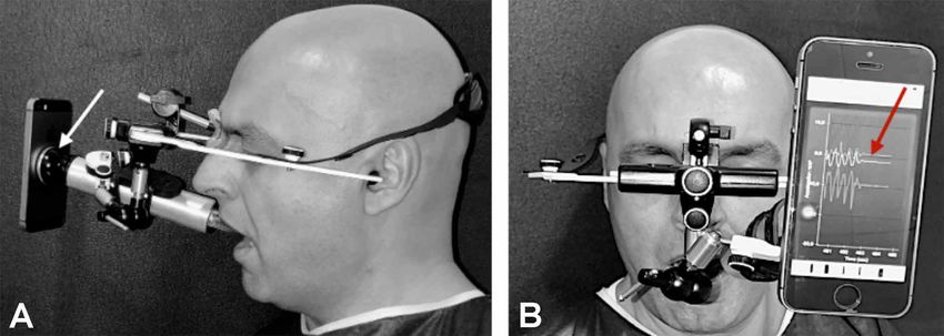
VIEIRA, J. C. F. Hands free technique: new tool possibility for image capture of sublingual microcirculation with handheld vital microspopy – HVM. Int. J. Odontostomat. 15(1):181-188, 2021.
However, there is a logistical challenge for The aim of this work was to develop a tool that
acquiring the image of sublingual microcirculation, allows healthcare professional to operate hands-free,
considering that HVM is distanced from the base by a using Handheld Vital Microscopy or similar.
cable, keeping the camera stable with the hands in the
sublingual area in the correct position, and fixing the
view of microcirculation on the computer screen. And, MATERIAL AND METHOD
at the same time, with the other hand, clicking on the
computer keyboard to start, while recording the video
that shows precise results, which is not an easy task. The focus of this study is to develop a tool
(Massey & Shapiro). that allows healthcare professionals to use the
unprecedented Hands Free technique (Figs. 1 and
The acceptable vídeo duration, without moving 2). For this, a replica of an aluminum HVM (Fig. 3,
the microcirculation image, in the usual technique 4 and 5) was created, with a diameter of 23 mm,
(Hands On) is 4 to 20 seconds, which is already length 220 mm and weight 120 g, similar to IDF
considered laborious (Ince et al., 2018), once the (Aykut et al.). The choice of IDF as a replica was
duration and stability of the video images go together made because it is the latest generation of
(van Elteren et al., 2015). microscope, in addition to being considered supe-
rior to its predecessor SDF (Gilbert-Kawai et al.,
The justification for this study emerged in the 2016).
Second Consensus on the evaluation of sublingual
microcirculation in critically ill patients: results of a task To measure the displacement of HVM repli-
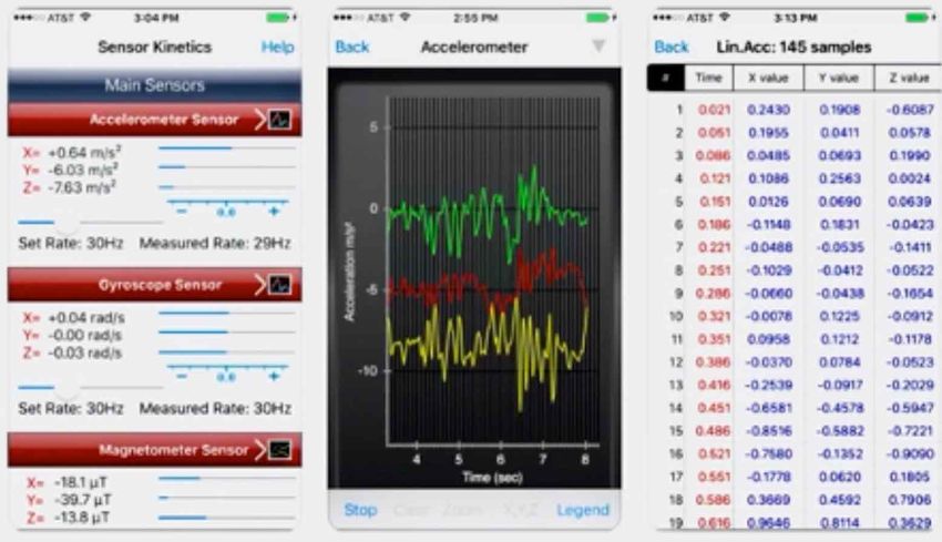
force of European Society for Intensive Care Medici- ca, the Sensor Kinetcs application was used on
ne. In future perspectives, the study mentions the need the cell phone. It will capture possible changes in
to create a tool in which the operator can monitor their movement, every second (Fig. 6). The
patient's microcirculation for a longer period. accelerometer was the sensor chosen to test the
acceleration along a certain axis. The sensor
Challenge: How will the doctor be able to keep HVM measures the displacements caused by the
stable for a long time, holding it with his/her hands? In operator, when holding HVM replica, using the
this scenario, the focus of this study was to develop a conventional or Hands On technique, versus the
support tool to enable the use of HVM in Hands Free Hands Free technique (Fig. 1).
technique. It seems to be a resource that could innovate
and improve the capture of sublingual image in humans To carry out such an experiment, a magnetic
for longer periods, thus enhancing the knowledge in base was attached to the back of the replica, where
microcirculation analysis. the cell phone was fixed (Fig. 7A).
Fig. 1. Drawing (A) of operator with HVM (M) using usual hand technique. Operator (B) using the
new support device for Hands Free technique, and observing the patient's microcirculatory flow in
real time for a longer period of time.
182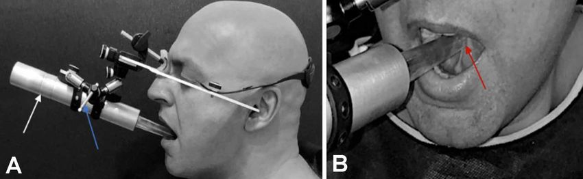
VIEIRA, J. C. F. Hands free technique: new tool possibility for image capture of sublingual microcirculation with handheld vital microspopy – HVM. Int. J. Odontostomat. 15(1):181-188, 2021.

Fig. 2. Comprises a facial arch (A) characterized by receiving a flat support (1), preferably elliptical in shape, provided with
a concave central recess (2) that configures a seat for a sublingual microscope (M), which houses on said central concave
recess (2) of the support (1); said support (1) is provided with two locking straps (3) that project from the side (4) of the
support (1), surrounding the outer perimeter of sublingual microscope body (M), and said locking straps (3) they are long
enough to surround the entire outer perimeter of the microscope (M) and reach locking pins (5) that protrude from the flat
surface (6) of the support (1); the locking straps (3) have elasticity and are provided with a sequence of holes (7) that
communicate with the locking pins (5) and promote the attachment of the microscope (M) to the support (1); the support (1)
receives a perpendicular adapter (19) fixed by a screw (20) to the hole (21), said perpendicular adapter (9) has a rectangular
seat (8) with a side slot (22), which receives and houses the cylindrical axis (9) of a lower articulated arm (10) of the facial
arch (A), said axis (9) has the flattened end containing a transverse screw with handle (11); the flat end of cylindrical shaft (9)
is fixed using the clamping knob (11) which is transversely attached to the slot in the rectangular seat (8) of the perpendicular
adapter (19) fixed on the flat surface (6) of the support (1 ); the extra-oral accessory has a second means of fixation,
composed of an adjustable headband (12) attached to the facial arch device (A) on the stems (13) of the auricles, and said
stems (13) have holes (14) that they receive fixing screws (15), adding said cranial strap (12) to the structure of the facial
arch device (A), the cranial strap (12) being adjustable and provided with a rotating element (16) to adjust the tension around
the head of the patient who acts together with the nasal support (24) coated with a silicone cushioning layer (25); once the
microscope (M) is attached to the support (1) through the locking straps (3) and pins (5), and the whole is attached to the
facial arch device (A) by fixing the axis (9) of the articulated arm lower (10) to the support (1), the data reading position is
selected in patient's sublingual region and the locking means (17) of the lower (10) and upper (18) articulated arms is
activated, locking the position of the set. This work generated a patent application number BR102018077323.

                                                                                                 Fig. 4. Replica of HVM (white arrow), adjustable rubber
Fig. 3. Support tool tested on a dummy for the Hands Free                                        clamps for HVM (blue arrow) and disposable plastic cover,
technique.                                                                                       similar to the original (red arrow).

                                                                                                                                                                                     183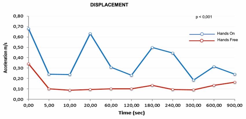
VIEIRA, J. C. F. Hands free technique: new tool possibility for image capture of sublingual microcirculation with handheld vital microspopy – HVM. Int. J. Odontostomat. 15(1):181-188, 2021.

Fig. 5. (A) Replica of HVM (white arrow) in position, after adjustment and locking (blue arrow) of the articulated arm, simulating
the Hands Free technique. Sublingual area in greatest increase (B), observe the tip of disposable plastic cap against the
desired area (red arrow)

           Fig. 6. Example of application showing the x, y, z axes: on the left, in the center, the accelerometer graphs,
           and numerical data on the right.

       The numerical data are on the x, y and z axes,                                                  There was a normal distribution, whose
where 5 measurements were performed for each                                                     quantitative variables of the main outcome were
technique in all periods. The chosen periods were 0,                                             determined using Kolmogorov-Smirnov test (KS)
5, 10, 20, 60, 120, 180, 240, 300, 600, 900 seconds.
                                                                                                        The purpose is to compare the displacements
      For statistical analysis, the following softwares                                          around each of the three axes (x, y and z), in HANDS ON
were used: SPSS V20, Minitab 16 and Excel Office                                                 versus HANDS FREE scenarios at specific times. As the
2010.                                                                                            data are paired, that is, the same subject is his own
                                                                                                 research and control, T-Student test was used. At each
      For this work, a significance level of 0.05 (5%)                                           selected time, the first 10 observations were collected
was defined. The confidence intervals adopted in this                                            and compared. Finally, the displacements of the three
work were constructed with a 95% statistical level.                                              axes, in modules, were added for a general measure.

184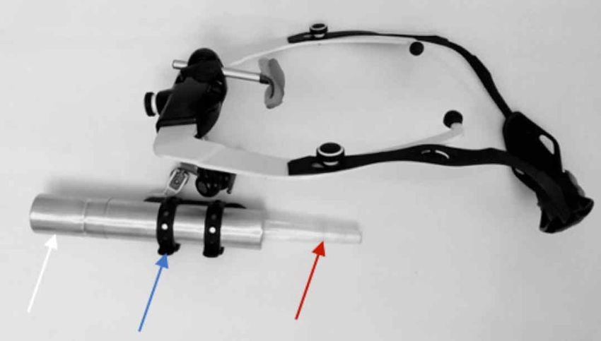
VIEIRA, J. C. F. Hands free technique: new tool possibility for image capture of sublingual microcirculation with handheld vital microspopy – HVM. Int. J. Odontostomat. 15(1):181-188, 2021.

Fig. 7. (A) Cell phone attached to a magnetic device installed at the rear of HVM replica (white arrow) to measure displacements.
(B) Cell phone screen displays initial changes to x, y, z axes and then the same stabilized axes (red).

RESULTS                                                                                          Positive points:

                                                                                                 1. After adjusting the arc opening (P, M, G), adjusting
        The results show that it is perfectly feasible to                                           the nasal support and stabilization strap on the user's
create a device to support the use of Hands Free                                                    head, the whole set remains stable (Fig. 3)
technique. The facial arch has been used in the dental                                           2. Enables the attachment of any HVM or similar, as
field for years, which ensures safety in clinical use (Fig.                                         the rubber straps are adjustable (Fig. 4).
5A).                                                                                             3. Lockable articulated arms allow the operator to
                                                                                                    choose the best sublingual area (Fig. 5A).
       In calculating the total displacement on the three                                        4. The user can move the neck smoothly and HVM
axes (Table I; Fig. 8), Hands Free technique was always                                             replica tends to be stable
less than the average in the situation of Hands On                                               5. Hands free technique has a smaller displacement
technique. The biggest difference occurs in 20 seconds,                                             than the Hands On technique, with a statistically
where the average was 0.63124 Hands Free and                                                        significant difference.
0.09509 Hands On (p-value
VIEIRA, J. C. F. Hands free technique: new tool possibility for image capture of sublingual microcirculation with handheld vital microspopy – HVM. Int. J. Odontostomat. 15(1):181-188, 2021.
Table I. Calculations of displacement of the three x, y z axes data obtained by Sensor Kintcs application.
tip of HVM on the tissue to be analyzed (Fig. 5B); (Spaide et al., 2015). With standardization in the cap-
which is likely to decrease the pressure artifacts on ture of microcirculatory image, such artifacts would
the microcirculatory flow. Common artifact in be avoided, which would ensure an adequate analysis
capturing the microcirculatory image (Ince et al., of blood flow (Massey & Shapiro). Therefore, it is
2018). necessary to take into account that the use of any
7. Facial arch and autoclavable ear supports monitoring device to measure the physiological va-
riables, as well as the use of measurements generated
Negative points: by HVM, requires skill and training of the operator
(Ince et al.).
1. The support system for Hands Free technique must
be adjusted sparingly to find the best fit. This requires In the case of HVM, the user holds the video
the operator's sensitivity to adjustments of nasal and microscope with their hands and manually adjusts the
ear support to avoid creating discomfort. focus and lighting. He must keep the tip of the
2. Non-autoclavable items: 3,12, 25 (Fig. 2). Requires microscope firm - around less than 0.1 mm /s of late-
cleaning and disinfection ral movement - when touching the tissue, and without
applying pressure to avoid obstruction of the flow
(Massey et al., 2013). Meanwhile, the operator
DISCUSSION monitors the operation on a video monitor.
With this logistical challenge, some studies that
The methods of image capture existing in the evaluated the sublingual microcirculatory flow in
real world are not perfect, as they are subject to extra critically ill patients showed that, in 240 clips analyzed,
elements, missing elements, or even translation errors 86 obtained pressure artifacts and only 74 showed
that cause defects - the so-called image artifacts excellent quality (Sallisalmi et al., 2012). The study
186VIEIRA, J. C. F. Hands free technique: new tool possibility for image capture of sublingual microcirculation with handheld vital microspopy – HVM. Int. J. Odontostomat. 15(1):181-188, 2021.
on the impact of video quality on the sublingual “37. Technology should be developed to allow
microcirculatory image in critically ill patients, analyzed stable measurements to be made for longer periods of
2455 videos; of these, only 56% were considered time to allow continuous measurement during, for
acceptable (Damiani et al., 2017). The article that example, a therapeutic maneuver allowing observation
analyzed the efficiency of two software for the analysis of single vessels response before and after
of microcirculation discarded 356 of 440 videos intervention”.
analyzed, as they are not of excellent quality. Studies
show that the software's analysis accuracy is directly Such evidence indicates that this study may
related to image quality. And therefore, to obtain contribute to the development of an unprecedented
acceptable results, the prerequisite is high-quality soft- technique called Hands Free. It is a tool (Fig. 3) that
ware (Carsetti et al., 2017). aims to couple HVM (SDF, IDF or similar) to increase
stability for a longer period, increasing the time by 75
Some technical options have been proposed times, or more, compared to current capture - 4
in order to reduce image artifacts. A vacuum tip was seconds - by the technique usual Hands on (Ince et
created to suck the mucosal surface to reduce al.). The limitations of this work refer to the use of a
pressure artifacts and increase stability (Balestra et HVM replica . Further studies will be necessary
al., 2010). But in practice, it is scarcely used (Massey comparing the benefits of Hands Free technique offered
& Shapiro). Another device that contributes to to health professional, with the sublingual
digitization operations during the recording of images microcirculatory image generated by original HVM in
is the pedal controls that replace the manual click on patients.
the computer (Massey & Shapiro). This operation is
not considered hands free because the operator's Microcirculation has been studied in the
hands remain occupied with the camera. With that, it pathophysiology of sepsis, heart failure and
is evident that image artifacts are the great villain in hypovolemia (Donati et al., 2013). Therefore,
the capture and analysis of microcirculatory videos, information about microcirculatory perfusion would be
and an obstacle in the research of sublingual of great interest in intensive care to predict results and
microcirculation. A recent article points to hope for a potentially guide therapy.
future improvement in the capture of such images.
The technique employs optical coherence tomography
(OCTA) angiography (Hessler et al., 2020). This new CONCLUSION
method makes it possible to avoid pressure artifacts;
but, as known, it is also subject to other types of image
artifacts (Spaide et al.). This study resulted in the development of a
device that will benefit HVM stabilization technique
After 10 years of the First Consensus (De (SDF, IDF or similar). It was designed to assist
Backer et al., 2007), more than 600 clinical and expe- healthcare professionals and reduce image artifacts
rimental articles with HVM have been published. A generated by the usual technique. Such a device allows
task force from the European Society for Critical Care the operator a longer time to stabilize HVM, for a longer
Medicine recently published the Second Consensus period, thanks to unprecedented Hands Free
on the assessment of sublingual microcirculation in technique, which allows future research aiming at
critically ill patients. therapeutic maneuvers in patients with serious
diseases. More tests will be needed to validate the use
Necessary statements were carried out for the of this system with the capture of microcirculation.
acquisition and interpretation of images acquired from
the sublingual microcirculation (Ince et al.). In the same
Consensus, in future perspectives, two sentences VIEIRA, J. C. F. Técnica de manos libres: nueva posi-
justified the intention to develop the tool with support bilidad de herramienta para capturar imágenes de
from HVM: microcirculación sublingual con microscopía vital por-
tátil – HVM. Int. J. Odontostomat. 15(1):181-188, 2021.
"35. Tools should be developed to make
pressure-artifact-free measurements and allow single- RESUMEN: Las nuevas técnicas de imagen
spot measurements to be made during a therapeutic microcirculatoria permitieron la observación directa de
maneuver". la microcirculación junto a la cama del paciente. Este
187VIEIRA, J. C. F. Hands free technique: new tool possibility for image capture of sublingual microcirculation with handheld vital microspopy – HVM. Int. J. Odontostomat. 15(1):181-188, 2021.
estudio sin precedentes presenta un nuevo dispositi- Goedhart, P. T.; Khalilzada, M.; Bezemer, R.; Merza, J. & Ince, C.
Sidestream Dark Field (SDF) imaging: a novel stroboscopic LED
vo que ayuda al operador con la técnica manos libres.
ring-based imaging modality for clinical assessment of the
Con este fin, se desarrolló una réplica de la Microscopía microcirculation. Opt. Express, 15(23):15101-14, 2007.
Vital Portátil para simular el método de captura de la Groner, W.; Winkelman, J. W.; Harris, A. G.; Ince, C.; Bouma, G. J.;
imagen en el área sublingual, el sitio más utilizado para Messmer, K. & Nadeau, R. G. Orthogonal polarization spectral
imaging: a new method for study of the microcirculation. Nat. Med.,
evaluar la microcirculación en pacientes críticos. Lo-
5(10):1209-12, 1999.
gramos una reducción en el desplazamiento de la ré- Hessler, M.; Nelis, P.; Ertmer, C.; Alnawaiseh, M.; Lehmann, F.; Schmidt,
plica del microscopio con el método de manos libres. C.; Kampmeier, T. G.; Rehberg, S. W.; Arnemann, P. H. & Rovas,
La consecuencia inmediata es un aumento en la esta- A. Optical coherence tomography angiography as a novel approach
to contactless evaluation of sublingual microcirculation: A proof of
bilidad de la réplica de HVM en 75 veces, o más, du-
principle study. Sci. Rep., 10:5408, 2020.
rante los 4 segundos actuales, durante su contacto Ince, C. The microcirculation is the motor of sepsis. Crit. Care, 9(Suppl.
con el tejido sublingual. El dispositivo también ofrece 4):S13-9, 2005.
un mejor control de la presión de la punta de la réplica Ince, C.; Boerma, E. C.; Cecconi, M.; De Backer, D.; Shapiro, N. I.;
Duranteau, J.; Pinsky, M. R.; Artigas, A.; Teboul, J. L.; Reiss, I. K.
de HVM sobre el área sublingual. Los resultados de-
M.; et al. Second consensus on the assessment of sublingual
mostraron que la técnica de manos libres, que opera microcirculation in critically ill patients: results from a task force of
en la misma área sublingual durante 900 segundos, the European Society of Intensive Care Medicine. Intensive Care
debería permitir futuras investigaciones destinadas a Med., 44(3):281-99, 2018.
Massey, M. J. & Shapiro, N. I. A guide to human in vivo microcirculatory
maniobras terapéuticas en pacientes con enfermeda-
flow image analysis. Crit. Care, 20:35, 2016.
des graves. Massey, M. J.; Larochelle, E.; Najarro, G.; Karmacharla, A.; Arnold,
R.; Trzeciak, S.; Angus, D. C. & Shapiro, N. I. The microcirculation
PALABRAS CLAVE: técnica manos libres, es- image quality score: development and preliminary evaluation of a
proposed approach to grading quality of image acquisition for
tabilizador, imagen HVM, artefactos, sublingual,
bedside videomicroscopy. J. Crit. Care, 28(6):913-7, 2013.
microcirculación, imagen SDF, imagen IDF. Sallisalmi, M.; Oksala, N.; Pettilä, V. & Tenhunen, J. Evaluation of
sublingual microcirculatory blood flow in the critically ill. Acta
Anaesthesiol. Scand., 56(3):298-306, 2012.
Sherman, H.; Klausner, S. & Cook, W. A. Incident dark-field illumination:
REFERENCES
a new method for microcirculatory study. Angiology, 22(5):295-303,
1971.
Spaide, R. F.; Fujimoto, J. G. & Waheed, N. K. Image artifacts in optical
Aykut, G.; Veenstra, G.; Scorcella, C.; Ince, C. & Boerma, C. Cytocam- coherence tomography angiography. Retina, 35(11):2163-80, 2015.
IDF (incident dark field illumination) imaging for bedside monitoring Trzeciak, S. & Rivers, E. P. Clinical manifestations of disordered
of the microcirculation. Intensive Care Med. Exp., 3(1):40, 2015. microcirculatory perfusion in severe sepsis. Crit. Care, 9(Suppl.
Balestra, G. M.; Bezemer, R.; Boerma, E. C.; Yong, Z. Y.; Sjauw, K. D.; 4):S20-6, 2005.
Engstrom, A. E.; Koopmans, M. & Ince, C. Improvement of Tsai, A. G.; Johnson, P. C. & Intaglietta, M. Oxygen gradients in the
sidestream dark field imaging with an image acquisition stabilizer. microcirculation. Physiol. Rev., 83(3):933-63, 2003.
BMC Med. Imaging, 13:10-15, 2010. van Elteren, H. A.; Ince, C.; Tibboel, D.; Reiss, I. K. M. & de Jonge, R.
Carsetti, A.; Aya, H. D.; Pierantozzi, S.; Bazurro, S.; Donati, A.; Rhodes, C. J. Cutaneous microcirculation in preterm neonates: comparison
A. & Cecconi, M. Ability and efficiency of an automatic analysis between sidestreamdark field (SDF) and incident dark field (IDF)
software to measure microvascular parameters. J. Clin. Monit. imaging. J. Clin. Monit. Comput., 29(5):543-8, 2015.
Comput., 31(4):669-76, 2017.
Damiani, E.; Ince, C.; Scorcella, C.; Domizi, R.; Carsetti, A.; Mininno,
N.; Pierantozzi, S.; Adrario, E.; Romano, R.; Pelaia, P.; et al. Impact
of microcirculatory video quality on the evaluation of sublingual
microcirculation in critically ill patients. J. Clin. Monit. Comput., Corresponding author:
31(5):981-8, 2017. José Custódio Feres Vieira
De Backer, D.; Hollenberg, S.; Boerma, C.; Goedhart, P.; Büchele, G.; Programa de Pós Graduação
Ospina-Tascon, G.; Dobbe, I. & Ince, C. How to evaluate the
PPG Ciência Cirúrgica Interdisciplinar
microcirculation: report of a round table conference. Crit. Care,
11(5):R101, 2007.
Universidade Federal de São Paulo (Unifesp)
Donati, A.; Domizi, R.; Damiani, E.; Adrario, E.; Pelaia, P. & Ince, C. São Paulo
From macrohemodynamic to the microcirculation. Crit. Care Res. BRASIL
Pract., 2013:892710, 2013.
Elbers, P. W. G. & Ince, C. Mechanisms of critical illness--classifying
microcirculatory flow abnormalities in distributive shock. Crit. Care, E-mail: custodio.vieira@unifesp.br
10(4):221, 2006.
Gilbert-Kawai, E.; Coppel, J.; Bountziouka, V.; Ince, C.; Martin, D. &
Caudwell Xtreme Everest and Xtreme Everest 2 Research Groups.
A comparison of the quality of image acquisition between the
incident dark field and sidestream dark field video-microscopes.
BMC Med. Imaging, 16:10, 2016.
188You can also read