Health Policy Advisory Committee on Technology - Technology Brief Argus II Retinal Prosthesis System for peripheral retinal degeneration November 2013
←
→
Page content transcription
If your browser does not render page correctly, please read the page content below

Health Policy Advisory Committee on
                      Technology

                                      Technology Brief

   Argus II® Retinal Prosthesis System for peripheral retinal
                         degeneration

                                       November 2013

Argus II® Retinal Prosthesis: November 2013                 1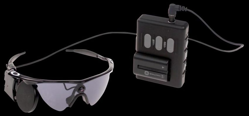
© State of Queensland (Queensland Department of Health) 2013

This work is licensed under a Creative Commons Attribution Non-Commercial No Derivatives 3.0
Australia licence. In essence, you are free to copy and communicate the work in its current form for
non-commercial purposes, as long as you attribute the authors and abide by the licence terms. You
may not alter or adapt the work in any way.
To view a copy of this licence, visit http://creativecommons.org/licenses/by-nc-nd/3.0/au/.
For further information, contact the HealthPACT Secretariat at:
HealthPACT Secretariat
c/o Clinical Access and Redesign Unit, Health Service and Clinical Innovation Division
Department of Health, Queensland
Level 13, Block 7
Royal Brisbane and Women’s Hospital
HERSTON QLD 4029
Postal Address: GPO Box 48, Brisbane Qld 4001
Email: HealthPACT@health.qld.gov.au             Telephone: +61 7 3646 9100
For permissions beyond the scope of this licence contact: Intellectual Property Officer, Queensland
Health, GPO Box 48, Brisbane Qld 4001, email ip_officer@health.qld.gov.au, phone (07) 3234 1479.
Electronic copies can be obtained from: http://www.health.qld.gov.au/healthpact
DISCLAIMER: This brief is published with the intention of providing information of interest. It is
based on information available at the time of research and cannot be expected to cover any
developments arising from subsequent improvements to health technologies. This brief is based on
a limited literature search and is not a definitive statement on the safety, effectiveness or cost-
effectiveness of the health technology covered.
The State of Queensland acting through Queensland Health (“Queensland Health”) does not
guarantee the accuracy, currency or completeness of the information in this brief. Information may
contain or summarise the views of others, and not necessarily reflect the views of Queensland
Health.
This brief is not intended to be used as medical advice and it is not intended to be used to diagnose,
treat, cure or prevent any disease, nor should it be used for therapeutic purposes or as a substitute
for a health professional's advice. It must not be relied upon without verification from authoritative
sources. Queensland Health does not accept any liability, including for any injury, loss or damage,
incurred by use of or reliance on the information.
This brief was commissioned by Queensland Health, in its role as the Secretariat of the Health Policy
Advisory Committee on Technology (HealthPACT). The production of this brief was overseen by
HealthPACT. HealthPACT comprises representatives from health departments in all States and
Territories, the Australian and New Zealand governments and MSAC. It is a sub-committee of the
Australian Health Ministers’ Advisory Council (AHMAC), reporting to AHMAC’s Hospitals Principal
Committee (HPC). AHMAC supports HealthPACT through funding.
This brief was prepared by Dr Meegan Vandepeer from the Australian Safety and Efficacy Register of
New Interventional Procedures – Surgical (ASERNIP-S).

Argus II® Retinal Prosthesis: November 2013                                                       2
Technology, Company and Licensing
Register ID                        WP173
Technology name                    Argus® II Retinal Prosthesis
Patient indication                 For use in blind adults (25 years or older) with severe to
                                   profound retinitis pigmentosa and bare light or no light
                                   perception in both eyes

Description of the technology
The Argus II® Retinal Prosthesis System is designed to restore some functional vision to
people who are blind. The system consists of surgically implanted and external equipment.
The external component consists of a miniature video camera and transmitter mounted on
a pair of glasses, as well as a small computer (video processor unit) and battery which are
worn on a belt or shoulder strap (Figure 1). The implanted portion, which is designed to go
in a single eye (typically the worse seeing), consists of a receiving and transmitting coil and
an electronic case which are fixed to the outside of the sclera (white of the eye), and an
electrode array (60 electrodes) that is surgically attached to the surface of the retina. The
electrode array is connected to the electronic case by a ribbon cable that goes through the
sclera.1 The camera housed within the glasses captures images which are sent to the video
processing unit and converted to electronic signals. These signals are then sent to the
transmitter coil on the glasses. The implanted receiving coil wirelessly receives these data
and sends the signals via the ribbon cable to the implanted electrode array which emits
small pulses of electricity to stimulate the remaining bipolar or ganglion cells in the retina.
These electrical signals pass through the optic nerve to the brain allowing light sensation
which patients learn to interpret as visual patterns.2

                          ®
Figure 1         Argus II Retinal Prosthesis System. Copyright © 2013 Second Sight Medical
                 Products, Inc.

Argus II® Retinal Prosthesis: November 2013                                                     1
Implantation of the Argus II® Retinal Prosthesis System can be performed by a single
surgeon in a two-hour outpatient procedure done under general anaesthesia.3 Details
regarding the surgery are as follows. At the start of the implant procedure, antibiotics and
steroids are administered by intravenous injection. In phakic eyes (eyes with a lens), the lens
is removed. An incision is made of the conjunctiva surrounding the cornea exposing the
underlying sclera and extra-ocular muscles (peritomy). The transmitting coil is placed on the
eyeball and centred under the lateral rectus muscle. The electronics package is centred in
the superior temporal quadrant of the eye, and the implant is then fixed to the eye with
sutures. The vitreous gel in the middle of the eye is removed (vitrectomy), along with any
epiretinal membrane in the area where the surgeon intends to tack the array. The
microelectrode array is inserted through an incision in the sclera (sclerotomy) and affixed to
the retina in the macula with a retinal tack. The extraocular portion of the ribbon cable is
then sutured to the sclera and all incisions are sutured. A transplanted scleral tissue graft (or
alternative) is sutured over the electronics package to reduce the likelihood of conjunctival
irritation. At the end of surgery, steroids, antibiotics and an anaesthetic are injected under
the conjunctiva. For approximately two weeks after the operation, patients are given
antibiotics (oral and eye drops), steroids (oral and eye drops) and atropine to dilate the
pupil.1 The implant is activated one week after surgery.3
Patients with the following characteristics are eligible for the procedure:
at least 25 years old
bare light or no light perception in both eyes (if there is no light perception, the eye
must respond to electrical stimulation)
previous ability to see objects, shapes and lines
implanted eye must have an artificial lens or no lens at all
the patient must be willing and able to follow the recommended schedule of clinical
follow-up, device programming and visual rehabilitation.4
Contraindications for the Argus II® Retinal Prosthesis System are:
an eye disease or condition that could prevent the device from working
an eye structure or condition that could make it difficult to successfully implant the
device or recover following surgery
eye diseases or conditions that make it difficult for the doctor to see inside the eye
inability to undergo general anaesthesia
inability to take the recommended antibiotic and steroid medications required prior
to and following surgery
presence of metallic or active device implants in the head
presence of a disease or condition that prevents a patient from giving informed
consent
Argus II® Retinal Prosthesis: November 2013 2any disease or condition that prevents medical follow-ups or having the video
processing unit programmed
constant eye rubbing.4
In addition to the above indications and contraindications, the company that produces the
Argus II® Retinal Prosthesis System provides information outlining general precautions,
precautions regarding other medical procedures, possible interference from other
electronic devices, air travel, general travel and international use, electromagnetic
interference and adverse events.4
Company or developer
Second Sight Medical Products, Inc. (Sylmar, CA, United States of America).
Reason for assessment
The Argus II® Retinal Prosthesis System is a novel device that can assist blind people with
severe to profound retinitis pigmentosa and bare or no light perception in both eyes. There
is currently no cure for this condition and no treatment that will stop or slow its progression.
Stage of development in Australia
Yet to emerge Established
Experimental Established but changed indication
or modification of technique
Investigational Should be taken out of use
Nearly established
Licensing, reimbursement and other approval
The Argus® II Retinal Prosthesis System received a CE mark in February 2011. It also received
a Humanitarian Device Exemption from the United States Food and Drug Administration
(FDA) in February 2013 on the proviso that two post-approval studies are conducted, one of
which must provide 10-year follow-up data on patients who have received the device.5 The
Argus® II Retinal Prosthesis System is available at two National Institute for Health Research
centres in the United Kingdom. In Germany and Italy, two to four patients per month are
currently receiving the device.2
Australian Therapeutic Goods Administration approval
Yes ARTG number (s) Not applicable
No
Not applicable
Argus II® Retinal Prosthesis: November 2013 3Technology type Device Technology use Assistive Patient Indication and Setting Disease description and associated mortality and morbidity Peripheral degeneration of the retina is caused by many diseases, the most common being a family of diseases known as retinitis pigmentosa (RP). RP is a heterogeneous group of inherited retinal disorders characterised by progressive damage to and loss of the light receptor (photoreceptor) cells in the retina, with subsequent degeneration of the retinal pigment epithelium.6 The retina contains two types of photoreceptors: rods and cones. Rods are responsible for peripheral vision and vision in low light, while cones provide vision in bright light, including colour vision. In most cases, the rods degenerate first, followed by the cones. Consequently, the first symptom patients experience is impaired night vision; loss of daytime vision occurs later in life.7 There are two forms of RP: syndromic and non-syndromic. The non-syndromic form, in which the signs and symptoms of RP are limited to loss of vision, is the most common. In the less common syndromic forms of RP, one or more other organs are affected as well. The typical non-syndromic form of RP slowly progresses over several decades, although some patients experience rapid onset over two decades or a slow disease progression that never leads to blindness. In the early stages of the disease, the main symptom is loss of the ability to see at night (night blindness) or in very low light. This symptom is often experienced in childhood, but may also appear during the second decade of life or later. In the middle stage of the disease, night blindness is obvious and results in difficulty driving and walking at night. Patients also become aware of a loss in peripheral vision in daylight (tunnel vision). Other symptoms include light intolerance (photophobia), colour vision deficiency (dyschromatopsia) and difficulty reading. In many cases, these severe vision problems do not occur until early adulthood. In the advanced stage of the disease, patients can no longer move independently as a result of peripheral vision loss, reading is difficult and photophobia is intense. Some individuals may retain limited central vision whereas others experience complete vision loss.8 The rate and degree of disease progression varies among individuals. Complications associated with RP include cataracts and macular oedema.7 Clinical diagnosis of RP is based on the presence and progressive worsening of night blindness and peripheral visual field defects, lesions in the fundus, and hypovolted electroretinogram traces. Currently there is no therapy that stops the progression of the disease or restores lost vision. Argus II® Retinal Prosthesis: November 2013 4
Number of patients
No New Zealand data could be found on the incidence or prevalence of RP, the most
common of the retinal degenerative diseases. It is estimated that one in every 3,000
Australians is affected by RP and that five to seven per cent of newly diagnosed blindness in
Western countries is attributable to this family of diseases.9, 10 According to a report on
vision loss in Australia from the Centre for Eye Research Australia (CERA), RP was the cause
of 769 cases of blindness (1.5%) in people older than 40 years in 2004.11
Speciality                         Ophthalmology
Technology setting                 Specialist hospital

Impact

Alternative and/or complementary technology
The Argus II® Retinal Prosthesis System is neither an alternative nor a complementary
technology. It is the world’s first and only United States FDA approved (humanitarian device
exemption) device for restoring some functional vision in people suffering from blindness as
a result of severe to profound RP.

Current technology
There is currently no cure for RP, the most common cause of peripheral retinal
degeneration, and there are no treatments that stop the condition from gradually
worsening over time.12 Therapy aimed at slowing the degenerative process includes
protecting the eyes from sunlight (wearing dark glasses outdoors), taking vitamins A and E
to protect the photoreceptors and treating complications. Psychological and genetic
counselling is used to help patients cope with the social and psychological impact of
blindness.7

Diffusion of technology in Australia
The Argus II® Retinal Prosthesis System is not listed on the Australian Register of
Therapeutic Goods and is yet to emerge in Australia.

Argus II® Retinal Prosthesis: November 2013                                              5
International utilisation
Country Level of Use
Trials underway or Limited use Widely diffused
completed
United States of America
Mexico
France
Switzerland
Germany
Italy
UK
Cost infrastructure and economic consequences
The cost1 of the device is estimated to be A$112,724. Additional costs estimated by the
company include A$10,513 for surgery and A$6,104 for follow-up.2 However, actual follow-
up costs may be significantly higher as they would include weekly training in a specialised
setting by specialised staff for a 6 to 12 month period.
Ethical, cultural or religious considerations
No ethical, cultural or religious considerations were identified in the published literature.
Evidence and Policy
Safety and effectiveness
Three studies were included in this Technology Brief. All three studies included the same
group of 30 patients enrolled in a worldwide, multicentre (10 centres), phase II clinical trial
(NCT00407602) sponsored by Second Sight Medical Products, Inc.13 All patients received the
Argus II® Retinal Prosthesis System and all patients acted as their own control, that is
outcomes were compared in each patient with the device activated and deactivated. The
trial, which is no longer recruiting, is expected to be completed in August 2019. The median
age of the 30 patients was 57 years (range 27–77) at the time of implantation and 30 per
cent of patients were women. All patients were followed up for a minimum of six months,
with some patients having follow-up data up to 2.7 years after surgery. Inclusion criteria for
patients entering the trial were as follows:
a confirmed history of RP (all centres) or outer retinal degeneration (France, Mexico,
Switzerland and the United Kingdom only), with remaining visual acuity of bare light
perception (all centres) or visual acuity of 2.3 logarithm of Minimum Angle
1
Costs converted from British pounds to Australian dollars where £1= A$1.6956 (source ozforex.com.au, 19 August 2013)
Argus II® Retinal Prosthesis: November 2013 6Resolution (logMAR) or worse in both eyes (France, Mexico, Switzerland and the
United Kingdom only);
functional ganglion cells and optic nerve;
a history of former useful form vision in the worse-seeing eye;
at least 18 (France, Mexico, and the United Kingdom) or 25 (Switzerland and the
United States of America) years old;
resides within two (Mexico, the United Kingdom and the United States of America)
or three hours (France and Switzerland) travel time (by ground transport) of the
investigational site; and
willing and able to comply with the protocol testing and follow-up requirements.
Patients were excluded from the trial if they had the following:
disease of the optic nerve or ocular surface;
diseases or conditions that affect retinal function, prevent adequate visualisation of
the retina or prevent adequate performance of the physical examination;
an ocular condition that predisposes the patient to eye rubbing;
any disease or condition that prevents understanding or communication of informed
consent, study demands, and testing protocols;
pregnancy;
another active implanted device;
conjunctival thinning;
any health concern that would preclude anaesthesia;
unrealistic expectations of the implant;
known allergy or contraindication to anticipated preoperative, intraoperative or
postoperative medications;
conditions likely to limit life to less than one year from the time of screening;
diseases or conditions that impede the ability to implant the device or would prevent
the system from functioning for the duration of the study; or
an axial eye length less than 20.5 mm or more than 26.0 mm in the implanted eye.13
Ahuja and Behrend, 201314; Humayun et al, 20121
Results for the 30 patients from the phase II clinical trial (NCT00407602) were reported in
the studies by Ahuja and Behrend (2013)14 and Humayun et al (2012)1 and are discussed
together here. Outcomes from these studies included safety (serious and non-serious
adverse events), outpatient use of the device and tests on direction of motion, real-world
utility (door and line tests), full-field light stimulus light threshold, square localisation, visual
acuity and the ability to read letters. Descriptions of tests on efficacy are provided in Table
1.
Argus II® Retinal Prosthesis: November 2013 7®
Table 1 Tests conducted to assess the efficacy of the Argus II Retinal Prosthesis
14 1
System conducted by Ahuja and Behrend (2013) and Humayun et al (2012)
Test name Description of test Number of patients who
completed the test and
time (post-implantation)
when test was conducted
Direction of Patients were required to draw a line on a touch screen n=28, time=ns
motion indicating the direction of motion of a white bar which swept
across the screen at random angles ranging from 0 to 360 .
Door test Patients were required to find a door on the other side of a Baseline (n=29), 3 months
room. (n=29), 6 months (n=30), 12
months (n=14), 18 months
(n=13), 24 months (n=8)
Line test Patients were required to follow a white line on the floor. Baseline (n=30), 3 months
(n=27), 6 months (n=29), 12
months (n=14), 18 months
(n=13), 24 months (n=8)
Square Patients were required to touch the centre of a square (2.3 x n=27, time=ns
localisation 2.3 inches) displayed in random locations on a monitor after
being given an auditory prompt. Accuracy was determined by
calculating the mean distance between individual responses
and the square’s centre.
Visual acuity Visual acuity was tested by presenting square-wave gratings of n=ns, time=ns
varying spatial frequency in one of four directions (horizontal,
vertical, diagonal to the upper right or diagonal to the upper
left) for five seconds.
Reading letters Three different tests were conducted: letter recognition, letter Test 1 (group A: n=24,
size reduction and word reading. Tests increased in difficulty group B: n=22, group C:
and only those who performed well in one test moved on to the n=21)
next.
Test 2 (n=7)
Test 1 (letter recognition): Patients had to identify letters in a
force-choice closed-set test. There were three different test Test 3 (n=4)
groups. Group A consisted of the letters L,T, E, J, F, H, I and Time=ns for all tests
U. Group B consisted of the letters A, Z, Q, V, N, W, O, C, D,
and M. Group C consisted of the letters K, R, G, X, B, Y, S,
and P. Letters were presented four times in random order.
Test 2 (letter size reduction): Patients who correctly identified
at least 50% of the letters in 60 seconds in all letter groups in
test 1 were included in test 2. Patients were required to identify
letters randomly presented from a closed set. Letters were
grouped in lines of 5 that reduced in size equivalent to one log
unit to mimic standard acuity testing. The test was stopped
when the time limit expired, five incorrect responses were
given in single line, or the patients were unable to guess at the
end of the forced-choice test. Testing was carried out twice for
each patient on separate days. The total number of letters
correctly identified and the size of the smallest correct letter
were recorded.
Test 3 (word recognition): Patients who performed best in test
2 were included in test 3. Patients were presented with two-,
three- and four-letter words and given the equivalent time to
guess them.
Full-field light Patient’s residual native light perception (without the use of the n=ns, time=2 years post-
threshold prosthesis) was measured before and 2 years after implantation
implantation
ns: not stated
Argus II® Retinal Prosthesis: November 2013 8Serious adverse events were defined according to the International Organization for Standardization (ISO) 14155 standard15 as medical occurrences that: caused death, were life threatening, caused permanent impairment of a body function or permanent damage to body structure, necessitated medical or surgical intervention to preclude such impairment or damage, or required hospitalisation or prolonged hospitalisation. Non-serious adverse events were defined as those related to the device or surgery that did not require surgical intervention (resolved with topical or oral medications or did not require treatment). Safety A total of 17 device- or surgery-related serious adverse events (SAEs) were reported (Table 2). Eighty-two per cent of SAEs occurred within the first six months after implantation and 70 per cent occurred within the first three months. Seventy per cent of patients did not experience any SAEs. Conjunctival erosion and dehiscence, which occurred in three and two patients respectively, were treated with additional sutures and/or placement of additional graft tissue in four patients and removal of the device due to damage in one patient. Culture-negative presumed inflammation of the aqueous and vitreous cavity inside the eye (endophthalmitis), which occurred in three patients, was resolved with intravitreal, subconjunctival, topical and systemic antibiotics. The first incidence occurred in the immediate postoperative period. The second and third incidences developed approximately five and eight weeks postoperatively. None of the presumed endophthalmitis cases required device removal. No incidences of presumed endophthalmitis occurred after a protocol change was implemented that included the routine use of intraoperative broad-spectrum antibiotics, a reduction in the number of observers, stricter sterile techniques during implant and the use of a temporary sleeve to cover the array region before introducing it intraocularly. All three cases of low intra-eye pressure (hypotony), two of which occurred within the first six months post-implantation and the third at one year, were successfully resolved. The patient who developed hypotony at one year post-implantation had the device removed because of device migration. With respect to the other two patients, one was treated with intraocular silicone oil tamponade, while the other, who had an associated rhegmatogenous retinal detachment requiring repair, was later also treated with silicone oil tamponade. The two cases of retinal detachment, which occurred between the 5- and 6-month post- implantation period, required surgical intervention. One was associated with hypotony, as described above, while the other resulted from blunt trauma to the implanted eye. The retinal detachment was successfully repaired with vitrectomy, partial removal of the retina (retinectomy) and silicone oil. Argus II® Retinal Prosthesis: November 2013 9
In two patients, the electrode array had to be re-tacked to the retina shortly after surgery.
In both cases, the reattachment was successful.
Table 2            Serious adverse events (device or surgery related) reported following surgery
                                          ®                           1
                   to implant the Argus II Retinal Prosthesis System.
Serious Adverse Event                                       Number of patients with event (%)

                                                                            (N=30)

Conjunctival dehiscence                                                      3 (10)

Conjunctival erosion                                                         2 (7)

Presumed endophthalmitis                                                     3 (10)

Hypotony                                                                     3 (10)

Re-tack                                                                      2 (7)

Retinal detachment –                                                         1 (3)
rhegmatogenous

Retinal detachment – tractional                                              1 (3)

Retinal tear                                                                 1 (3)

Uveitis – inflammatory                                                       1 (3)

Patients were followed up for a minimum of 6 months and up to 2.7 years

The types of non-serious adverse events and the number of patients in which they occurred
are listed in Table 3. The exact number of patients who experienced each event was not
reported other than that 10 patients developed conjunctival oedema that lasted longer than
what is typically seen postoperatively. This was the most frequently reported non-serious
adverse event.
Table 3            Non-serious events (device or surgery related) reported following surgery to
                                       ®                           1
                   implant the Argus II Retinal Prosthesis System.
Number of patients who                    Types of non-serious adverse events
experienced the events listed*
(N=30)
                                                                 †
10                                        Conjunctival oedema
5–7                                       Intraocular inflammation, hypotony without significant choroidal
                                          detachment, suture irritation and ocular pain
2–3                                       Inflammatory conjunctivitis, corneal filaments, epiretinal membrane,
                                          high intraocular pressure controlled by topical anti-glaucoma
                                          medication, epiphora, mild hyphema, inflammatory uveitis and mild
                                          vitreous haemorrhage.
1                                         Limited conjunctival dehiscence, corneal abrasion, mild peripheral
                                          corneal vascularisation, cystoid macular oedema, decrease in light
                                          perception, dry eye, transient headache, iris vessel engorgement that
                                          receded after surgery to resuture the sclerotomy (to treat hypotony), a
                                          stable tractional retinal detachment, transient nausea, transient
                                          increased nystagmus, scleritis, and transient vertigo
Patients were followed up for a minimum of 6 months and up to 2.7 years.
* The exact number of patients who experienced each event could not be deduced from the data provided in the study
†
 Considered to be more extensive or longer lasting than what is typically seen postoperatively

Argus II® Retinal Prosthesis: November 2013                                                                          10
Efficacy As of March 2010, 29 of the 30 patients had used the system at home for an average of 15.8 months. One patient’s device was removed due to migration1 (discussed above in safety). At 2-years’ follow-up, there was no significant difference in full-field stimulus light threshold before and after surgery in either the implanted eye or the fellow eye in any patient (p>0.2).14 Tests on visual acuity, where 20/20 vision is equivalent to 0.00 logMAR and the higher the logMAR the worse the vision, demonstrated that no patients had a recordable visual acuity prior to implantation or with the system off after implantation. Seven patients had a measurable acuity below the 2.9 logMAR test limit with the system on. The best result achieved was a score of 1.8 logMAR. With respect to the door and line test, patients performed significantly better with the system on than with the system off at all follow-up time points except at the 12-month time point for the door test (p
second experiment or whose system-off performance was comparable to the average
system-on performance could move onto the third test. The paths the patients were
required to follow, in order of testing, included right-angle tests, mixed-angle single-turn
tests and two-turn tests. Patients had training with the system on prior to each experiment.
The study did not state the time after surgery when the tests were conducted.
Efficacy
The Argus II® Retinal Prosthesis significantly improved the patients fine hand movements.
On average, across all subjects and trials, prosthesis use significantly reduced the error in
tracing by 60 per cent (p
®
Table 6            Registered clinical trials underway for the Argus II Retinal Prosthesis System.

Trial Identifier   Trial         Interventions   N    Study design   Outcomes                      Estimated
and site           status                                                                          completion
details                                                                                            date

NCT00279500        Ongoing,      Argus retinal   6    Non-           Medical and surgical          December
Single centre      not           prosthesis           randomised     risk, ability to resolve      2014
(United States)    recruiting    (first               comparative    multiple percepts and
                                 generation                          patterns, optimize
                                 device)                             effectiveness of
                                                                     stimulus parameters

NCT00407602        Ongoing,             ®
                                 Argus II        30   Non-           Visual acuity, safety,        July 2014
                   not           retinal              randomised     activities of daily living,
Multicentre        recruiting
(France,                         prosthesis           comparative    quality of life,
Mexico,                                                              orientation and
Switzerland,                                                         mobility, spatial vision,
United                                                               stability of implant,
Kingdom,                                                             system functionality
United States)

NCT01860092        Not yet              ®
                                 Argus II        53   Case series    Safety, visual function,      August
                   open for      retinal                             functional vision,            2018
Countries not      recruitment
specified                        prosthesis                          device reliability
                                 implantation

NCT01490827        Currently            ®
                                 Argus II        45   Case series    Nature and rate of            May 2016
                   recruiting    retinal                             adverse events, visual
Multicentre
(Germany,                        prosthesis                          function
Italy)

Searched on 13 August 2013.

Other issues
There are several conflicts of interest in the three studies included in this Brief. All three
studies were derived from the same clinical trial sponsored by Second Sight Medical
Products, Inc., the manufacturer of the Argus II® Retinal Prosthesis System. Both authors of
Barry and Dagnelie (2012)16 disclosed affiliations with Second Sight Medical Products, Inc.
The first author of Ahuja and Behrend (2013)14 worked for Second Sight Medical Products,
Inc. and later served as a consultant, and employees and consultants of Second Sight
Medical Products, Inc. were involved with the design, data collection and analysis of this
study. Finally, several of the authors of Humayun et al (2012)1 have affiliations with the
device manufacturer ranging from financial interests, stock options, receiving consulting
fees, being employees of the company or being currently or previously employed by an
institution that receives funding from Second Sight Medical Products, Inc.
There are several companies and academic institutions that are at various stages in the
development of retinal prostheses with the aim of restoring light perception in blind
individuals. Other devices being developed include: the Learning Retinal Implant (Intelligent
Medical Implants AG, Zug, Switzerland), the Alpha IMS (Retina Implant AG, Reutlingen,

Argus II® Retinal Prosthesis: November 2013                                                                 13
Germany), the EPIRET3 retinal implant (Philipps University, Marburg, Germany), the Microelectrode-STS (Osaka University, Osaka, Japan) and the Tubingen Retinal Implant (University of Tubingen, Tubingen, Germany).17 Of these devices, the Alpha IMS is CE marked; however, none have received United States FDA approval. Summary of findings The evidence on the safety and effectiveness of the Argus® II Retinal Prosthesis System considered in this Technology Brief is of a low level, being derived from the same group of 30 patients enrolled in the same phase two clinical trial. Given that the device is in its initial stage of assessment and the studies included in this Technology Brief are derived from the first clinical trial to assess the safety and efficacy of the device, the initial results are promising. All 30 patients who received the device have been using it at home, outside an outpatient clinical setting. In addition, patients performed significantly better in a range of tests concerned with orientation, navigation, guidance of fine hand movement and letter recognition when the system was on compared to when it was off, demonstrating that the prosthesis significantly improves visual acuity. Although, overall, a significant improvement in test results was observed with the system on compared to when it was off, there was a wide range in ability observed among the patients which indicates that the system may have more benefit for some patients than others. With respect to safety, the most common serious adverse events reported included conjunctival dehiscence and erosion, hypotony, retinal detachment and presumed endophthalmitis. The finding of no significant difference in full-field light stimulus light threshold at two years post-implantation between implanted and fellow eyes supports the notion that the Argus® II implant has no harmful effect on remaining photoreceptor function, at least over the time period that was investigated in these studies (maximum follow-up of 2.7 years). HealthPACT assessment Although the results from the studies included in this Technology Brief were promising, they were all derived from the same multicentre, phase II clinical trial of only 30 patients. HealthPACT therefore recommend that this technology be monitored for 24-months, in which time additional evidence may become available. Number of studies included All evidence included for assessment in this Technology Brief has been assessed according to the revised NHMRC levels of evidence. A document summarising these levels may be accessed via the HealthPACT web site. Total number of studies 3 Argus II® Retinal Prosthesis: November 2013 14
Search criteria to be used (MeSH terms)
Retinitis pigmentosa (MESH) AND visual prosthesis (MESH) OR retinitis pigmentosa AND
retinal prosthesis
References
1.      Humayun, M. S., Dorn, J. D. et al (2012). 'Interim results from the international trial
        of Second Sight's visual prosthesis', Ophthalmology,119(4), 779-788.
2.      Horizon Scanning Centre (2012). Argus II Retinal Prosthesis System for peripheral
        retinal degeneration [Internet]. National Institute for Health Research. Available
        from: http://www.hsc.nihr.ac.uk/topics/argus-iiandtrade-retinal-prosthesis-system-
        for-per/ [Accessed 13 August 2013].
3.      Wiltz, C. (2013). Argus II Bionic Eye Gives Second Sight to the Blind [Internet]. UBM
        Canon. Available from: http://www.mddionline.com/article/argus-ii-bionic-eye-
        gives-second-sight-blind [Accessed 8 October 2013].
4.      Second Sight Medical Products, Inc. (2013). Important safety information [Internet].
        Second Sight Mecial Products, Inc. Available from: http://2-sight.eu/en/important-
        safety-information-en-pf [Accessed 9 September 2013].
5.      Food and Drug Administration (2013). Argus II Retinal Prosthesis System - H110002
        [Internet]. U.S. Department of Health and Human Services. Available from:
        http://www.fda.gov/MedicalDevices/ProductsandMedicalProcedures/DeviceApprov
        alsandClearances/Recently-ApprovedDevices/ucm343162.htm [Accessed 9
        September 2013].
6.      Shintani, K., Shechtman, D. L. & Gurwood, A. S. (2009). 'Review and update: current
        treatment trends for patients with retinitis pigmentosa', Optometry,80(7), 384-401.
7.      Hamel, C. (2006). 'Retinitis pigmentosa', Orphanet journal of rare diseases,1, 40.
8.      Australia and New Zealand Horizon Scanning Network (2010). Horizon Scanning
        Prioritising Summary. Retinal implants to restore light perception in individuals
        blinded by retinitis pigmentosa [Internet]. Department of Health and Ageing.
        Available from:
        http://www.horizonscanning.gov.au/internet/horizon/publishing.nsf/Content/C8A5
        BA60BD01A93ECA257757000A2015/$File/PS%20retinal_implants%20June%202010.
        pdf [Accessed 20 August 2013].
9.      Better Health Channel (2013). Eyes - retinitis pigmentosa [Internet]. State
        Government of Victoria. Available from:
        http://www.betterhealth.vic.gov.au/bhcv2/bhcarticles.nsf/pages/Eye_conditions_re
        tinitis_pigmentosa [Accessed 9 September 2013].
10.     Roessler, G., Laube, T. et al (2009). 'Implantation and explantation of a wireless
        epiretinal retina implant device: observations during the EPIRET3 prospective clinical
        trial', Invest Ophthalmol Vis Sci,50(6), 3003-3008.
11.     Centre for Eye Research Australia (2004). Clear Insight. The economic impact and
        cost of vision loss in Australia [Internet]. Centre for Eye Research Australia. Available
        from: http://www.cera.org.au/uploads/CERA_clearinsight_overview.pdf [Accessed
        19 August 2013].

Argus II® Retinal Prosthesis: November 2013                                                  15
12. Horizon Scanning Centre (2013). Alpha IMS for blind retinitis pigmentosa [Internet].
National Institute for Health Research. Available from:
http://www.hsc.nihr.ac.uk/topics/alpha-ims-for-blind-retinitis-pigmentosa/
[Accessed 19 August 2013].
13. U.S. National Institute of Health (2013). Argus II Retinal Stimulation System Feasiblity
Protocol [Internet]. U.S. National Institute of Health. Available from:
http://clinicaltrials.gov/show/NCT00407602 [Accessed 16 September 2013].
14. Ahuja, A. K. & Behrend, M. R. (2013). 'The Argus™ II retinal prosthesis: Factors
affecting patient selection for implantation', Progress in Retinal and Eye Research,36,
1-23.
15. International Organisation for Standardisation (2013). ISO 14155:2011 Clinical
investigation of medical devices for human subjects - good clinical practice [Internet].
International Organisation for Standardisation. Available from:
http://www.iso.org/iso/home/store/catalogue_tc/catalogue_detail.htm?csnumber=
45557 [Accessed 8 October 2013].
16. Barry, M. P. & Dagnelie, G. (2012). 'Use of the Argus II retinal prosthesis to improve
visual guidance of fine hand movements', Investigative Ophthalmology and Visual
Science,53(9), 5095-5101.
17. BlueCross BlueShield of Kansas City (2013). Retinal Prosthesis [Internet]. BlueCross
BlueShield of Kansas City. Available from:
https://www.bcbskc.com/Public/Uploads/Medical_Policies/Other/07-
13_9_Retinal_Prosthesis.pdf [Accessed 23 September 2013].
Argus II® Retinal Prosthesis: November 2013 16You can also read