HHS Public Access Author manuscript Clin Chim Acta. Author manuscript; available in PMC 2022 June 01 - CDC stacks
←
→
Page content transcription
If your browser does not render page correctly, please read the page content below
HHS Public Access
                                Author manuscript
                                Clin Chim Acta. Author manuscript; available in PMC 2022 June 01.
Author Manuscript

                    Published in final edited form as:
                     Clin Chim Acta. 2021 June ; 517: 171–197. doi:10.1016/j.cca.2021.03.002.

                    Recommendations on the measurement and the clinical use of
                    vitamin D metabolites and vitamin D binding protein – A position
                    paper from the IFCC Committee on bone metabolism
                    Konstantinos Makrisa,b,*, Harjit P Bhattoac, Etienne Cavalierd, Karen Phinneye, Christopher
                    T. Semposf, Candice Z. Ulmerg, Samuel D. Vasikaranh, Hubert Vesperg, Annemieke C.
                    Heijboeri
Author Manuscript

                    aClinicalBiochemistry Department, KAT General Hospital, 14561 Athens, Greece bLaboratory for
                    Research of the Musculoskeletal System “Th. Garofalidis”, Medical School, University of Athens,
                    Athens, Greece cDepartment of Laboratory Medicine, Faculty of Medicine, University of
                    Debrecen, Debrecen, Hungary dDepartment of Clinical Chemistry, University of Liège, CHU de
                    Liège, Domaine du Sart-Tilman, B-4000 Liège, Belgium eBiomolecular Measurement Division,
                    National Institute of Standards and Technology, Gaithersburg, MD, USA fCoordinator, Vitamin D
                    Standardization Program (VDSP), Havre de Grace, MD 21078, USA gClinical Chemistry Branch,
                    Division of Laboratory Sciences, National Center for Environmental Health, Centers for Disease
                    Control and Prevention, Atlanta, GA, USA hPathWest Laboratory Medicine, Fiona Stanley
                    Hospital, Murdoch, WA, Australia iDepartment of Clinical Chemistry, Endocrine Laboratory,
                    Amsterdam Gastroenterology Endocrinology & Metabolism, Vrije Universiteit Amsterdam and
Author Manuscript

                    University of Amsterdam, Amsterdam UMC, Amsterdam, Netherlands

                    Abstract
                         Vitamin D, an important hormone with a central role in calcium and phosphate homeostasis, is
                         required for bone and muscle development as well as preservation of musculoskeletal function.
                         The most abundant vitamin D metabolite is 25-hydroxyvitamin D [25(OH)D], which is currently
                         considered the best marker to evaluate overall vitamin D status. 25(OH)D is therefore the most
                         commonly measured metabolite in clinical practice. However, several other metabolites, although
                         not broadly measured, are useful in certain clinical situations. Vitamin D and all its metabolites are
                         circulating in blood bound to vitamin D binding protein, (VDBP). This highly polymorphic
                         protein is not only the major transport protein which, along with albumin, binds over 99% of the
Author Manuscript

                    *
                     Corresponding author at: Clinical Biochemistry Department, KAT General Hospital, 2 Nikis Street, 14561 Kifissia, Athens, Greece.
                    kostas.makris.km@gmail.com (K. Makris).
                    CRediT authorship contribution statement
                    Konstantinos Makris: Conceptualization, Writing - original draft. Harjit P Bhattoa: Writing - review & editing. Etienne Cavalier:
                    Supervision, Writing - review & editing. Karen Phinney: Writing - review & editing. Christopher T. Sempos: Writing - review &
                    editing. Candice Z. Ulmer: Writing - review & editing. Samuel D. Vasikaran: Writing - review & editing. Hubert Vesper: Writing -
                    review & editing. Annemieke C. Heijboer: Conceptualization, Writing - original draft.
                    Declaration of Competing Interest
                    Konstantinos Makris, Harjit P Bhattoa, Karen Phinney, Christopher T Sempos, Candice Z. Ulmer, Samuel D Vasikaran, Hubert Vesper,
                    and Annemieke C Heijboer declare no conflict of interest. Etienne Cavalier is a consultant for DiaSorin, IDS, Fujirebio, bioMérieux,
                    Nittobo, and Menarini.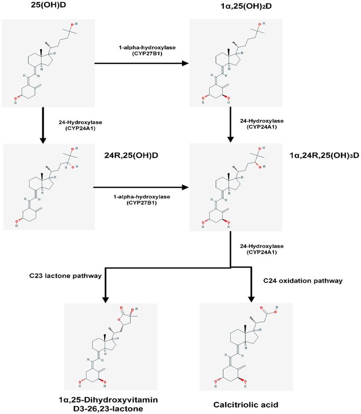
Makris et al. Page 2
circulating vitamin D metabolites, but also participates in the transport of the 25(OH)D into the
Author Manuscript
cell via a megalin/cubilin complex.
The accurate measurement of 25(OH)D has proved a difficult task. Although a reference method
and standardization program are available for 25(OH)D, the other vitamin D metabolites still lack
this. Interpretation of results, creation of clinical supplementation, and generation of therapeutic
guidelines require not only accurate measurements of vitamin D metabolites, but also the accurate
measurements of several other “molecules” related with bone metabolism.
IFCC understood this priority and a committee has been established with the task to support and
continue the standardization processes of vitamin D metabolites along with other bone-related
biomarkers.
In this review, we present the position of this IFCC Committee on Bone Metabolism on the latest
developments concerning the measurement and standardization of vitamin D metabolites and its
Author Manuscript
binding protein, as well as clinical indications for their measurement and interpretation of the
results.
Keywords
Vitamin D; 25-hydroxyvitamin D; Liquid chromatography; Mass spectrometry; Immunoassays;
Standardization; Vitamin D Standardization Program; Vitamin D binding protein; 1α,25-
dihydroxyvitamin D; 24,25-dihydroxyvitamin D
1. Introduction
Vitamin D is an important hormone required for bone and muscle development as well as the
Author Manuscript
preservation of musculoskeletal function. Due to its central role in calcium and phosphate
homeostasis, it plays an important role in bone metabolism.[1] Moreover, a number of non-
skeletal diseases have been associated with a vitamin D deficiency, including cancer,
cardiovascular disease, diabetes, immune dysfunction, etc.[2,3] Although genetic, molecular,
and animal studies suggest that vitamin D signaling has many extraskeletal effects, and
observational studies in human subjects, also suggest that poor vitamin D status is associated
with nearly all diseases, results of randomized controlled trials and Mendelian
randomization studies are mixed. Well designed basic and clinical studies are needed with
larger numbers of patients as well as well-designed randomized clinical trials, with baseline
vitamin D determination and accurate monitoring to establish a cause-effect relationship
between vitamin D deficiency and some diseases.[4,5]
1.1. Sources and production of vitamin D
Author Manuscript
Vitamin D is a fat-soluble secosteroid that is extensively metabolized in the human body.
Over the last 40 years, its synthesis and metabolism have been elucidated and more than 50
metabolites of vitamin D have been discovered.[6-8] However, to date, researchers have
been able to develop measurement procedures for only a few of them (Table 1).
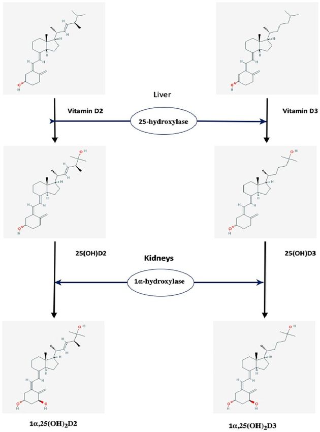
Vitamin D exists in two major forms, vitamin D2 (or ergocalciferol) and vitamin D3 (or
cholecalciferol), which exhibit only minor differences in their structure. (Fig. 1). As a
Clin Chim Acta. Author manuscript; available in PMC 2022 June 01.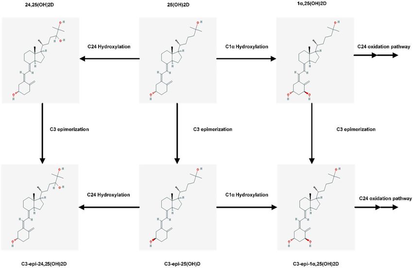
Makris et al.                                                                                               Page 3

                                      consequence, vitamin D2 and D3 have different molecular weights of 396.65 g/mol and
Author Manuscript

                                      384.64 g/mol, respectively.[9] These differences in the chemical structure of vitamin D2
                                      contribute to its lower affinity for vitamin D binding protein (VDBP), thus resulting in faster
                                      clearance from blood, a limited conversion to 25 hydroxyvitamin D [25(OH)D], and an
                                      altered catabolism by 24-hydroxyase (CYP24A1).[10-12] A recent meta-analysis found that
                                      vitamin D3 is more potent at raising serum 25(OH)D concentrations than is vitamin D2.
                                      Hence, vitamin D3 could potentially become the preferred choice for supplementation.[13]

                                      Vitamin D3 is synthesized from 7-dehydrocholesterol (7-DHC) in the skin by UVB radiation
                                      while vitamin D2 is derived from plant/yeast by irradiation of ergosterol (Figs. 2 and 3).
                                      [14,15] In humans the main sources of vitamin D (e.g., D2 and/or D3), are sunlight, diet, and
                                      supplements. However, most foods (except for fatty fish) contain low levels of vitamin D
                                      unless fortified (Table 2). Exposure of human skin to solar UVB radiation (wavelengths
                                      290–315 nm) leads to the conversion of 7-DHC to pre-vitamin D (pre-D) in the skin, which
Author Manuscript

                                      isomerizes to D3 in a non-catalytic, thermo-sensitive process.[16] Vitamin D3 production
                                      depends on the intensity of UV irradiation, which varies with season, latitude and altitude.
                                      [17] Skin pigmentation, sunscreen use, and clothing have been reported to affect the
                                      conversion of 7-DHC to vitamin D3.[18-20] Melanin in the skin blocks UVB from
                                      converting 7-DHC, thus limiting D3 production, as does extensive covering of the body with
                                      clothes and the use of sun-screen. A recent meta-analysis concluded that pigmented skin has
                                      less effective photoproduction of vitamin D and 25(OH)D. The quantity of sun exposure
                                      needed for dark-skinned, compared with light-skinned, people to achieve vitamin D
                                      sufficiency however remains uncertain.[21] However this view has been debated lately;
                                      Bogh et al., in an elegant study show that baseline vitamin D levels and total cholesterol
                                      levels are more important factors than skin pigmentation.[22,23]
Author Manuscript

                       1.2.     Vitamin D metabolism
                                      Vitamin D synthesized in the skin diffuses into the bloodstream where it is transported by
                                      vitamin D binding protein (VDBP) to the liver. Vitamin D from the diet is absorbed in the
                                      small intestine, incorporated into chylomicrons, which are released into the lymphatic
                                      system, and enters the venous blood where it binds to VDBP and lipoproteins before being
                                      transported to the liver. Vitamin D is essentially biologically inactive and must be converted
                                      to hydroxylated metabolites to gain hormonal activity. Its activation involves two
                                      hydroxylation steps (Fig. 4).[24]

                                      The first step occurs predominantly in the liver where vitamin D is hydroxylated at the C25
                                      position by the cytochrome p450 enzyme CYP2R1 (also called 25-hydroxylase) to 25-
                                      hydroxyvitamin D [25(OH) D]. There are several other CYP enzymes that are capable of 25-
Author Manuscript

                                      hydroxylation, namely CYP27A1, CYP3A4, CYP2D25, and perhaps others, but the
                                      CYP2R1 is emerging as the most critical enzyme for 25-hydroxylation.[25-27] This step is
                                      poorly regulated by any feedback mechanism in the context of the vitamin D endocrine
                                      system and it seems to be dependent primarily on the concentration of vitamin D [28].
                                      Hence, 25(OH)D levels increase in proportion to vitamin D intake and, plasma 25(OH)D
                                      levels are a good indicator of vitamin D status. The 25(OH)D produced in the liver is
                                      returned to circulation. Severe liver failure affects the function of the CYP2R1 enzyme.

                                          Clin Chim Acta. Author manuscript; available in PMC 2022 June 01.
Makris et al. Page 4
Moreover, loss-of-function mutations for the same enzyme are responsible for vitamin D-
Author Manuscript
dependent rickets (VDDR), type 1B (VDDR-1B) [29] as shown in Table 3.
The second step in vitamin D activation is the formation of 1α,25-dihydroxy vitamin D [or
calcitriol, 1α,25(OH)2D]. It occurs, under physiological conditions, mainly in the kidney by
another CYP450 enzyme, CYP27B1 or 1α-hydroxylase. This second hydroxylation takes
place at the carbon in the C1 position. Calcitriol binds again to VBDP and re-enters the
systemic circulation. It is now recognized that 1α-hydroxylase is expressed in many other
extrarenal tissues, including skin, brain, and colon and serves as an autocrine/paracrine
factor with cell specific functions (Table 4 and Fig. 5).[9,30-34].
The activity of renal 1α-hydroxylase is tightly controlled by 1α,25(OH)2D itself,
parathyroid hormone (PTH), fibroblast growth factor 23 (FGF23) and serum concentrations
of calcium (Ca) and phosphate (PO‐3
4 ). Under normal conditions, FGF23 acts on kidney
Author Manuscript
proximal tubular cells regulating phosphate excretion in order to maintain systemic
phosphate homeostasis. However, FGF23 can also influence the synthesis of calcitriol in the
proximal tubular cells by suppressing the expression of 1α-hydroxylase and increasing the
expression of 24-hydroxylase.[30,35] The activity of extrarenal 1α-hydroxylase is not
regulated by the same factors controlling its renal synthesis.[30] The kidney is the major
source for circulating 1α,25(OH)2D. Only in certain granulomatous diseases such as
sarcoidosis does the extrarenal tissue produces sufficient 1,25(OH)2D to contribute to the
circulating levels, which is generally associated with hypercalcemia.[36] Inactivating
mutations of this enzyme are responsible for vitamin D-dependent rickets (VDDR) type 1A
[VDDR-1A] [28,32,33,37] as shown in Table 3.
1.3. Catabolism
Author Manuscript
To retain calcitriol levels within the strict boundaries required for appropriate calcium
homeostasis and bone metabolism, both 1α,25(OH)2D and 25(OH)D may undergo further
hydroxylation by renal CYP24A1 (24-hydroxylase), leading to 1,24,25-trihydroxyvitamin D
[1,24,25(OH)3D] and 24R,25-dihydroxyvitamin D [24,25(OH)2D], respectively (Fig. 6).
Thus the main function of 24-hydroxylase is vitamin D inactivation, since [1] it limits the
amount of 1α,25(OH)2D3 in target tissues both by accelerating its catabolism to
1,24,25(OH)3D3 and ultimately in calcitroic acid or [2] by producing 24,25(OH)2D3 and
thus decreasing the pool of 25(OH)D3 available for 1 hydroxylation.[38]
CYP24A1 has been found in many tissues that express the vitamin D receptor. In the kidney,
it is found in the proximal and distal tubules. [39,40] The CYP24A1 gene is highly inducible
by 1α,25(OH)2D in all tissues in which it is found and it acts as a control mechanism to
Author Manuscript
prevent intoxication from 1α,25(OH)2D. [41] The importance of this feedback mechanism
was demonstrated when inactivating mutations of CYP24A1 reported in children and adults
with hypercalcemia.[29,42]
Another enzyme, CYP3A4, also plays a role in vitamin D catabolism. [43] This enzyme is
involved in drug metabolism, and is located in the liver and the intestine. Recently, a gain-of-
function mutation in CYP3A4 was described that leads to rickets with decreased serum
calcium and phosphate and elevated PTH and alkaline phosphatase (Table 3).[44] This is a
Clin Chim Acta. Author manuscript; available in PMC 2022 June 01.Makris et al. Page 5
distinct form of vitamin D dependent rickets (named type 3 vitamin D-dependent rickets or
Author Manuscript
VDDR3) since it does not involve a defect in synthesis of vitamin D metabolites but rather is
due to accelerated inactivation of vitamin D metabolites as CYP3A4 was found to inactivate
both 25(OH)D3 and 1,25(OH)2D, leading to vitamin D deficiency through accelerated
vitamin D metabolite inactivation (Table 3). [24,45] It is well known that CYP3A4 is
induced by certain drugs, such as rifampicin.[46,47] Thus, the induction of CYP3A4 gene
expression by certain drugs may enhance 25OHD and 1α,25(OH)2D3 catabolism.[43] and
hence modulate vitamin D effects in the body and could present as an alternative therapeutic
strategy to reduce serum levels of vitamin D metabolites in cases of patients with
inactivating mutations of CYP24A1. [48]
2. Measurement of vitamin D metabolites
Today, more than 50 vitamin D metabolites have been described and characterized, with
Author Manuscript
some of them exhibiting biological activity [6]. However, methods for measurement have
only been developed for five of them (vitamin D, 25(OH)D2 and 25(OH)D3, 1α,25(OH)2D,
24R,25(OH)2D, and C3-epi-25(OH)D) as shown in Table 1. These metabolites are present in
serum at concentrations that allow for their measurement with these methods.[49]
The above metabolites differ significantly in their biological activity. For example,
1α,25(OH)2D is five times more potent than vitamin D in its ability to regulate calcium
allowing for its extraction from the intestines and mobilization in bones. [50] A significant
factor that determines the biological activity of a metabolite is its affinity to the VDR.
Experimental studies have shown that 1α,25(OH)2D exhibits the highest affinity to the VDR
among all vitamin D metabolites [51], while the affinity of the rest of the metabolites is
significantly lower. For instance, 25OHD and 24,25(OH)2D exhibit approximately 900 and
Author Manuscript
5000 times lower affinity to the VDR, respectively, compared to that of 1α,25(OH)2D.[52]
2.1. Pre-analytical considerations
Five types of pre-analytical variability will be examined in detail in this section:
sample collection and handling
factors that relate to the individual (i.e., age, sex, ethnicity, and lifestyle)
environmental factors
disease factors and pregnancy
genetic factors
Author Manuscript
2.2. Sample collection and handling
Sample types and collection tubes: Serum and plasma (heparin and EDTA) can be
used for the measurement of vitamin D and its metabolites.[53] Serum is the preferred
matrix since it has the advantage of being free of anticoagulants used for plasma collection
such as EDTA, heparin, or citrate, which may interfere with their measurement, especially in
immunoassays. However even when serum is used, significant interferences can be observed
Clin Chim Acta. Author manuscript; available in PMC 2022 June 01.Makris et al. Page 6
with certain commercial assays, and even with High Performance Liquid Chromatography
Author Manuscript
(HPLC) and Liquid Chromatography tandem Mass Spectrometry (LC-MS/MS) methods
when tubes with serum clot activator and/or gel are used.[54-57] However, this type of
interference does not seem to apply to all commercial assays and methods as well as all
commercial collection tubes. [58] Vitamin D metabolites have been also detected and
measured in several body fluids and tissues including breast milk, urine, semen,
cerebrospinal fluid, synovial fluid, hair and in skin and muscle biopsies. However, these
matrices may require specific preanalytical protocols which are not standardized. Moreover,
standardization of analytical techniques and External Quality Assurance Schemes for the
measurement of vitamin D metabolites in these matrices are lacking.[59] Saliva has also
been explored in several studies and with assays based on different principles with often
inconsistent results. More recently, the technological advances in LC-MS/MS have made it
possible to measure 25(OH)D in dried blood spots (DBSs).[60] The utility of the
Author Manuscript
measurement of vitamin D metabolites in these matrices is limited to research setting.[59]
Sample stability: The stability of Vitamin D metabolites in clinical samples is a key
aspect for a reliable assessment of the results from epidemiological studies where the
samples usually are not tested on the day of collection. The majority of studies evaluating
the influence of storage conditions only studied 25(OH)D while the other metabolites have
been ignored or insufficiently studied.[53,61-67] These studies have shown that 25(OH)D is
stable in plasma and serum when samples are stored at room temperature (24 °C), 4 °C, or
frozen, as long as metabolites are not separated from their binding protein. There are studies
that claim that 25(OH)D can be stored without any significant loss in concentration for
several days at room temperature and for up to 3 years at −20 °C. In addition, no special
precautions are necessary during the transport of samples to the laboratory. In stored
samples, repeated cycles of freeze–thaw don’t seem to have any significant effect on
Author Manuscript
25(OH)D levels.[68] Attention is only needed when the samples have been already pre-
treated and vitamin D has been separated from its binding protein. Then, samples should be
kept in dark vials to avoid exposure to light and should be stored at < −70 °C. [53,61,69,70]
One study that examined the stability of 1α,25(OH)2D and 24,25(OH)2D concluded that
these two metabolites exhibit a lower stability in comparison to 25(OH) D upon storage,
with significantly decreased levels after 3 freeze–thaw cycles.[66] We must note here that
these stability studies present several limitations (i.e., a limited number of specimens
examined, chosen time intervals for storage, and lack of uniform definition of instability).
2.3. Environmental factors
Effect of season on 25(OH)D levels: UVB sunlight exposure, rather than diet, has been
reported as the main source of 25(OH)D for majority of the population.[71] Therefore,
Author Manuscript
levels of vitamin D are directly dependent upon exposure to UVB irradiation from the sun.
Several environmental factors such as latitude, altitude, season, and prevailing weather
conditions determine whether sunlight of a sufficient strength is available to stimulate the
conversion of 7-DHC in the skin to cholecalciferol (vitamin D3). This results in a 25(OH)D
seasonal variation and an effect based on the geographical location where the person lives
(distance from equator and altitude).[72,73] Generally, people that live in the northern
Clin Chim Acta. Author manuscript; available in PMC 2022 June 01.Makris et al. Page 7
hemisphere present the highest levels of 25(OH)D during the summer and autumn with
Author Manuscript
lower levels during winter and spring.[74-77]
2.4. Factors that relate to the individual
Age, sex, body fat, and lifestyle do have a, often small, effect on 25(OH)D levels.[78]
Age: It is known that age affects calcium and vitamin D metabolism. [1.] Calcium
absorption is reduced with age [2.] Intestinal resistance of calcium absorption to circulating
1,25(OH)2D increases with age. [3.] The ability of the older skin to produce vitamin D is
reduced [4.] VDR expression is also reduced with age. [5.] The ageing kidneys are less able
to produce 1α,25(OH)2D compared to younger kidneys. [6.] Substrate deficiency of vitamin
D increases with age.[79-82] Finally, older people are more home-bound and therefore less
exposed to sunshine and to outdoors activities compared to younger people.[83] Recent
Author Manuscript
studies, however, have shown that the effect of age on 25(OH)D levels is small. [75,84]
These studies included only subjects less than 75 years of age, which might explain the lack
of association between 25(OH)D levels and age.
Body mass index (BMI).—There is a consistent association in literature between
increasing BMI and lower serum 25(OH)D concentrations. Several studies have reported an
association between obesity (BMI greater than 30) and low serum 25(OH)D, 1α,25(OH)2D
concentrations, and high PTH concentrations.[85-88]
Adipose tissue might play a role in the low vitamin D levels observed in people with obesity.
[89-91] However, this relationship between obesity and low 25(OH)D levels, has not been
elucidated completely. Different mechanisms have been proposed to explain this inverse
association using behavioral factors such as a reduced exposure to sunlight due to less
Author Manuscript
outdoor physical activity and a low dietary intake of vitamin D enriched food.[92,93]
Moreover, decreased intestinal absorption, impaired hydroxylation in adipose tissue, and
25(OH)D accumulation in fat have been proposed to explain the hypovitaminosis in obesity.
[91] The fact that vitamin D is a fat soluble molecule led to the hypothesis that vitamin D is
sequestered in body fat depots, resulting in a lower bioavailability in the obese state.[91,94]
On the other hand, some studies have speculated that vitamin D deficiency itself could cause
obesity or even prevent weight loss.[91,94] Despite the well-established association between
obesity and vitamin D deficiency, few experimental studies have investigated the biological
bases involved in vitamin D metabolism in adipose tissue, with those studies that have
investigated this demonstrating inconsistent results. [95]
Sex.—Some studies have shown that men have higher levels of 25(OH) D, which are
Author Manuscript
independent of age, season, and race.[96-100] This could be explained by the fact that
women have relatively more body fat than men and store more fat in the gluteal-femoral
region, while men typically store more fat in the visceral (abdominal) depot.[101] On
average, men have 10–15% less fat content than women with the same BMI, thus having a
smaller reservoir to sequestrate vitamin D.[102-104] These differences in body fat amongst
genders might be an explanation for the difference between men and women in 25(OH)D
concentrations. However, these sex differences do not seem to be universal, as in several
Clin Chim Acta. Author manuscript; available in PMC 2022 June 01.Makris et al. Page 8
large studies, women showed either no significant differences or had higher levels of vitamin
Author Manuscript
D compared to men.[84,105-112]
Lifestyle.—Depending on the time of the day, duration of exposure, season, latitude, and
skin pigmentation, daily exposure of the skin to sunlight (i.e. arms or legs for 5–30 min) can
promote adequate endogenous synthesis of vitamin D3.[113] Outdoors activities allowing
for more exposure to sunlight,[114-118] and dressing habits (e.g., coverage of the body and
even the type of clothes), affect 25(OH)D levels.[119-121] Sunscreen use seems to have less
effect on 25(OH)D levels than earlier thought, although high SPF sunscreens have not been
studied. [122]
Ethnicity.—Although most studies are conducted in subjects of European descent, there are
studies that have shown that the levels of 25(OH)D differ according to ethnicity and skin
color. This seems logical since a darker skin color protects from exposure to UV irradiation
Author Manuscript
and increases the risk of vitamin D deficiency.[18,123] Vitamin D synthesis is highly
dependent on the concentration of melanin in the skin as melanin absorbs and takes care of
ultraviolet radiation (UVR), resulting in a less efficient conversion of 7-DHC to provitamin
D3.[124-127] Therefore, darker-skinned individuals will experience a slower vitamin D
synthesis than lighter-skinned individuals. This is more obvious and important at higher
latitudes where the intensity and duration of sunlight is limited. Metabolic differences based
on race/ethnicity may provide an additional explanation.[128]
2.5. Effect of disease and pregnancy
Effect of liver and kidney disease: The liver and the kidneys are the two most
important organs involved in the metabolism of vitamin D.
Author Manuscript
The liver is the organ where 25-hydroxylation of vitamin D occurs and the majority of
VDBP is synthesized.[129-132] In patients with liver disease, the prevalence of insufficiency
and deficiency ranges between 64 and 92%, which is much higher than in the general
population. Serum 25(OH)D is inversely related to the severity of liver disease.[133-135]
The high prevalence of vitamin D deficiency in this populations occurs regardless of the
etiology of liver disease.[136]
Synthetic liver dysfunction is not entirely responsible, as vitamin D deficiency is still highly
prevalent in those with non-cirrhotic liver disease.[133] 25(OH)D levels “normalize” after
oral or parenteral administration of vitamin D in patients with cirrhosis, indicating that 25-
hydroxylation is preserved in this patient population.[137] A recent study showed that in
patients with liver disease, 25-hydroxylase activity, although low compared to subjects
Author Manuscript
without liver disease, was relatively well-preserved and did not affect serum 25(OH)D
concentrations.[138]
Low vitamin D levels in chronic liver disease (CLD) may result from a variety of reasons
and mechanisms including: [1] reduced sun exposure and dietary intake, [2] intestinal
malabsorption of dietary vitamin D, [3] reduced endogenous production of VDBP and
albumin in the liver, which are both impaired in CLD and in the presence of cirrhosis, [4]
decreased hepatic hydroxylation of vitamin D to 25(OH)D and finally [5] increased
Clin Chim Acta. Author manuscript; available in PMC 2022 June 01.Makris et al. Page 9
catabolic removal of 25(OH)D.[139,140] Hence, when catabolism is increased, there will be
Author Manuscript
less 25(OH)D available for production of the active hormone.[139] Low total 25(OH)D
levels do not seem to disrupt its biological activity as long as unbound vitamin D levels are
maintained within a normal range.[141]
As VDBP has a single sterol-binding site and only 5% of the total circulating VDBP is
actually bound to a vitamin D metabolite at any time [142], liver function would have to be
severely impaired in order for low VDBP levels to have a significant role in 25(OH)D
deficiency in CLD. [143] However, although total 25(OH)D levels decrease as the severity
of CLD increases, PTH levels are not associated with total 25(OH)D levels.[144] Patients
with end stage liver disease and low total 25(OH)D levels maintain a normal serum
corrected calcium concentration and do not develop secondary hyperparathyroidism.[145]
The kidneys are essential not only for the conversion of 25(OH)D to 1α,25(OH)2D, but also
Author Manuscript
for the re-absorption of 25(OH)D from renal ultra filtrate for its recycling into circulation.
Normal renal function is also essential to maintain the endocrine actions of calcitriol, which
by itself contributes to maintaining the VDR in target tissues since it protects the receptor
from degradation by binding.[146]
In chronic kidney disease (CKD), less 1α,25(OH)2D is produced. The mechanisms involved
in the reduced calcitriol production during the course of CKD have been discussed in detail
elsewhere. [147] Impaired uptake of 25(OH)D by the kidneys seems to be the main cause of
1α,25(OH)2D deficiency.[148] Decreased kidney function and calcitriol deficiency lead to
hypocalcemia and are key contributors to secondary hyperparathyroidism (SHPT).[148]
This is more obvious among patients with end-stage renal disease where 1α,25(OH)2D is
almost undetectable. CKD is also characterized by low serum 25(OH)D levels. The main
Author Manuscript
causes and risk factors for vitamin D deficiency among CKD patients have also been
discussed in detail elsewhere.[148] The 25(OH)D levels for CKD patients are suggested to
be progressively low as renal function deteriorates. However, not all studies show that
25(OH)D insufficiency or deficiency in CKD patients is greater than in the general
population.[149,150] For CKD patients, vitamin D deficiency is a strong predictor of
accelerated renal disease and death. In addition, since the kidney is not the only site of
calcitriol production, the maintenance of sufficient 25(OH)D levels could be a possible
objective.[151] However, the best treatment approach and the best biomarker for follow-up
in CKD patients is still a debate.[152,153]
Kidney disease also disrupts vitamin D catabolism. Within the kidneys, 1α-hydroxylase and
24-hydroxylase are under hormonal regulation of FGF23 and PTH. FGF23 is responsible for
Author Manuscript
the reduced expression of 1α-hydroxylase in renal tubular cells and induces the expression
of 24-hydroxylase, which is responsible for the catabolism of vitamin D. PTH seems to
increase the expression of 1α-hydroxylase in renal tubular cells.[40,154]. CKD is
characterized by high levels of FGF23 and phosphorus. These increased phosphate levels
have been correlated with low concentrations of 1α,25(OH)2D, but it is not clear whether
this correlation is direct, induced by FGF23, or confounded with other factors. [40]
Moreover, other metabolic disturbances that are observed in patients with CKD such as
Clin Chim Acta. Author manuscript; available in PMC 2022 June 01.Makris et al. Page 10
diabetes, metabolic acidosis, and uremia are able to reduce the expression of CYP27B1.
Author Manuscript
[155-157] In the end, this results in the reduced production of 1α,25(OH)2D in patients with
CKD. Also, 24R,25(OH)2D seems to be lower in patients with CKD compared to healthy
subjects.[158] The net effect of FGF23 and PTH on vitamin D catabolism in CKD is
however still debated.
Systemic inflammatory response (SIR): As vitamin D is often linked to acute and
chronic inflammatory disease it is important to realize that 25(OH)D can act as a negative
acute phase reactant.[159] This was clearly shown in a study by Waldron et al, where
25(OH)D concentrations decreased after an elective orthopedic surgery leading to a systemic
inflammatory response with increased CRP levels. Also VDBP decreases after SIR however
cannot explain all of the decrease in 25(OH)D.
Pregnancy: Special attention must be given to pregnancy since several studies report low
Author Manuscript
levels of 25(OH)D in pregnant women. In a recent meta-analysis, it was reported that 54%
of pregnant women had levels of vitamin D below 50 nmol/L.[160] Moreover, several
studies have suggested that low levels of 25(OH)D during pregnancy are associated with an
increased risk of pre-eclampsia, gestational diabetes, and other pregnancy complications.
[161-164] However, the results from these studies that relate low levels of 25(OH)D during
pregnancy with adverse outcomes are conflicting.[165-167] These conflicting results are not
only due to possible methodological problems related to the study design, but also with the
methods used for 25(OH)D quantitation. In pregnancy, VDBP is known to be increased and
when 25(OH)D is measured with an immunoassay, its levels can be underestimated due to
incomplete dissociation of 25(OH)D from its binding protein. On the other hand, when a
HPLC or a LC-MS/MS method is used, the dissociation of 25(OH)D from its binding
protein is more complete due to the use of strong chemical solvents during sample
Author Manuscript
preparation.[168-170] These analytical problems cause significant assay variation and the
results from meta-analyses may be subject to error, especially when results are included
from studies based on certain immunoassay measurements or from unstandardized assays.
2.6. Genetic factors
Gene-environment interactions that could have an impact on various vitamin D-related
disorders have recently drawn the attention of several researchers.[171,172] For example, it
has been suggested that hypovitaminosis D occurs in the presence of specific gene variations
related to vitamin D metabolism. Therefore, individuals with specific vitamin D-related
genotypes may require specific personalized advice to optimize their vitamin D status. Data
from twin and family-based studies have demonstrated that circulating vitamin D
concentrations can be partially determined by genetic factors.[173,174] Moreover, it has
Author Manuscript
been shown that genetic variants (e.g., mutation) and alterations (e.g., deletion,
amplification, and inversion) in genes involved in the metabolism, catabolism, transport, or
even binding of vitamin D to its receptor might have an effect on vitamin D levels.[175]
However, the underlying genetic determinants of 25(OH)D plasma levels have not been fully
elucidated. Furthermore, the association between epigenetic modifications such as DNA
methylation and vitamin D levels have now been reported in several studies.[175]
Clin Chim Acta. Author manuscript; available in PMC 2022 June 01.Makris et al. Page 11
Linkage studies, studies involving candidate genes in the vitamin D metabolism pathway, as
Author Manuscript
well as genome wide association studies (GWAS) have shown human genetic variants to be
related to vitamin D status.
Single nucleotide polymorphisms: Candidate gene studies and GWAS have shown
that certain gene single nucleotide polymorphisms (SNP) involved in vitamin D metabolism
pathways (e.g., CYP2R1, CYP27B1, CYP24A1, DHCR7, the VDR, and GC) have an effect
on vitamin D levels as shown in Ref [175]. Vitamin D binding protein (VDBP) is discussed
in detail further down in this article, but, briefly, VDBP has two common SNPs (rs7041 and
rs4588), which results in three VDBP isotypes (Gc1f, Gc1s, and Gc2). These isotypes show
different binding affinity constants to 25(OH)D. This means that persons with different
SNPs have different total 25(OH)D concentrations while they might have the same
concentration of free 25(OH)D. These polymorphisms are distributed differently based on
ethnicity as shown in several studies and might affect the way we interpret the total
Author Manuscript
25(OH)D concentration.[176] The effect of these SNPs on the levels of circulating 25(OH)D
only account for 5% of its variability and is considered small compared to other
environmental factors that have a more significant effect on circulating 25(OH)D levels.
[32,177,178]. Therefore, their presence does not seem to have significant clinical value in
everyday practice if we consider that most laboratory assays present an analytical variability
of approximately 10%.
3. The measurement of 25(OH)D
3.1. Clinical relevance
The measurement of 25(OH)D is performed mainly for two reasons: [1] to determine the
nutritional status of vitamin D, and [2] to monitor the efficacy of supplementation. As
Author Manuscript
previously mentioned, vitamin D exists in two different forms and in order to adequately
monitor therapy, both types of vitamin D need to be accurately quantitated. In fact, the
accurate measurement of 25(OH)D for the assessment of vitamin D status has always been a
major goal of all clinical laboratories involved in the measurement of vitamin D metabolites.
25(OH)D is the metabolite of choice to determine vitamin D status for several reasons:
25(OH)D levels in the blood are higher than those of any other vitamin D metabolite. The
serum concentration of 25(OH)D is in the range of 25–200 nmol/L, which is 1000 times
higher than that of 1α,25(OH)2D, whose concentration is in the range of 50–150 pmol/L.
Majority of 25(OH)D is found in the systemic circulation, with limited distribution in less
accessible tissues (e.g., fat) [179].
Author Manuscript
It is well accepted that adequate levels of vitamin D are required to prevent nutritional
rickets and osteomalacia, both of which are characterized by low levels of 25(OH)D as
shown in Table 3. [9]
Several clinical studies have demonstrated that there is an association between serum levels
of 25(OH)D and several clinical outcomes such as bone mineralization, fracture risk, fall
risk, cancer, diabetes, and cardiovascular events. Meta-analyses and randomized control
trials demonstrated a positive dose–response relationship between vitamin D
Clin Chim Acta. Author manuscript; available in PMC 2022 June 01.Makris et al. Page 12
supplementation and fracture prevention, which could partly be attributed to fall reduction.
Author Manuscript
[180,181]
25(OH)D has a relatively long half-life (2–3 weeks) compared to that of 1α,25(OH)2D
(approximately 4–6 h), and, therefore, serum levels vary little within short periods of time
[41,182].
The hydroxylase enzymes that metabolize vitamin D to 25(OH)D in vivo behave according
to first-order reaction kinetics. This means that its rate of production is dependent on vitamin
D levels and, therefore, its level in systemic circulation is the best indicator of vitamin D
nutritional status [183].
Furthermore, 25(OH)D represents the sum of vitamin D intake and dermal production [184].
Serum levels of 25(OH)D are relatively stable and not affected by diet (i.e., calcium intake)
Author Manuscript
and lifestyle (i.e., sedative life or regular physical exercise), whereas 1α,25(OH)2D levels
are affected by all the latter [179,182].
Serum levels of 25(OH)D can determine if there is enough 25(OH)D for the extrarenal
tissues to produce 1α,25(OH)2D for autocrine or paracrine action. Recent data have revealed
that many of these tissues also contain CYP27B1, which is responsible for converting
25(OH)D to 1,25(OH)2D. Regulation of CYP27B1 in these non-renal tissues is generally
different from that in the kidney and may be more substrate-dependent. This finding has led
to the concept that the maintenance of adequate 25OHD levels in the blood is required for
vitamin D regulation of a large number of physiologic functions beyond those of the classic
actions involved in bone mineral metabolism. Measurement of 1α,25(OH)2D does not
provide this information since its extrarenal production does not contribute much to the
Author Manuscript
systemic load [52,185].
3.2. Methods of measurement
25(OH)D can be measured using various methods including immunoassays, which are
mainly used, protein-binding assays, HPLC-UV, or LC-MS/MS.[59,183,186-188]
3.3. Analytical variability and the standardization of the 25(OH)D assays
Serum total 25(OH)D, [the sum of 25(OH)D2 and 25(OH)D3], is considered to be the best
biological marker of an individual’s vitamin D status as described above. However, in
clinical guidelines, differences exist in the definition deficiency, insufficiency, and
sufficiency, creating a great deal of controversy [189,190]. The most critical factor that
confounds efforts to develop consensus clinical and nutritional public health guidelines for
Author Manuscript
interpreting serum 25(OH)D concentrations is the substantial variability that existed (and
still exists) in many assays that have been used over the years to measure 25(OH)D in
clinical research studies [191]. The lack of assay standardization is the main source of bias,
making it impossible to pool research results in order to develop consensus cut-off points
[192].
Clin Chim Acta. Author manuscript; available in PMC 2022 June 01.Makris et al. Page 13
To overcome these problems, the Vitamin D Standardization Program (VDSP) was
Author Manuscript
established in 2010 by the Office of Dietary Supplements, National Institutes of Health, as
an international collaborative effort with the National Institute of Standards and Technology
(NIST), the Centers for Disease Control and Prevention (CDC), Ghent University in
Belgium, the American Association for Clinical Chemistry (AACC), the IFCC, and national
health and nutrition surveys from Australia, Canada, Germany, Ireland, Mexico, South
Korea, United Kingdom, and the USA [193]
A reference measurement procedure (RMP) is now available. [194,195] NIST first
developed a RMP based on ID-LC-MS/MS for the determination of 25(OH)D2 and
25(OH)D3 in human serum [195]. This method is now recognized by the Joint Committee
for Traceability in Laboratory Medicine (JCTLM) as a RMP. Later, Stepman et al. (at Ghent
University, Belgium) also described an ID-LC-MS/MS method for the determination of
25(OH)D2 and 25(OH)D3, which was also recognized as an RMP for 25(OH)D by the
Author Manuscript
JCTLM [196]. Finally, the CDC developed an ID-LC-MS/MS method that was subsequently
recognized by the JCTLM as an RMP for 25(OH)D [197]. These three methods are currently
the only JCTLM-recognized RMPs for the determination of 25(OH)D2 and 25(OH)D3and
make it possible to standardize 25(OH)D measurements. As standardization is crucial as cut-
off values for deficiency, insufficiency, and sufficiency are used internationally, the Centers
for Disease Control (CDC) started an international Vitamin D standardization certification
program (VDSCP).[193] This led to an impressive improvement in the number of
standardized 25(OH)D assays.
The achievements of VDSP and of the institutions that supported the objectives of the VDSP
were significant: it resulted in a reference measurement system that is the backbone for
standardizing 25(OH)D measurements in current and future assay systems [198,199]. The
Author Manuscript
components of this reference measurement system include: [1] the gold standard RMPs, [2]
NIST Standard Reference Materials (SRMs), [3] the VDSCP (developed, implemented and
executed by the CDC), [4] the accuracy-based performance testing or external quality
assessment schemes (PT/EQA) conducted by the College of American Pathologists (CAP)
and the Vitamin D External Quality Assessment Scheme (DEQAS) [195,200,5] methods for
retrospective standardization of studies completed in the past, and [6] a set of laboratory
performance guidelines for both reference laboratories (those that govern the RMPs) and for
routine laboratories developed by the VDSP [200].
The VDSP adopted the assay performance criteria for 25(OH)D that were developed by
Stockl et al. for different types of labs and assays [200]. According to these criteria, for
RMPs, the limits for total CV and mean bias should be less than or equal to 5% and less than
Author Manuscript
or equal to 1.7%, respectively. For routine clinical laboratories, the limits for total CV and
mean bias are not so strict and should be less than or equal to 10% and 5%, respectively.
However even today, several immunoassays suffer from other analytical issues that lead to
continuing problems with the quality of 25(OH)D measurements. These immunoassays
show patient or matrix dependent deviations, for instance in pregnant women, patients on
intensive care, hemodialysis patients, osteoporotic patients, and patients with liver failure.
Sera from these patient groups behave differently in these immunoassays than in LC-
Clin Chim Acta. Author manuscript; available in PMC 2022 June 01.Makris et al. Page 14
MS/MS assays. [168,201-205] One of the causes is a vitamin D binding protein (DBP)
Author Manuscript
dependency in the immunoassay, but other causes are still unknown. Furthermore,
immunoassays often demonstrate difficulties with the measurement of 25(OH)D3 and
25(OH)D2.[204,206,207] Where LC-MS/MS methods can separate 25(OH)D3 and
25(OH)D2, immunoassays make use of antibodies with a different affinity for 25(OH)D3 and
25(OH)D2 and therefore often over- or underestimate measured 25(OH)D depending on
whether the subject uses 25(OH)D2. In addition, some immunoassays suffer from cross
reactivity with other vitamin D metabolites such as 24,25(OH)2D. [208,209] On the other
hand, LC-MS/MS assays are not always designed to separate the epimer of 25(OH)D. This
can lead to falsely high 25(OH)D concentrations, especially in young children.[209-211]
Most immunoassays do not show cross reactivity with the epimer.
The above-mentioned issues, including the different affinities for 25(OH)D3 and 25(OH)D2,
cross reactivity with 24,25(OH)2D, and matrix and/or patient-dependent biological
Author Manuscript
variations make these currently available immunoassays difficult for standardization and
accurate measurements.
3.4. Recommendations
1. Preanalytical recommendations:
• 25(OH)D is a very stable analyte, which can be measured in serum and plasma.
• There is no clear evidence that other prenalytical factors should be taken into
account with regard to blood withdrawal. However they have impact on the
measured value of 25(OH)D and the results should be interpreted accordingly
2. Analytical recommendations:
Author Manuscript
• All clinical and research laboratories are encouraged to participate in an accuracy
based external quality assessment scheme (i.e., CAP or DEQAS). The providers
of these schemes should regularly perform commutability studies to ensure that
the results they provide correspond to the clinical results obtained with the
different assays
• Manufacturers as well as research and reference laboratories are encouraged to
participate in the Vitamin D Standardization-Certification Program (VDSCP)
• Many immunoassays suffer from patient and pregnancy dependent deviations
(due to patient related specific changes in the composition of the serum which
influence the assays) and manufacturers should improve these assays. Well-
characterized and standardized LC-MS/MS methods are currently the only
Author Manuscript
methods which are able to measure 25(OH)D in all serum samples, regardless of
the nature of the sample.
• In published studies the absolute levels of total 25(OH)D should be interpreted
with caution and the standardization status of the assay used should be taken into
account. In meta-analyses only studies that have been used standardized assays
should be included and those that a retrospective standardization has been
performed according to VDSP methods.
Clin Chim Acta. Author manuscript; available in PMC 2022 June 01.Makris et al. Page 15
3. Post-analytical recommendations:
Author Manuscript
• For results reporting of total 25(OH) preference should be given to SI units
(nmol/L) as opposed to mass units (ng/mL).
Further research:
• Revision of the assay performance criteria
• Is 25(OH) the optimal marker for determining vitamin D status?
• Find consensus on the reference values (or target values) to report with clinical
samples
4. The measurement of 1,25(OH)2D
4.1. Clinical relevance
Author Manuscript
Although 1α,25(OH)2D is the active form of vitamin D, its measurement does not provide
any additional value in determining an individual’s vitamin D status. Its measurement thus,
should be limited in serious clinical conditions such as hypo- or hypercalcemia.[179,212]
Three types of conditions can influence 1α,25(OH)2D disorders such as [1] disorders
regarding CYP27B1, [2] disorders in the VDR, and [3] disorders in the extrarenal
production of 1α,25(OH)2D. For example, vitamin D dependent rickets type 1 or pseudo-
vitamin D deficiency rickets is a genetic disorder leading to a CYP27B1 deficiency, which
causes hypocalcemia and early onset rickets.[213]
Also X-linked hypophosphatemia, autosomal dominant hypophosphatemic rickets,
autosomal recessive hypophosphatemic rickets 1 – 3, tumor induced osteomalacia, and other
Author Manuscript
rare disorders leading to FGF23-mediated hypophosphatemia all led to inhibition of the
CYP27B1 gene, abnormally low 1α,25(OH)2D concentrations, and eventually osteomalacia
or rickets [see Table 5 and for detailed review see references [214,215]]. Additionally, there
are some rare disorders that may manifest as FGF23-mediated hypophosphatemia. These
include, osteoglophonic dysplasia, McCune–Albright syndrome, epidermal nevus syndrome,
neurofibromatosis, hypophosphatemic rickets with hyperparathyroidism, and Jansen
metaphyseal chondrodysplasia. High FGF23 levels result in inhibition of CYP27B1 and
therefore, abnormally low 1α,25(OH)2D concentrations (Table 5).
High 1α,25(OH)2D concentrations can be seen in disorders associated with the VDR such as
hereditary vitamin D resistant rickets (or vitamin D dependent rickets type 2). Despite high
1α,25(OH)2D concentrations, hypocalcemia and rickets appear as the VDR does not detect
vitamin D (see Table 3). [216] Also, in diseases with excessive and uncontrolled extrarenal
Author Manuscript
production of active vitamin D, high concentrations of 1α,25(OH)2D are detected. Examples
of these disorder types include sarcoidosis, tuberculosis, rheumatoid arthritis, inflammatory
bowel disease, and lymphoproliferative disorders (Table 6). These disorders often result in a
low bone mineral density/osteomalacia.[36,217-219] This extrarenal 1α-hydroxylation by
local CYP27B1 is not controlled by PTH, FGFG23, phosphate, or 1α,25(OH)2D itself, but
is rather regulated by local factors such as IFN-γ and IL15, and is dependent on the
availability of substrate.[36,220] When the locally produced 1α,25(OH)2D concentration is
Clin Chim Acta. Author manuscript; available in PMC 2022 June 01.Makris et al. Page 16
too high, it may escape the confinements of the intracellular space, spill over to the systemic
Author Manuscript
circulation, and raise blood concentrations to abnormally high levels.[219] In the case of
hypo- or hypercalcemia or hypophosphatemia, the measurement of 1α,25(OH)2D plays an
important role in the verification of absence or onset of these conditions.
4.2. Analytical methods
As serum 1,25(OH)2D concentrations are very low (pmol/L), it is very difficult to measure
this analyte. Over the few past decades 1,25(OH)2D was often measured using manual
competitive protein binding assays or radioimmunoassays. Many of these
radioimmunoassays encountered problems with specificity, as cross-reactivity with other
vitamin D metabolites influenced the results.[221] Recently automated immunoassays
became commercially available, which appear to perform better regarding cross reactivity.
However, these methods cannot separate 1,25(OH)2D2 from 1,25(OH)2D3, which might be
Author Manuscript
problematic in countries with D2 supplementation.[222,223] In addition, over the last few
years, several laboratories have developed LC-MS/MS methods to measure 1,25(OH)2D.
The sensitivity issue with these LC-MS/MS methods can be resolved by 2D
chromatography, derivatization of the vitamin D molecule, and/or immunopurification. [212]
This last option is not only advantageous for the sensitivity, but also regarding specificity.
LC-MS/MS methods without immunopurification might suffer from isobaric interferences
as it is difficult to separate 1β-25-dihydroxyvitamin D3 from its epimer.[224] Therefore, not
all LC-MS/MS methods fully agree in method comparison studies. [225]
Unfortunately, no reference methods exist for measuring 1,25(OH)2D. Without reference
method standardization, an evaluation of the quality of currently available methods is not
possible. Absent of standardization, the determination of reference values is subjective to the
method used.
Author Manuscript
4.3. Recommendations
• Measurement of 1α,25(OH)2D is recommended in the investigation of
– Hypercalcemia
– Calcipenic rickets/osteomalacia
– Differentiation of phosphopenic rickets between those that are/are not
FGF23 mediated
• Measurement of 1α,25(OH)2D is not recommended for the monitoring of
chronic kidney disease patients
Author Manuscript
• Further research is needed to ensure the development of a reference method for
1α,25(OH)2D, standardization of currently available methods, and development
of reference values in adults and children traceable to the reference method
5. The measurement of 24,25(OH)2D
The dihydroxylated metabolite of vitamin D, 24R,25(OH)2D, has attracted much attention
recently. It is the main product and first step in the process of vitamin D catabolism. This
Clin Chim Acta. Author manuscript; available in PMC 2022 June 01.Makris et al. Page 17
process is mediated by the enzyme 24-hydroxylase (CYP24A1). This metabolite exists in
Author Manuscript
two molecular forms: 24R,25(OH)2D and 24S,25(OH)2D. They are named R or S depending
on the spatial position of the carbon C24 and hydroxyl group. As the 24S isomer is not
present in humans, this review focuses on the 24R isomer, and from now on when we refer
to 24,25(OH)2D, we mean the R isomer.[32,226]
5.1. Clinical relevance
24,25(OH)2D circulates in a concentration range of nmol/L and has a half-life of about 7
days. These characteristics make this metabolite attractive for quantitation and a candidate
biomarker of vitamin D catabolism.[158,227] The amount of circulating 24,25(OH)2D
depends on the amount of its predecessor 25(OH)D and the activity of CYP24A1. The
expression of CYP24A1 is upregulated by 1α,25(OH)2D, and FGF23 and is downregulated
by PTH. Moreover, it is partly regulated by VDR activity.[228,229]
Author Manuscript
Consequently, if there are sufficient levels of biologically active vitamin D and the
expression of CYP24A1 is adequate, then calculating the ratio 25(OH)D/24,25(OH)2D (also
called vitamin D metabolite ratio or VMR) is a good indication of the catabolic clearance by
CYP24A. If we also take into account that the production of 24,25(OH)2D is dependent on
25(OH)D and on the expression of CYP24A1, then the absolute concentration of
24,25(OH)2D or the VMR may be a better indicator of vitamin D sufficiency than 25(OH)D
alone since it is not affected by race. [230] In addition, several studies have suggested that
24,25(OH)2D has effects of its own [231-235], and that human bone cells and human
mesenchymal stem cells (hMSCs) metabolize 25(OH)D3 into both 1α,25(OH)2D3 and
24R,25(OH)2D3.[236-238] These results demonstrate the ability of bone cells to convert
25(OH)D3 in vitro, indicate the importance of systemic and tissue-specific 24,25(OH)2D3
actions, suggest a role in osteoblastic differentiation, and enhance the concept that the
Author Manuscript
hydroxylation of 25(OH)D3 leads to 2 bioactive forms of vitamin D3, 24,25(OH)2D3 and
1α,25(OH)2D3, each with its own unique functions. [239] Moreover these studies
demonstrated that 24,25(OH)2D3 is an active form of vitamin D3 with an essential role in
osteoblast maturation, Ca2+ mineralization, gene expression, and the regulation of
cytochrome P450 expression, resulting in decreased 1α,25(OH)2D3 biosynthesis.[239]
These data suggest a direct role in bone cells—in particular, in osteoblasts. It should also be
noted that 24-hydroxylation is the first step of a degradation cascade. Therefore, the
biologically-active levels of 24,25D3 or 1,24,25D3 fully depend on the velocity of the
subsequent steps in the degradation pathway.[240] It is of no surprise the biological
significance of 24-hydroxylase has been continuously discussed because of its dual role first
as a catalytic enzyme initiating the side chain catabolism of both 25(OH)D3 and more
importantly 1α,25(OH)2D3 in target tissues and second as an enzyme with a synthetic
Author Manuscript
capacity since, in some situations, it is activated to produce 24,25(OH)2D3.[241]
Chronic kidney disease (CKD) is characterized by a state of active vitamin D deficiency. In
contrast to the focus placed on the decreased renal production of 1α,25(OH)2D3, relatively
little attention has been paid to the potential role of altered vitamin D catabolism in CKD. In
healthy people, the concentrations of vitamin D metabolites in blood and target tissues
represent a balance of production and catabolism. CYP24A1 is the primary enzyme
Clin Chim Acta. Author manuscript; available in PMC 2022 June 01.You can also read