Histopathological atlas of desmoplastic reaction characterization in colorectal cancer - Oxford Academic Journals
←
→
Page content transcription
If your browser does not render page correctly, please read the page content below
Japanese Journal of Clinical Oncology, 2021, 51(6)1004–1012
                                                                                                      https://doi.org/10.1093/jjco/hyab040
                                                                                            Advance Access Publication Date: 15 April 2021
                                                                                                                           Technical Note

Technical Note

Histopathological atlas of desmoplastic reaction
characterization in colorectal cancer
Hideki Ueno1 ,*, Yoshiki Kajiwara1 , Yoich Ajioka2 , Tamotsu Sugai3 ,

                                                                                                                                                                 Downloaded from https://academic.oup.com/jjco/article/51/6/1004/6225738 by guest on 03 November 2021
Shigeki Sekine4 , Megumi Ishiguro5 , Atsuo Takashima6 and
Yukihide Kanemitsu7
1
 Department of Surgery, National Defense Medical College, Saitama, Japan, 2 Division of Molecular and Diagnostic
Pathology, Graduate School of Medical and Dental Sciences, Niigata University, Niigata, Japan, 3 Department of
Molecular Diagnostic Pathology, School of Medicine, Iwate Medical University, Iwate, Japan, 4 Division of
Pathology and Clinical Laboratories, National Cancer Center Hospital, Tokyo, Japan, 5 Department of Translational
Oncology, Tokyo Medical and Dental University Graduate School, Tokyo, Japan, 6 Gastrointestinal Medical Oncology
Division, National Cancer Center Hospital, Tokyo, Japan and 7 Department of Colorectal Surgery, National Cancer
Center Hospital, Tokyo, Japan
*For reprints and all correspondence: Hideki Ueno, Department of Surgery, National Defense Medical College, 3-2 Namiki,
Tokorozawa, Saitama 359-8513, Japan. E-mail: ueno_surg1@ndmc.ac.jp
Received 29 December 2020; Editorial Decision 25 February 2021; Accepted 25 February 2021

Abstract
Emergent scientific evidence indicates the central role of cancer-associated fibroblasts in determin-
ing whether the microenvironment of cancer works as friend or foe of the host; however, there is no
unified histological evaluation framework of fibrotic stroma in colorectal cancers. Myxoid stroma
and keloid-like collagen are site-specific histopathological features generated by cancer-associated
fibroblasts, which appear exclusively in the tumor front during desmoplastic reaction. On the
basis of these two stromal components, desmoplastic reaction is categorized into three patterns—
immature, intermediate and mature—using hematoxylin and eosin staining. In January 2020, a
prospective randomized clinical trial, JCOG1805, to elucidate the value of adjuvant chemotherapy
in stage II colorectal cancer patients with pathological risk factors of recurrence was launched
in Japan, in which intermediate/immature desmoplastic reaction is one of the four risk factors
selected as inclusion criteria. This paper covers the diagnostic criteria for the desmoplastic reaction
classification being used in the JCOG1805 study.
Key words: cancer stroma, cancer-associated fibroblasts, extracellular matrix, myxoid stroma, keloid-like hyalinized collagen

Introduction                                                                                Being different from advances in the field of cancer biological
A number of molecular biology studies have clarified the central                        studies, where genes closely associated with poor-prognosis subtypes
role of stromal environment in cancer development, and cancer-                          are shown to be expressed in the stroma rather than in cancer cells
associated fibroblasts (CAFs) have recently been regarded as key                        (3,4), tumor factors are still major determinants in clinical practice
players of modulating cancer microenvironment (1). Biological                           for colorectal cancer (CRC). A major example would be judging
behavior of at least part of CAFs is closely associated with epithelial–                the indication of adjuvant chemotherapy in stage II CRCs, wherein
mesenchymal transition (EMT) and cancer stemness via modulating                         patients are considered at high-risk of recurrence if they present
growth factor signaling and extracellular matrix remodeling, thereby                    at least one of the following clinical characteristics: lymph-node
promoting acquisition of proinvasion/metastasis phenotype and                           sampling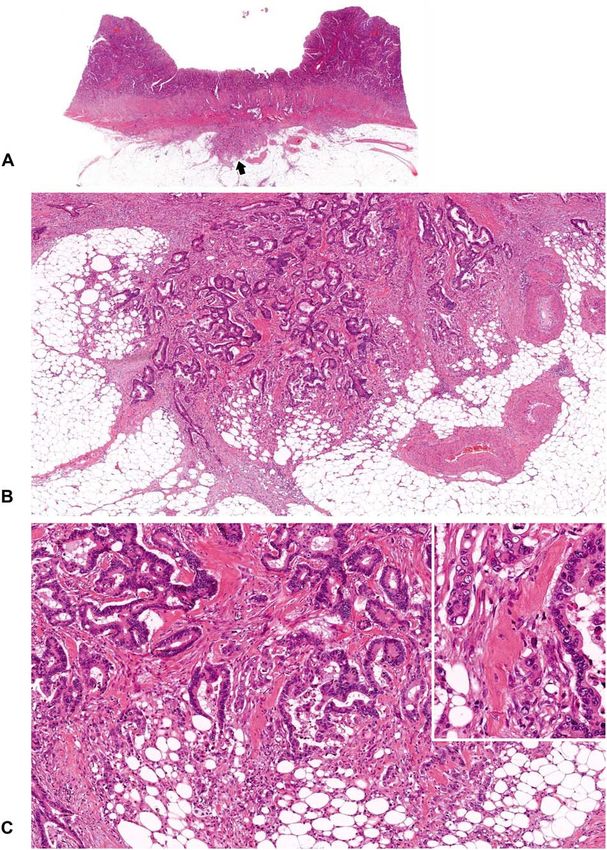
Jpn J Clin Oncol, 2021, Vol. 51, No. 6              1005

                                                                                                                                                             Downloaded from https://academic.oup.com/jjco/article/51/6/1004/6225738 by guest on 03 November 2021
Figure 1. Diagnostic criteria for the desmoplastic reaction (DR) classification.

perforation; or pT4 staging in international guidelines such as the               CRC patients at high-risk of developing recurrence according to T-
National Comprehensive Cancer Network (NCCN) guidelines(5)                        stage and three selected pathological factors (Pn, DR and BD)]—was
and European Society for Medical Oncology (ESMO) guidelines (6).                  initiated by the Japanese Clinical Oncology Group. In the process of
No stromal factors are included in the decision-making process of                 developing a study protocol, the evidence levels for prognostic values
the treatment. The lack of appropriate assessment criteria for cancer             of candidate risk factors, including those adopted in the NCCN
stroma may be a major reason for expanding the gap in the approach                and ESMO guidelines, had been reviewed; consequently, the DR
to understand the nature of individual tumors between the bench and               classification was selected, together with T-stage, perineural invasion,
the clinic.                                                                       tumor budding, as a prognostic factor to be used as an inclusion
    In recent years, an increasing number of studies have highlighted             criterion (Supplemental Fig. 1).
the higher prognostic value of the histological desmoplastic reaction                 Myxoid stroma and keloid-like, hyalinized collagen bundles are
(DR) classification over conventional risk factors in CRCs, includ-               two criteria for judging the DR pattern (11). These are site-specific,
ing two multicenter retrospective studies analyzing stage II CRC                  histopathological features produced by CAFs and exclusively appear-
populations in Japan (7) and the UK (8). The prognostic value                     ing at the front of the tumor. Their association with a dedifferentiated
of the DR characterization was also assessed in the prospective                   tumor phenotype, reduced distribution of immune cells, extracellular
randomized SACURA (surgical adjuvant chemotherapy with UFT                        matrix changes and high incidence of metastasis (12,13) suggests
for curatively resected stage II colon cancer) trial (9). As a result,            that underlying molecular mechanisms generating these features are
the DR classification was demonstrated to play an essential role in               essential for cancer development, such as EMT (14,15). This article
a prognostic model predicting relapse-free survival (RFS), and this               covers the diagnostic criteria of the DR classification described in the
factor was identified as the best single discriminator of RFS events,             diagnostic manual prepared for quality control in the JCOG1805. We
followed by T-stage, microsatellite instability status and budding                believe that the DR classification greatly contributes to validating
(10).                                                                             the link between cells’ acquired functions and tissues’ structural
    In January 2020, a prospective phase III trial—JCOG1805 [ran-                 changes and further provides new insights into a better histological
domized controlled study of adjuvant chemotherapy for stage II                    understanding of cancer microenvironment.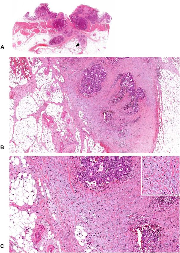
1006           Atlas of the DR categorization

                                                                                                                                                                    Downloaded from https://academic.oup.com/jjco/article/51/6/1004/6225738 by guest on 03 November 2021
Figure 2. Field in the primary tumor to be evaluated for the DR classification. Fibrotic cancer stroma outside the muscularis propria is evaluated on pathological
slides prepared in routine practice. Cancer stroma in the submucosa or those in the muscularis propria are excluded from the evaluation. Fibrotic stroma along
the leading edge of the primary tumors (desmoplastic front) is the area subject to intensive evaluation because both myxoid stroma and keloid-like hyalinized
collagen mostly appear in this lesion. When a tumor nodule exists in the fatty tissue attached to the primary tumor, the fibrotic stroma of the lesion should be
evaluated entirely for the DR classification. (A) Schema and (B) a whole slide image [Hematoxylin–eosin (H&E) stained] of the primary tumor. Area circled with
blue dotted line, desmoplastic front; brown dotted line, estimated lower border of the muscularis propria.

Evaluation criteria for the DR classification                                       (desmoplastic front), where myxoid stroma and keloid-like collagen
                                                                                   most often appear, is the highly important field to be evaluated.
Myxoid stroma and keloid-like collagen are two components of the
                                                                                   When tumor nodules exist in the fatty tissue attached to the primary
DR classification criteria, by which DR is histologically categorized
                                                                                   tumor, the fibrotic stroma of the nodule must be examined in its
into three patterns—immature, intermediate and mature. A summary
                                                                                   entirety. Areas of stroma with localized inflammation suspected of
of the diagnostic criteria is shown in Fig. 1.
                                                                                   being caused by external pathogens, such as that around microscopic
                                                                                   abscesses by intratumoral perforation, are not taken into considera-
                                                                                   tion (Fig. 3).
Site of cancer stroma to be evaluated
Hematoxylin–eosin (H&E) glass slides of the primary tumor pre-
pared in routine pathology practice are used to determine the DR                   Myxoid stroma
category. Fibrotic cancer stroma outside the muscularis propria layer              Myxoid stroma is defined as stroma accompanied by an amorphous
is the area to be evaluated (Fig. 2). Findings in the submucosa and                mucinous substance, which is typically a basophilic or amphophilic,
muscularis propria are not considered in classifying the DR pattern.               vacuolated extracellular material among the collagen fibers (Fig. 4).
The fibrotic stroma along the leading edge of the primary tumor                    The proportion of stromal mucin to cancer stroma varies depending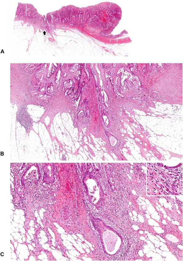
Jpn J Clin Oncol, 2021, Vol. 51, No. 6                    1007

                                                                                                                                                                     Downloaded from https://academic.oup.com/jjco/article/51/6/1004/6225738 by guest on 03 November 2021
                                                                                  Figure 4. Myxoid stroma and keloid-like hyalinized collagen bundles as two
                                                                                  elemental components to categorize DR pattern. The DR pattern is cate-
                                                                                  gorized as immature, intermediate, or mature, based on two site-specific
                                                                                  histological products of fibroblasts—myxoid stroma and hyalinized keloid-
Figure 3. Fibrotic area to be excluded from the DR classification evaluation.      like collagens—that appear exclusively at the desmoplastic front. (A) Myxoid
The DR pattern is evaluated at fibrotic stroma exclusively induced by tumor–       stroma: a fibrotic stroma with an amorphous mucinous stromal substance
stroma interaction. Acute inflammation such as microscopic abscess with            composed of a basophilic or amphophilic extracellular material. (B) Keloid-
intratumoral perforation can evoke a dense fibrosis, but this area should          like collagen bundles are thick, hyalinized bundles of hypocellular collagen
be distinguished from the desmoplastic reaction to be assessed for the DR         with bright eosinophilic hyalinization (arrow) (A, B: Original magnification,
classification. In figure (A), dense fibrotic tissue exists beyond the desmo-        ×40 objective; H&E staining).
plastic front (area surrounded by the triangle symbols), in which a remnant of
microscopic abscess (shown with an asterisk) is observed, thereby indicates
this fibrosis was induced by the external pathogens, rather than tumor–
stroma interaction. (B; ×1.5 objective) is photograph magnifying the part of
the extramural area indicated by the triangles in (A; whole slide image) and
(C; ×40 objective) is that magnifying that part of asterisk. All, H&E staining.

on the individual tumor. For the minimum amount of myxoid
stroma to be judged as immature, the microscopic field of a ×40
objective lens is used as a yardstick (Fig. 5). Generally, the myxoid
stroma exists in relatively limited areas at the desmoplastic front,
even in cases with an immature DR in the CRC (Fig. 6). This
is different from the situation in pancreatic cancer, a notoriously
aggressive desmoplastic tumor with a dismal prognosis, in which
myxoid stroma is more commonly and extensively observed. It is
not difficult to distinguish stromal mucin as a component of myxoid
stroma from stromal mucin leakage associated with mucinous cancer,
since the former is accompanied by other DR components including
fibroblasts, collagen fibers, inflammatory cells and endothelial cells
in stromal mucin (Fig. 7).

Keloid-like collagen
Keloid-like collagen is a thick bundle of hypocellular collagen with
bright eosinophilic hyalinization (Fig. 4). To differentiate it from              Figure 5. Extent of myxoid stroma area to be judged as in the immature
fragmentary collagen fiber, a 20-μm width can be used as a yardstick              DR category. For the minimum amount of myxoid stroma to be judged as
for the minimum thickness of hyalinized collagen (Fig. 8). Although               immature, the microscopic field of a ×40 objective lens is used as a yardstick.
                                                                                  (A) Although there is an area with stromal mucin, as circled with a black dotted
keloid-like collagen appears exclusively at the desmoplastic front, not
                                                                                  line, it does not completely fill the field of a ×40 objective lens, thereby does
in the superficial portion or center of the tumor, there is heterogeneity         not meet the criteria to place it in the immature category; (B–D) Increased
in terms of its anatomical distribution at the desmoplastic front. Some           stromal mucin forms the area of myxoid stroma, which fully occupies the
keloid-like collagen is distributed near tumor nests (Fig. 9), and other          field of a ×40 objective lens with various proportions of stromal mucin-to-
keloid-like collagen is found at the most desmoplastic front, which is            collagen fibers. These DR lesions are classified in the immature category. (A–
slightly distant from the tumor nests (Fig. 10).                                  D) Microscopic fields of a ×40 objective lens (field number, 20; H&E staining).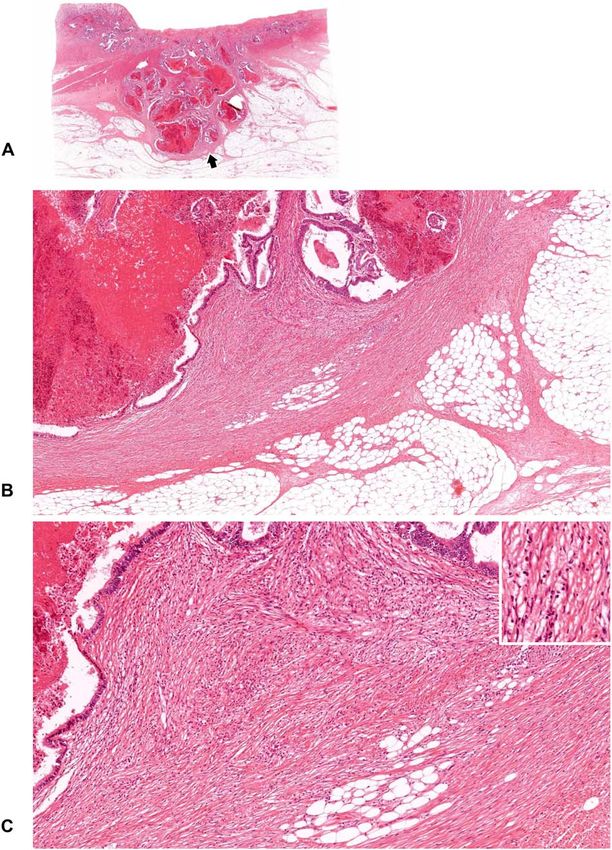
1008 Atlas of the DR categorization
Downloaded from https://academic.oup.com/jjco/article/51/6/1004/6225738 by guest on 03 November 2021
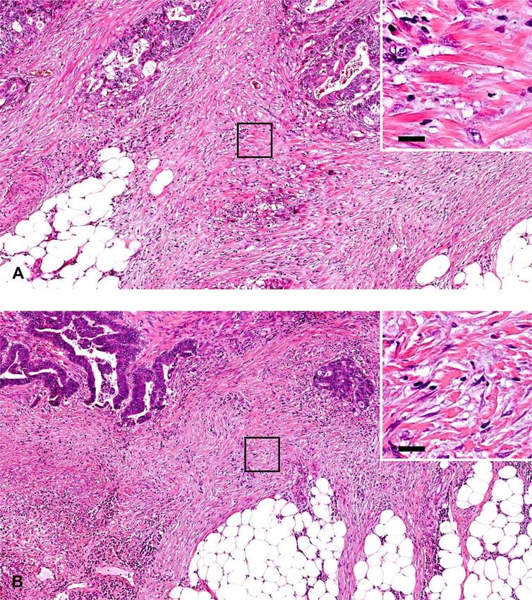
Figure 6. A tumor with immature DR (1). An amorphous and vacuolated, Figure 7. A tumor with immature DR (2). An amorphous, vacuolated,
basophilic, myxoid stroma is observed in some areas within the desmoplastic amphophilic myxoid material extends to the desmoplastic front. Note that
front. Note that there is an intratumoral heterogeneity in the DR pattern, and although the tumor cells produce abundant extracellular mucin, myxoid
in the case of colorectal cancer, myxoid stroma generally occupies a limited stroma can be differentiated from the mucin pool in the stroma. The mucin
area of the desmoplastic front. (B; ×4 objective) and (C; ×10 objective) are leakage is devoid of desmoplastic reaction, whereas the myxoid stroma
photographs magnifying that part of the desmoplastic front indicated by the is intermingled with other desmoplastic components such as fibroblasts,
arrow in (A; whole slide image). Insert in (C), ×20 objective. All, H&E staining. collagen fibers, inflammatory cells and endothelial cells. (B; ×4 objective)
and (C; ×10 objective) are magnified photographs of the portion of the
desmoplastic front indicated by the arrow in (A; whole slide image). Insert
in (C), ×20 objective. All, H&E staining.
Diagnostic flow of DR category
The flowchart of categorizing DR is shown in Fig. 1. Extramural
stroma of the primary tumor is first scanned at low or middle-
Discussion
power magnification to search for myxoid stroma and keloid-like The roles of cancer stroma are complex and multifaceted and can-
collagen. The category of ‘immature’ is designated for the pattern not be defined simply as helpful or harmful to tumors (16,17). In
of DR accompanying myxoid stroma. In cases without myxoid general, tumor stroma is thought to act in a direction of supporting
stroma that satisfy the criteria for the immature DR category, the and promoting the growth of cancer, but it is reported that some
DR pattern is categorized as ‘intermediate’ if the fibrotic stroma stromal components can act to restrain tumor growth (18,19). The
contains keloid-like collagen. Only one focus with such a collagen potential of tumor growth and metastasis may be determined by
bundle is enough for the classification as intermediate. Note that how tumor cells orchestrate stromal cells for their benefit through
DR pattern of the coexistence of the myxoid stroma which fully various preexisting molecular mechanisms for tissue remodeling. In
occupies the field of a ×40 objective lens and keloid-like collagen this regard, although there have been some attempts to categorize
is classified as immature category. DR is classified as ‘mature’ if cancer stroma on the basis of its amount (20,21) or the percentage
there is no myxoid stroma or keloid-like collagen in the extramu- of stroma in a tumor (22,23), it would be difficult to distinguish
ral stroma. Mature DR is typically characterized by fine, multi- between ‘prohost DR’ or ‘protumor DR’ with these morphometric
layered, mature collagen fibers, which may develop to encompass approaches.
tumor nests or develop radially to form the most desmoplastic front Myxoid stroma and keloid-like hyalinized collagen bundles are
(Fig. 11). Immune-cell infiltration status does not affect the DR histological features of fibrotic stroma, supposedly generated by
categorization. Figure 12 exhibits the finding of a desmoplastic front specific subgroups of activated fibroblasts, given that these are not
of a tumor having conspicuous immune cells, in which no myxoid universally observed in the field of DR. These are not malignant
stroma or keloid-like collagen existed, thereby being diagnosed as neoplasm-specific, and they can be generated in other contexts, for
mature DR. example, inflammatory responses to infections, pathogens or soft
Jpn J Clin Oncol, 2021, Vol. 51, No. 6                    1009

                                                                                                                                                                    Downloaded from https://academic.oup.com/jjco/article/51/6/1004/6225738 by guest on 03 November 2021
Figure 8. Size of hyalinized collagen bundles to be judged as keloid-like
collagen, the criterion of the intermediate DR category. Keloid-like collagen
is a thick, hyalinized, collagen bundle, and is recognized at low or middle
magnification. A yardstick of 20 μm can be used for the minimum width of the
bundle to be judged as keloid-like collagen. In cases (A) and (B), fragmentary
collagen fibers are observed at the desmoplastic front, but they are not judged
as keloid-like collagen because the thickness of the fibers is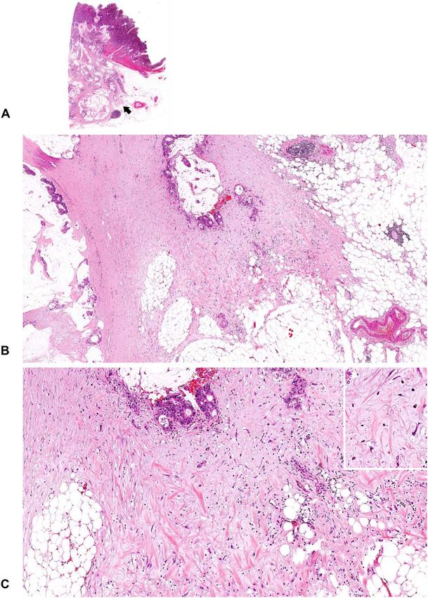
1010           Atlas of the DR categorization

                                                                                                                                                                     Downloaded from https://academic.oup.com/jjco/article/51/6/1004/6225738 by guest on 03 November 2021
                                                                                   Figure 11. A tumor with mature DR (1). Cancer nests are encompassed by
Figure 10. A tumor with intermediate DR (2). Keloid-like collagen bundles
                                                                                   fine, mature collagen fibers, which are developing in a multilayer form. No
appear at the leading edge of the desmoplastic stroma. (B; ×4 objective) and
                                                                                   myxoid stroma or keloid-like collagen bundles are seen. (B; ×4 objective) and
(C; ×10 objective) are magnifications of the area of the desmoplastic front as
                                                                                   (C; ×10 objective) are magnifications of the area of the desmoplastic front as
indicated by the arrow in (A) (whole slide image). Insert in (C), ×20 objective.
                                                                                   indicated by the arrow in (A; whole slide image). Insert in (C), ×20 objective.
All, H&E staining.
                                                                                   All, H&E staining.

validated in a prospective multicenter study [SACURA trial; (10)].                 key clues to understanding molecular mechanisms of favorable clin-
The JCOG1805 will become the first RCT to determine clinical                       ical outcomes associated with mature DR. Rhim et al. have demon-
values of pathological risk factors, including a stromal factor, as                strated that reduced stromal desmoplasia by modulating the hedge-
treatment indicators in high-risk stage II CRC patients.                           hog signaling pathway is associated with undifferentiated histol-
    Future studies will elucidate the underlying molecular back-                   ogy and increased proliferation and metastasis of cancer cells (18).
ground of adverse prognostic results in tumors with non-mature                     Özdemir et al. have observed that myofibroblast depletion leads
DR, which is highly suggestive of a link to the protumor function                  to immunosuppression and increased tumor invasion in transgenic
of the cancer microenvironment that switches indolent tumor cells                  mice with delete αSMA+ myofibroblasts in pancreatic cancer (19).
to cells with a high potential for growth and metastasis. Most                     These findings obviously indicate that some CAF populations and the
likely, one would be transforming growth factor-β (TGF-β) family                   microenvironment induced by them act as a potent barrier for tumor
signaling. For example, thick, hyalinized collagen bundles, which                  growth. In this regard, the heterogeneity of the CAF population may
are observed not only in tumors with intermediate DR but in                        be expressed morphologically as the differential DR pattern, which
most tumors classified as immature DR, are typically observed in                   is associated with tumor differentiation and immune surveillance
keloids, scars that invade adjacent healthy tissue and rarely regress              status. Specifically, mature DR is characterized by a high incidence
over time. As compared with normal fibroblasts, keloid fibrob-                     of well differentiated carcinoma without the dedifferentiation phe-
lasts are associated with increased expression in various growth                   notype (11,13,26) or the conspicuous lymphocytes (11,13,39). The
factors, including TGF-β (36). Since TGF-β is a well-established                   issues to be resolved in the future are to verify the hypothesis that the
inducer of EMT (37), and, reportedly, TGF-β levels in CRC are                      CAF population is different between the mature and non-mature DR
robust predictors of disease relapse in CRC (38), it is presumed that              patterns; the former act as a prohost by restraining tumors, whereas
prometastatic program induced by TGF-β exists in the microenvi-                    the latter act as a protumor by supporting tumor cell growth and
ronment of non-mature DR tumors with keloid-like collagen bun-                     metastasis.
dles.                                                                                  In conclusion, the DR pattern of the desmoplastic front
    In contrast, some previous biological studies showing that some                may be a new stromal prognostic factor to be reported in
stromal components can act to restrain tumor growth may include                    routine pathology practice for CRC patients. Increasing evidence
Jpn J Clin Oncol, 2021, Vol. 51, No. 6                      1011

                                                                                  References
                                                                                  1.    Chen X, Song E. Turning foes to friends: targeting cancer-associated
                                                                                        fibroblasts. Nat Rev Drug Discov. 2019;18:99–115.
                                                                                  2.    Erdogan B, Webb DJ. Cancer-associated fibroblasts modulate growth
                                                                                        factor signaling and extracellular matrix remodeling to regulate tumor
                                                                                        metastasis. Biochem Soc Trans. 2017;45:229–36.
                                                                                  3.    Isella C, Terrasi A, Bellomo SE, et al. Stromal contribution to the colorectal
                                                                                        cancer transcriptome. Nat Genet. 2015;47:312–9.
                                                                                  4.    Calon A, Lonardo E, Berenguer-Llergo A, et al. Stromal gene expres-
                                                                                        sion defines poor-prognosis subtypes in colorectal cancer. Nat Genetics.
                                                                                        2015;47:320–9.
                                                                                  5.    National Comprehensive Cancer Network. NCCN Clinical Practice
                                                                                        Guidelines in Oncology; Colon Cancer Version 3. 2020; (4 June 2020,
                                                                                        date last accessed)..
                                                                                  6.    Labianca R, Nordlinger B, Beretta GD, et al. Early colon cancer: ESMO

                                                                                                                                                                         Downloaded from https://academic.oup.com/jjco/article/51/6/1004/6225738 by guest on 03 November 2021
                                                                                        clinical practice guidelines for diagnosis, treatment, and follow-up. Ann
                                                                                        Oncol 2013;24:vi64–72.
                                                                                  7.    Ueno H, Kanemitsu Y, Sekine S, et al. A multicenter study of the prognostic
                                                                                        value of desmoplastic reaction categorization in stage II colorectal cancer.
                                                                                        Am J Surg Pathol. 2019;43:1015–22.
                                                                                  8.    Nearchou IP, Kajiwara Y, Mochizuki S, Harrison DJ, Caie PD, Ueno H.
                                                                                        Novel internationally verified method reports desmoplastic reaction as the
                                                                                        most significant prognostic feature for disease-specific survival in stage II
                                                                                        colorectal cancer. Am J Surg Pathol. 2019;43:1239–48.
                                                                                  9.    Ishiguro M, Mochizuki H, Tomita N, et al. Study protocol of the SACURA
                                                                                        trial: a randomized phase III trial of efficacy and safety of UFT as adjuvant
                                                                                        chemotherapy for stage II colon cancer. BMC Cancer. 2012;12:281.
                                                                                  10.   Ueno H, Ishiguro M, Nakatani E, et al. Prognostic value of desmoplastic
                                                                                        reaction characterization in stage II colon cancer: prospective validation
                                                                                        in a phase III study (SACURA trial). Br J Cancer. 2021;124:1088–1097.
                                                                                  11.   Ueno H, Jones AM, Wilkinson KH, Jass JR, Talbot IC. Histological
                                                                                        categorisation of fibrotic cancer stroma in advanced rectal cancer. Gut.
                                                                                        2004;53:581–6.
Figure 12. A tumor with mature DR (2). Desmoplastic stroma with relatively        12.   Ueno H, Jones A, Jass JR, Talbot IC. Clinicopathological significance of
abundant lymphocyte infiltration is developing at the leading edge of the                the ’keloid-like’ collagen and myxoid stroma in advanced rectal cancer.
tumor. Neither myxoid stroma nor keloid-like collagen bundles are observed,             Histopathology. 2002;40:327–34.
and the DR pattern is classified as the mature category. (B; ×4 objective) and     13.   Ueno H, Shinto E, Shimazaki H, et al. Histological categorization of
(C; ×10 objective) are magnifications of the area of the desmoplastic front as           desmoplastic reaction: its relevance to the colorectal cancer microenviron-
indicated by the arrow in (A; whole slide image). Insert in (C), ×20 objective.         ment and prognosis. Ann Surg Oncol. 2015;22:1504–12.
All, H&E staining.                                                                14.   Thiery JP, Acloque H, Huang RYJ, Nieto MA. Epithelial-mesenchymal
                                                                                        transitions in development and disease. Cell. 2009;139:871–90.
                                                                                  15.   Ueno H, Shinto E, Kajiwara Y, et al. Prognostic impact of histological
                                                                                        categorisation of epithelial-mesenchymal transition in colorectal cancer.
indicates that it is certainly an essential prognostic determinant                      Br J Cancer. 2014;111:2082–90.
independent of the anatomical extent of disease or conventional                   16.   Mueller MM, Fusenig NE. Friends or foes-bipolar effects of the tumour
tumor factors, and its predictive value for chemotherapy efficacy                       stroma in cancer. Nat Rev Cancer. 2004;4:839–49.
                                                                                  17.   Ronca R, Van Ginderachter JA, Turtoi A. Paracrine interactions of cancer-
will be established by the JCOG1805 trial, which is the first
                                                                                        associated fibroblasts, macrophages and endothelial cells: tumor allies and
RCT incorporating the stromal factor as an inclusion criterion to
                                                                                        foes. Curr Opin Oncol. 2018;30:45–53.
determine the value of adjuvant chemotherapy in CRCs. Further,                    18.   Rhim AD, Oberstein PE, Thomas DH, et al. Stromal elements act to
molecular investigation focusing on the DR classification would                         restrain, rather than support, pancreatic ductal adenocarcinoma. Cancer
lead to better understanding of the heterogeneity of CAFs and                           Cell. 2014;25:735–47.
to the development of strategies for personalized CRC manage-                     19.   Özdemir BC, Pentcheva-Hoang T, Carstens JL, et al. Depletion of
ment.                                                                                   carcinoma-associated fibroblasts and fibrosis induces immunosuppres-
                                                                                        sion and accelerates pancreas cancer with reduced survival. Cancer Cell.
                                                                                        2014;25:719–34.
                                                                                  20.   Halvorsen TB, Seim EVA. Association between invasiveness, inflamma-
Funding                                                                                 tory reaction, desmoplasia and survival in colorectal cancer. J Clin Pathol.
This work was supported by the Practical Research for Innovative                        1989;42:162–6.
Cancer Control from Japan Agency for Medical Research and Devel-                  21.   Japanese Society for Cancer of the Colon and Rectum. Japanese Classifica-
                                                                                        tion of Colorectal Carcinoma (second English edition). Tokyo: Kanehara
opment (AMED) under Grant Number jp20ck0106584.
                                                                                        & Co., Ltd., 2009.
                                                                                  22.   Sis B, Sarioglu S, Sokmen S, Sakar M, Kupelioglu A, Fuzun M. Desmo-
                                                                                        plasia measured by computer assisted image analysis: an independent
Conflict of interest statement                                                           prognostic marker in colorectal carcinoma. J Clin Pathol. 2005;58:
                                                                                        32–8.
The authors declare that there are no conflicts of interest.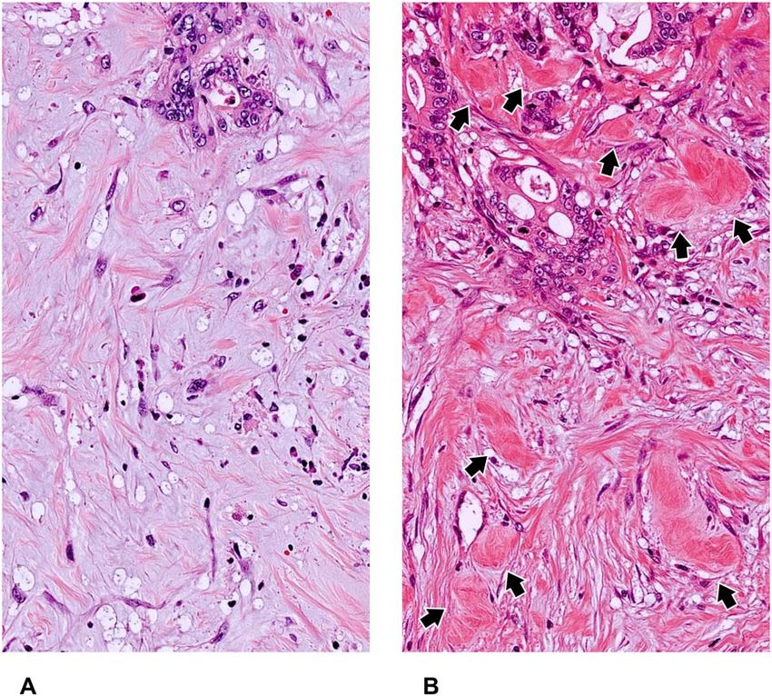
1012            Atlas of the DR categorization

23. Mesker WE, Junggeburt JMC, Szuhai K, et al. The carcinoma-stromal               31. Wang LM, Silva MA, D’Costa Z, et al. The prognostic role of
    ratio of colon carcinoma is an independent factor for survival com-                 desmoplastic stroma in pancreatic ductal adenocarcinoma. Oncotarget.
    pared to lymph node status and tumor stage. Cell Oncol. 2007;29:                    2016;7:4183–94.
    387–98.                                                                         32. Hernández-Ruiz E, Hernández-Muñoz I, Masferrer E, et al. A myxoid
24. Ueno H, Konishi T, Ishikawa Y, et al. Histological categorization of                fibrotic reaction pattern is associated with metastatic risk in cutaneous
    fibrotic cancer stroma in the primary tumor is an independent prog-                 squamous cell carcinoma. Acta Derm Venereol. 2019;99:89–94.
    nostic index in resectable colorectal liver metastasis. Am J Surg Pathol.       33. Cao L, Sun P-L, He Y, Yao M, Gao H. Desmoplastic reaction and
    2014;38:1380–6.                                                                     tumor budding in cervical squamous cell carcinoma are prognostic fac-
25. Zippi M, Toma GD, Minervini G, et al. Desmoplasia influenced recurrence             tors for distant metastasis: a retrospective study. Cancer Manag Res.
    of disease and mortality in stage III colorectal cancer within five years           2020;12:137–44.
    after surgery and adjuvant therapy. Saudi J Gastroenterol. 2017;23:             34. Venook AP, Niedzwiecki D, Lopatin M, et al. Biologic determinants of
    39–44.                                                                              tumor recurrence in stage II colon cancer: validation study of the 12-gene
26. Ueno H, Kanemitsu Y, Sekine S, et al. Desmoplastic pattern at the tumor             recurrence score in cancer and leukemia group B (CALGB) 9581. J Clin
    front defines poor-prognosis subtypes of colorectal cancer. Am J Surg               Oncol. 2013;31:1775–81.
    Pathol. 2017;41:1506–12.                                                        35. Japanese Society for Cancer of the Colon and Rectum. Japanese Classi-

                                                                                                                                                                     Downloaded from https://academic.oup.com/jjco/article/51/6/1004/6225738 by guest on 03 November 2021
27. Ueno H, Sekine S, Oshiro T, et al. Disentangling the prognostic heterogene-         fication of Colorectal, Appendiceal, and Anal Carcinoma (third English
    ity of stage III colorectal cancer through histologic stromal categorization.       edition). Tokyo: Kanehara & Co., Ltd., 2019.
    Surgery. 2018;163:777–83.                                                       36. Bran GM, Goessler UR, Hormann K, Riedel F, Sadick H. Keloids: current
28. Konishi T, Shimada Y, Lee LH, et al. Poorly differentiated clusters predict         concepts of pathogenesis (review). Int J Mol Med. 2009;24:283–93.
    colon cancer recurrence: an in-depth comparative analysis of invasive-          37. Lamoulle S, Xu J, Derynck R. Molecular mechanisms of epithelial–
    front prognostic markers. Am J Surg Pathol. 2018;42:705–14.                         mesenchymal transition. Nat Rev Mol Cell Biol. 2014;15:178–96.
29. Ao T, Kajiwara Y, Yonemura K, et al. Prognostic significance of histolog-       38. Calon A, Espinet E, Palomo-Ponce S, et al. Dependency of colorectal
    ical categorization of desmoplastic reaction in colorectal liver metastases.        cancer on a TGF-beta-driven programme in stromal cells for metastasis
    Virchows Arch. 2019;475:341–8.                                                      initiation. Cancer Cell. 2012;22:571–84.
30. Ao T, Kajiwara Y, Yonemura K, et al. Morphological consistency of               39. Ueno H, Shinto E, Hashiguchi Y, et al. In rectal cancer, the type of
    desmoplastic reactions between the primary colorectal cancer lesion and             desmoplastic response after preoperative chemoradiotherapy is associated
    associated metastatic lesions. Virchows Arch. 2020;477:47–55.                       with prognosis. Virchows Arch. 2015;466:655–63.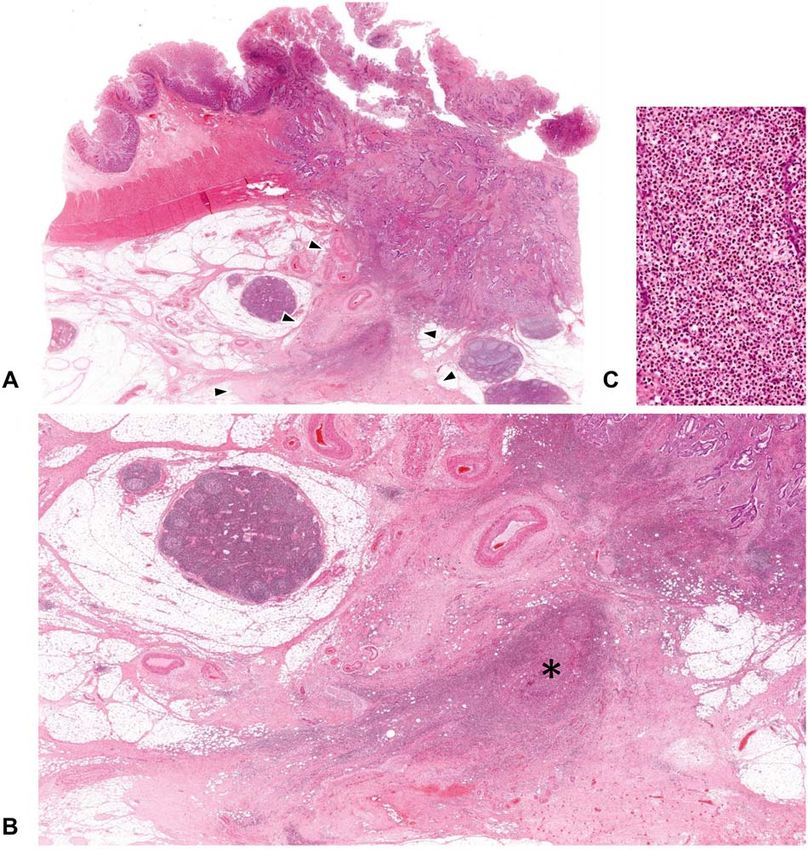
You can also read